Clinical Trial
. 2014 Feb 5;9(2):e87629.
doi: 10.1371/journal.pone.0087629. eCollection 2014.

[¹⁸F]FDG positron emission tomography within two weeks of starting erlotinib therapy can predict response in non-small cell lung cancer patients

Affiliations
Clinical Trial

[¹⁸F]FDG positron emission tomography within two weeks of starting erlotinib therapy can predict response in non-small cell lung cancer patients

Mammar Hachemi et al. PLoS One. .

Abstract

Purpose: The aim of this prospective study was to evaluate whether [¹⁸F]FDG-PET/CT, performed within two weeks of starting erlotinib therapy can predict tumor response defined by RECIST 1.1 criteria after 8 weeks of treatment in patients with inoperable (stage IIIA to IV) non-small cell lung cancer patients.

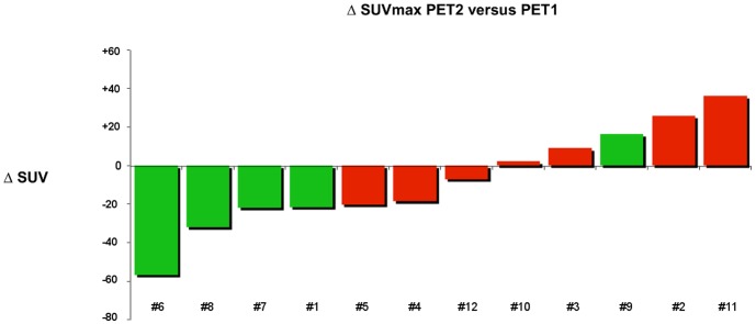
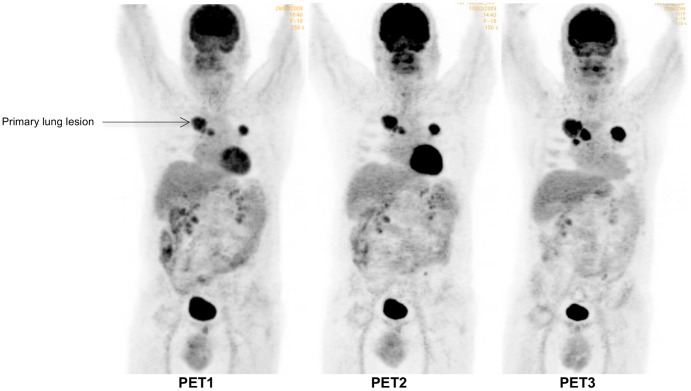
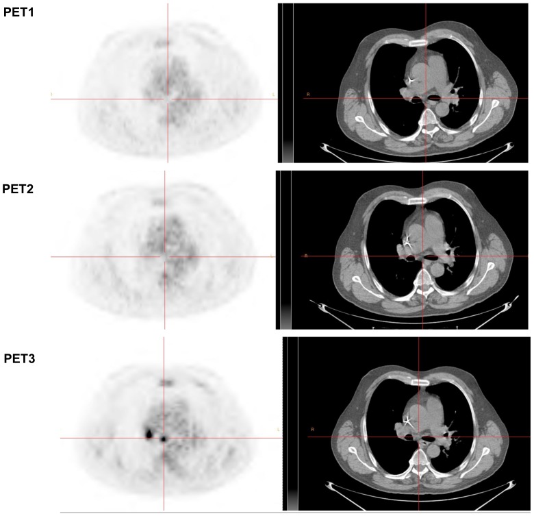
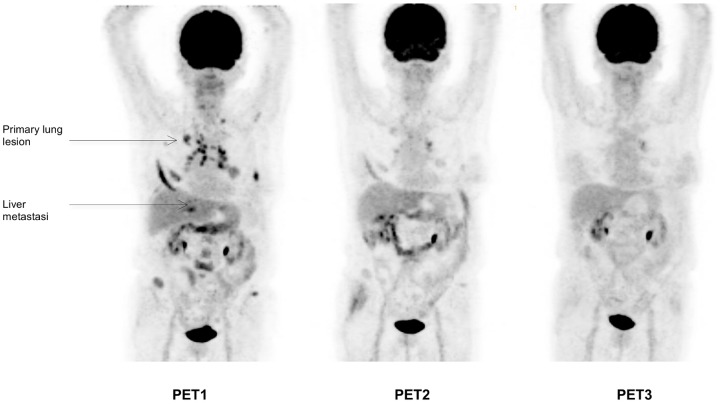
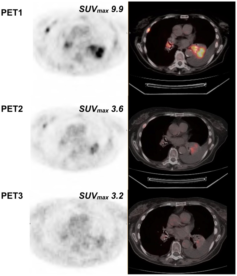
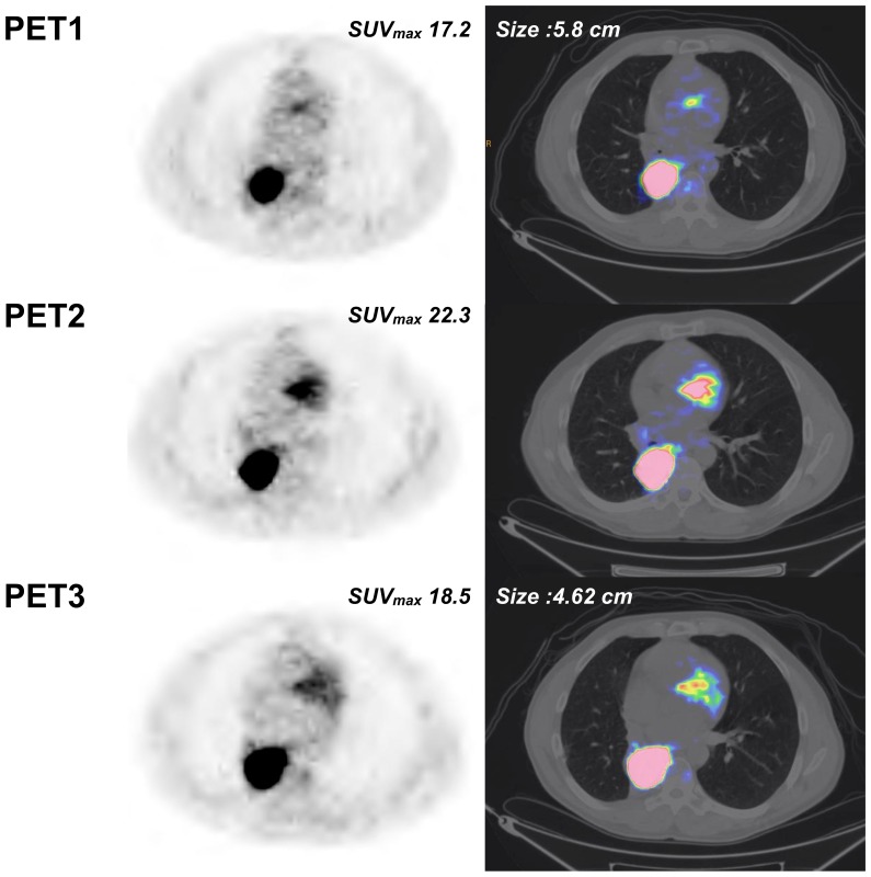
Patients and methods: Three [¹⁸F]FDG-PET/CT scans were acquired in 12 patients before (5±4 days) and after 9±3 days (early PET) and 60±6 days (late PET) of erlotinib therapy. Conventional evaluation, including at least chest CT (baseline versus after 8 weeks of treatment), was performed according to RECIST 1.1 criteria. Change in [¹⁸F]FDG uptake was compared with conventional response, progression-free survival (PFS), and overall survival (OS).

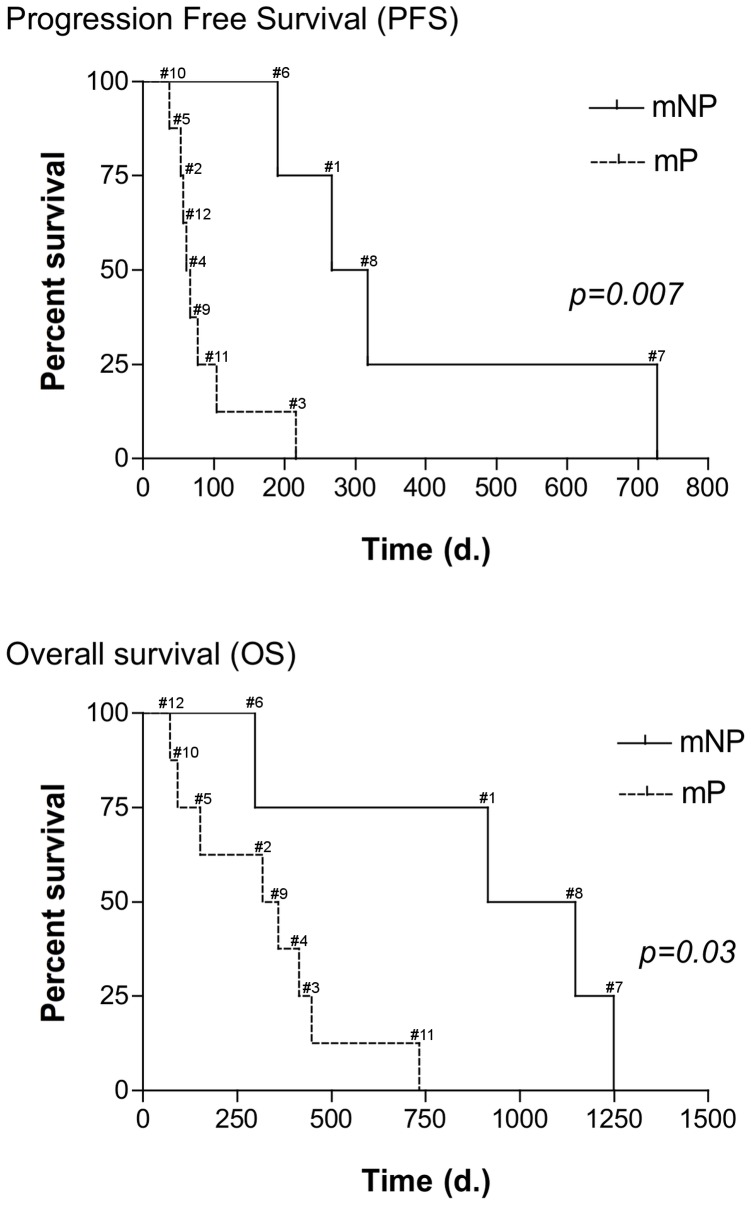
Results: By using ROC analysis, the Area Under the Curve for prediction of metabolic non-progressive disease (mNP) by early PET was 0.86 (95% CI, 0.62 to 1.1; P = 0.04) at a cut-off of 21.6% reduction in maximum Standardized Uptake Value (SUVmax). This correctly classified 11/12 patients (7 with true progressive disease; 4 with true non-progressive disease; 1 with false progressive disease). Non-progressive disease after 8 weeks of treatment according to RECIST 1.1 criteria was significantly more frequent in patients classified mNP (P = 0.01, Fisher's exact test). mNP patients showed prolonged PFS (HR = 0.27; 95% CI, 0.04 to 0.59; P<0.01) and OS (HR = 0.34; 95% CI, 0.06 to 0.84; P = 0.03). Late PET analysis provided concordant results.

Conclusion: Morphologic response, PFS and OS survival in non-small cell lung cancer patients can be predicted by [¹⁸F]FDG-PET/CT scan within 2 weeks after starting erlotinib therapy.


Conflict of interest statement

Competing Interests: The authors have declared that no competing interests exist.

Figures

Figure 1
Figure 1. Percentage change in SUVmax on 18F-FDG PET/CT (cut-off: −21.6%) within 2 weeks of starting erlotinib therapy in relation to conventional imaging response.
Each red or green bar represents a patient NP or P, respectively.
Figure 2
Figure 2. Example of a progressive patient on PET (mP) and conventional imaging.
Progressive patient with right upper lobe NSCLC associated with médiastinal lymphadenopathy, lung and bone metastases (patient #2). Sum of the SUVmax of the 5 most hypermetabolic lesions (2 lung lesions, 2 mediastinal lymph nodes, one hilar lesion) were 35.2, 44.3 (+26%) and 59.9 (+70%) for PET1, PET2 (% versus PET1) and PET3 (% versus PET1), respectively. Based on a SUVmax cut-off value of −21.6, the patient was classified as mP on PET2, in accordance with RECIST evaluation on CT scan (performed 57 days after starting erlotinib). mP was confirmed on PET3 with the appearance of a new lesion (subcarinal adenopathy) and a 70% increase of SUVmax.
Figure 3
Figure 3. New subcarinal adenopathy on PET3 (same patient as Figure 2).
Figure 4
Figure 4. Example of an mNP patient.
Non-progressive patient with right upper lobe NSCLC associated with mediastinal lymphadenopathy, lung, liver and bone metastases (patient #6). Sum of the SUVmax of the 5 most hypermetabolic lesions (2 lung lesions, 2 mediastinal lymph nodes, one liver lesion) were 45.6, 19.7 (−56.7%) and 12.7 (−72%) for PET1, PET2 (% versus PET1) and PET3 (% versus PET1), respectively. Based on a SUVmax cut-off value of −21.6, the patient was classified as mNP on PET2, in accordance with RECIST evaluation on CT scan (performed 58 days after starting erlotinib). mNP was confirmed on PET3 with almost complete extinction of the various lesions and a 72% decrease of SUVmax.
Figure 5
Figure 5. Example of left lower lobe pulmonary target lesion (same patient as Figure 4).
Figure 6
Figure 6. Example of a patient with discordant PET2 and conventional imaging.
Patient with right upper lobe NSCLC associated with subcarinal lymphadenopathy and ipsilateral lung metastasis (patient #9). Sum of the SUVmax of the most hypermetabolic lesions (2 lung lesions, 1 mediastinal lymph node) were 25.2, 29.3 (+16.3%) and 23.8 (−5.4%) for PET1, PET2 (% versus PET1) and PET3 (% versus PET1), respectively. Based on a SUVmax cut-off value of −21.6, the patient was classified as mP on PET2, in contrast with RECIST evaluation on CT scan (performed 71 days after starting erlotinib). This patient was subsequently reclassified as mNP on PET3 in accordance with RECIST evaluation with a 5.4% decrease of SUVmax (cut-off: 18.5%).
Figure 7
Figure 7. Kaplan-Meier estimates of PFS and OS.
No statistically significant difference (P = 0.007) in PFS was observed between metabolic non-progressive (mNP) patients (median PFS, 292 days ; range, 190–727) and metabolic (mP) progressive patients (median PFS, 64 days ; range: 37–216). Improved PFS in non-progressive patients was associated with prolonged OS (mNP; n = 4; median OS: 1031 days ; 296 to 1249 days versus mP; n = 8 ; 337, 5 days ; 71 to 734 days) (HR, 0.34; 95% CI, 0.06 to 0.84; P = 0.03).

References

    1. Ferlay J, Parkin DM, Steliarova-Foucher E (2010) Estimates of cancer incidence and mortality in Europe in 2008. Eur J Cancer 46: 765–781. - PubMed
    1. Jemal A, Bray F, Center MM, Ferlay J, Ward E, et al. (2011) Global cancer statistics. CA Cancer J Clin 61: 69–90. - PubMed
    1. Chemotherapy in non-small cell lung cancer: a meta-analysis using updated data on individual patients from 52 randomised clinical trials. Non-small Cell Lung Cancer Collaborative Group. BMJ 311: 899–909. - PMC - PubMed
    1. Schiller JH, Harrington D, Belani CP, Langer C, Sandler A, et al. (2002) Comparison of four chemotherapy regimens for advanced non-small-cell lung cancer. N Engl J Med 346: 92–98. - PubMed
    1. Lynch TJ, Bell DW, Sordella R, Gurubhagavatula S, Okimoto RA, et al. (2004) Activating mutations in the epidermal growth factor receptor underlying responsiveness of non-small-cell lung cancer to gefitinib. N Engl J Med 350: 2129–2139. - PubMed

Publication types

MeSH terms