. 2014 Feb 4;9(2):e87858.
doi: 10.1371/journal.pone.0087858. eCollection 2014.

Loss of Igfbp7 causes precocious involution in lactating mouse mammary gland

Affiliations

Loss of Igfbp7 causes precocious involution in lactating mouse mammary gland

Sumanta Chatterjee et al. PLoS One. .

Abstract

Background: Insulin like growth factors (IGFs) and their binding proteins (IGFBPs) are secreted peptides that play major roles in regulating the normal development and maturation of mammary gland. While Igfbp7 has been shown to decrease breast tumor growth, its role in regulating the normal mammary gland development has not been studied. To this end, we generated Igfbp7-null mice and examined the development and maturation of mammary glands in the virgin, pregnant and lactating animals.

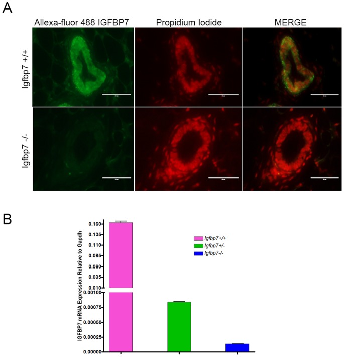
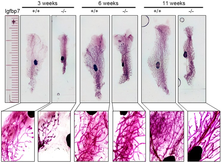
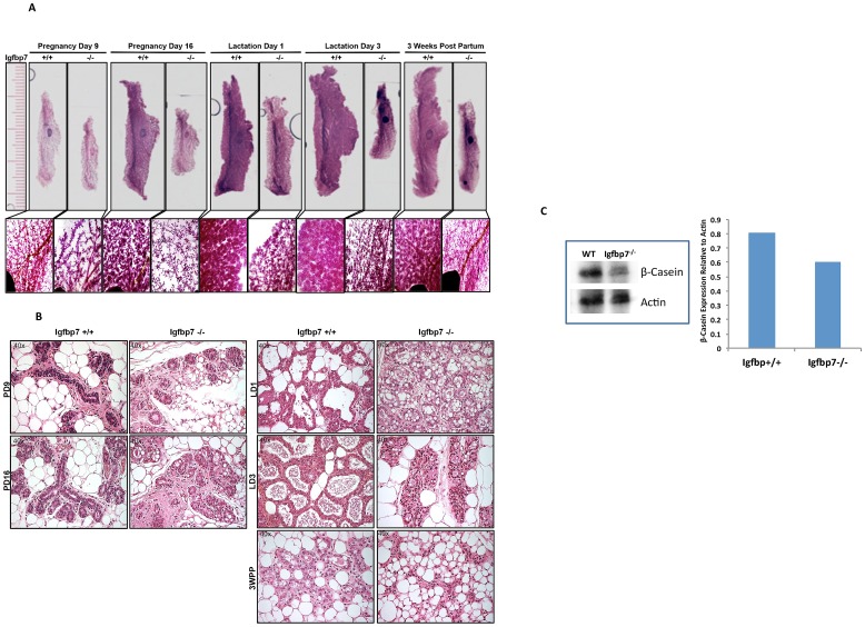
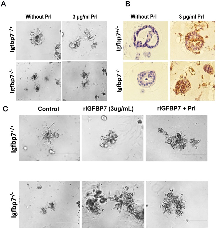
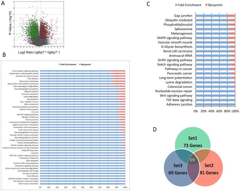
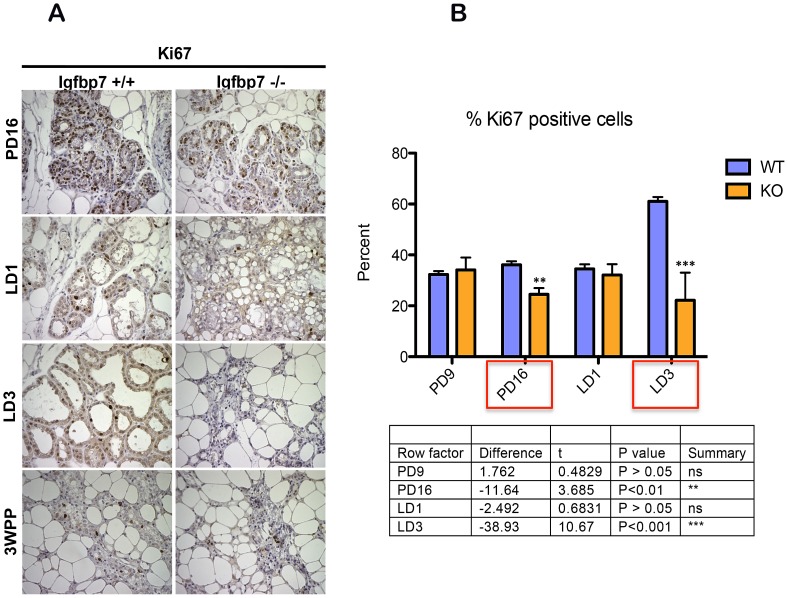
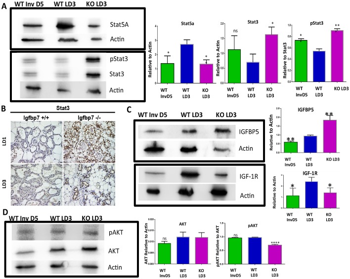
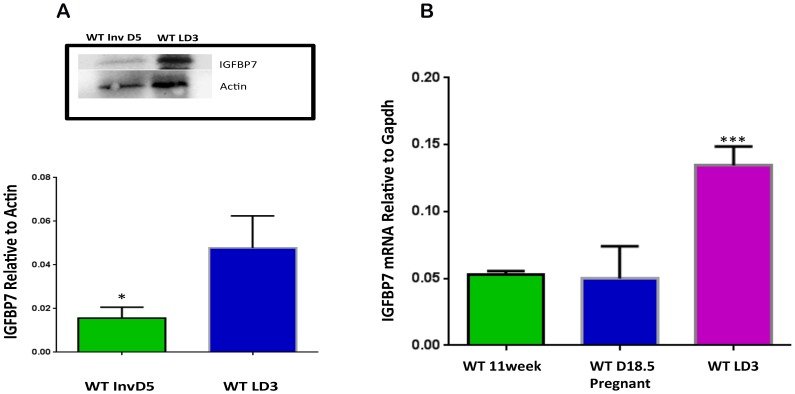
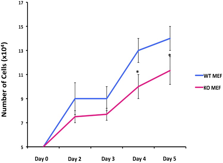
Results: We report here that loss of Igfbp7 significantly retards mammary gland development in the virgin animals. More significantly, the pregnant Igfpb7-null glands contained fewer alveolar structures and that during lactation these glands exhibit the morphological changes that are associated with involution. The transcriptome profile of the Igfbp7-null glands on the lactation day 3 revealed a distinct involution-related gene signature compared to the lactating WT glands. Interestingly, we found that the lactating Igfbp7-null glands exhibit increased expression of Stat3 and enhanced activation of (phosphorylated) Stat3, combined with decreased expression of Stat5 suggesting that the absence of Igfbp7 accelerates the onset of involution. We also found that in absence of Igfpb7, the lactating glands contain increased Igfbp5 protein along with decreased expression of IGF-1 Receptor and Akt activation. Finally, we show that during the normal course of involution, Igfbp7 expression is significantly decreased in the mammary gland.

Conclusion: Our data suggest that loss of Igfbp7 induces precocious involution possibly through diminished cell survival signals. Our findings identify Igfbp7 as major regulator of involution in the mammary gland.


Conflict of interest statement

Competing Interests: The authors have declared that no competing interests exist.

Figures

Figure 1
Figure 1. Igfbp7−/− mice do not show expression of Igfbp7 mRNA or protein.
(A) Inguinal mammary glands from 11 week old virgin female mice were extracted from the wild-type mice (WT) or the knockout animals (Igfbp7−/−) and where fixed and stained with an antibody specific to Igfbp7 (green fluorescent color) and with propidium iodide (red color) to distinguish the nucleus of cells. As seen, Igfbp7−/− glands do not show detectable expression of Igfbp7 protein. (B) The Igfbp7 transcript expression was measured using quantitative real time PCR. RNA was extracted from the 11-week-old inguinal WT or Igfbp7−/− female mice and turned into cDNA. The transcript expression of Igfbp7 was quantified relative to the Gapdh transcript levels. As can be seen the Igfbp7 transcripts are at limit detection in the Igfbp7−/− glands. Each data point is the average of 3 independent experiments.
Figure 2
Figure 2. Mammary gland development is retarded in the virgin and adult Igfbp7−/− female mice.
The inguinal glands from the wild-type (+/+) or the Igfbp7-null mice (−/−) were removed, fixed and stained with carmine alum during the different stages of the mammary gland development. Whole mount slides were visualized at 2X magnification and a ruler was photographed at the same magnification as reference (longer marks are in centimeters and shorter marks are in millimeters). The insert pictures were obtained at 4X the magnification. As can be seen, the Igfbp7−/− glands are smaller and less dense at 3-week or 11-week old, pregnant and lactating glands compared to the wild-type glands.
Figure 3
Figure 3. Loss of Igfbp7 decreases the alveolar density of the mammary gland during the pregnancy and lactation.
(A) Whole mount slides were prepared from inguinal glands extracted from the wild-type (+/+) or the Igfbp7-null (−/−) female mice during pregnancy days (PD) 9 and 16 and lactation days (LD) 1 and 3 as well as 3 weeks port partum (3WPP). The slides were photographed at 2x the magnification and a ruler was also photographed as a point of reference (the longer marks are in centimeters and the shorter marks are in millimeters). The insert pictures were obtained at 4x the magnification to allow more detailed analysis of the mammary structures. As demonstrated, loss of Igfbp7 leads to marked decrease in the alveolar densities in the mammary glands at each of the developmental time points studied. (B) H&E stained sections were prepared from formalin fixed paraffin-embedded mammary gland on the PD6, PD16, LD1, LD3, and 3WPP. As can be seen, alveolar development is completely defective in the Igfbp7−/− glands during pregnancy and lactation. The black arrows point to typical lobular structures in the WT or the Igfbp7−/− glands. Compared to the WT, the Igfbp7−/− glands contain smaller and partially closed alveolar sacks. Interestingly on the lactation day 3 the alveolar structures in the Igfbp7−/− glands resemble the WT glands at 3WPP. (C) Western blot analysis showing decreased β-casein expression in the Igfbp7−/− glands. Total protein was extracted from WT or Igfbp7−/− glands on the lactation day 3, and size fractionated through western blots and the β-casein protein expression was determined and quantified relative to β-actin. One representative Western Blot is shown and the graph shows average of 2 independent protein extracts. As can be seen Igfbp7−/− glands contain slightly less β-casein protein.
Figure 4
Figure 4. Igfbp7−/− mammary epithelial cells show defective alveolar differentiation in matrigel.
Inguinal glands from 11-week old virgin WT (Igfbp7+/+) or Igfbp7−/− mice were extracted and made into single-cell suspension were placed in matrigel cultures for 5 days. (A) To induce alveolar development, matrigel cultures were treated with 30 ng/mL of Prolactin (Prl) for two days. As can be seen WT cells developed alveolar structures that in the presence of Prl differentiated into multi-lobular structures. Interestingly however, the Igfbp7−/− cells formed much smaller alveolar structures that failed to develop into multi-lobular structure in the presence of Prl. (B) To examine if in presence of Prl, the WT or the Igfbp7−/− cells can differentiate into milk-producing cells, the matrigel cultures were fixed and sectioned, and the expression of β-casein protein was determined immunohistochemically (brown staining). As shown, WT and Igfbp7−/− cells can differentiate into milk-producing cells. (C) To examine if recombinant IGFBP7 (rIGFBP7) can restore the lobular differentiation ability of the Igfbp7−/− mammary cells, matrigel cultures were set up as in (A) and rIGFBP7 (3 µg/mL) was added to the matrigel cultures for 2 days. As shown, the addition of rIGFBP7 is sufficient to cause the Igfbp7−/− mammary cells to form multi-lobular structures similar to the WT cells. The addition of rIGFBP7 and Prl did not yield any synergistic affects. These data suggest that alveolar differentiation defects may be due cell autonomous effects of Igfbp7 loss.
Figure 5
Figure 5. The Igfbp7−/− transcriptome is enriched for involution-associate gene signature.
(A) The transcriptome profiles of the Wild-Type (WT) and the Igfbp7−/− glands on the lactation day 3 were analyzed using the RNA-Seq technique. Differentially expressed transcripts were identified by imposing a minimum of 1.5 fold change in the transcript expression level and a P-value of 0.05 or lower and are depicted on a Volcano plot. (B and C) The differentially expressed transcripts in the Igfbp7−/− mammary epithelial cells (MECs) are categorized into the different KEGG Signaling Pathways using the DAVID Bioinformatics Resources. The fold enrichment scores are calculated based on the number of differentially regulated genes that belong to a particular KEGG signaling pathway out of the total gene set. The Benjamini statistics is used to identify the statistically significant gene enrichment for specific KEGG signaling pathways. Using this analysis the down regulated transcripts were found to be enriched in extracellular matrix receptor interaction, focal adhesion and cell adhesion molecules. The up regulated transcripts in the Igfpb7 −/− MECs showed enrichment for the WNT, TGF beta and adherens junction proteins. (D) To examine if the differentially regulated transcripts in the Igfbp7 −/− MECs show enrichment for involution-related genes, we first obtained a common involution gene signature by comparing 3 publically available transcriptome profile of involuting glands. Set 1 is from , Set 2 is from , Set 3 is from . The Ven diagram depicts a 72 gene-set that is common among all 3 data sets.
Figure 6
Figure 6. Igfbp7−/− glands show decreased proliferation index.
(A) Inguinal mammary glands were isolated from wild-type (Igfbp7+/+) or the Igfbp7 −/− mice on pregnancy days 9 (PD9) and 16 (PD16) as well as on lactation days 1 (LD1) and 3 (LD3) and at 3 weeks postpartum (3WPP). Sections from the formalin fixed and paraffin embedded glands were used to detect the expression of Ki-67 protein by Immunohistochemical stains. As can be seen, the Igfbp7 −/− glands show decreased number of cells positive for the expression of Ki-67. (B) The proliferation index of the WT and the Igfbp7 −/− glands are determined by examining the nuclear expression of Ki-67 protein in over 600 nuclei in the slide sections. The proliferation index is calculated by obtaining the percentage of Ki-67 nuclei positive cells in each section. The average values obtained from 3 different fields and the standard deviations are plotted again the developmental stage of the mammary glands. P-values were obtained by performing two-tailed T-tests and provided in a table under the graph.
Figure 7
Figure 7. Lactating Igfbp7−/− glands exhibit accelerated involution.
To determine if Igfbp7 −/− glands exhibit molecular changes that are the hallmark of involution process we prepared protein extracts from the Wild Type (WT) or the Igfbp7 −/− glands on lactation day 3 (WT LD3, KO LD3 respectively) or from WT lactating glands were weaned for 5 days (WT Inv D5) to induce post-lactational involution. The expression of Stat5a, Stat3, phospho Stat3 (pStat3), AKT, pAKT, Igfbp5, and IGF-1R proteins was determined by Western Blots. (A, C–D) Representative Western Blots are shown. The protein expression levels for Stat5a and Stat3, AKT, Igfbp5, and IGF-1R have been normalized to beta actin expression while the expression of pStat3, pAKT have been normalized to total corresponding protein expression in each sample. The bar graphs show the average expression obtained from of 3 independent protein extracts and the statistical significance was calculated based on two-tailed t-test (*P = <0.05, **p<0.005, ***P<0.0005). As shown, lactating KO LD3 glands show elevated Stat3, pSTat3, and Igfbp5, along with decreased Stat5a, IGF-1R, and pAKT; indicating that Igbp7 −/− glands are undergoing involution. (B) Immunohistochemical staining was used to detect the expression of Stat3 in tissue sections obtained from WT or Igbp7 −/−glands on lactation days 1 or 3. As shown, expression of Stat3 can be detected as early as the first day of lactation in the Igbp7 −/− glands while WT glands show detectable expression of Stat3.
Figure 8
Figure 8. Igfbp7 expression is substantially decreased during involution in the mammary gland.
(A) To ascertain if expression of Igfbp7 correlates with involution, protein extracts were prepared from the Wild Type glands on the lactation day 3 (WT LD3) or involuting WT glands (weaned for 5 days, WT Inv D5) and the expression of Igfbp7 protein was examined using Western blots. A representative blot is shown and average Igfbp7 expression was obtained from 3 independent protein extracts and depicted in the bar graph. As shown, Igfbp7 expression is dramatically decreased during involution (*P<0.05). (B) Igfbp7 −/− transcript expression was examined using qPCR assays using RNA extracted from Wild Type 11 week-old virgin (WT 11Week), day 18.5 pregnant (WT D18.5 pregnant) or on lactation day 3 glands. The average transcript expressions obtained from 3 independent RNA samples are shown. While Igfbp7 expression in the mammary gland does not show significant change during pregnancy, its expression is substantially increased during lactation. Statistical analysis was done through two-tailed t-test (***p<0.0005).
Figure 9
Figure 9. Igfbp7 −/− fibroblasts show decreased proliferation potential.
To determine if Igfbp7 can affect cell proliferation in normal cells, fibroblasts were derived from the WT or Igfbp7 −/− embryos (WT MEF or KO MEF) and their expansion in tissue culture was examined. 5x10⁴-starting cells were placed in parallel cultures (day 0) and cell numbers on the indicated days were determined. Average cell counts obtained from 3 independent fibroblast lines were plotted against the day of collection. As can be seen, starting from day 4 the Igfbp7 −/− fibroblast show statistically decreased expansion potential (two tailed t-test, *P<0.05).

Similar articles

Cited by

References

    1. Hennighausen L, Robinson GW (2001) Signaling pathways in mammary gland development. Dev Cell 1: 467–475. - PubMed
    1. Bocchinfuso WP, Korach KS (1997) Mammary gland development and tumorigenesis in estrogen receptor knockout mice. J Mammary Gland Biol Neoplasia 2: 323–334. - PubMed
    1. Howlin J, McBryan J, Martin F (2006) Pubertal mammary gland development: insights from mouse models. J Mammary Gland Biol Neoplasia 11: 283–297. - PubMed
    1. Humphreys RC, Lydon JP, O'Malley BW, Rosen JM (1997) Use of PRKO mice to study the role of progesterone in mammary gland development. J Mammary Gland Biol Neoplasia 2: 343–354. - PubMed
    1. Brisken C, Park S, Vass T, Lydon JP, O'Malley BW, et al. (1998) A paracrine role for the epithelial progesterone receptor in mammary gland development. Proc Natl Acad Sci U S A 95: 5076–5081. - PMC - PubMed

Publication types

MeSH terms

Substances