. 2014 Feb 4;9(2):e88005.
doi: 10.1371/journal.pone.0088005. eCollection 2014.

Role of docosahexaenoic acid treatment in improving liver histology in pediatric nonalcoholic fatty liver disease

Affiliations

Role of docosahexaenoic acid treatment in improving liver histology in pediatric nonalcoholic fatty liver disease

Valerio Nobili et al. PLoS One. .

Abstract

Introduction: Nonalcoholic fatty liver disease (NAFLD) is one of the most important causes of liver-related morbidity and mortality in children. Recently, we have reported the effects of docosahexaenoic acid (DHA), the major dietary long-chain polyunsaturated fatty acids, in children with NAFLD. DHA exerts a potent anti-inflammatory activity through the G protein-coupled receptor (GPR)120. Our aim was to investigate in pediatric NAFLD the mechanisms underlying the effects of DHA administration on histo-pathological aspects, GPR120 expression, hepatic progenitor cell activation and macrophage pool.

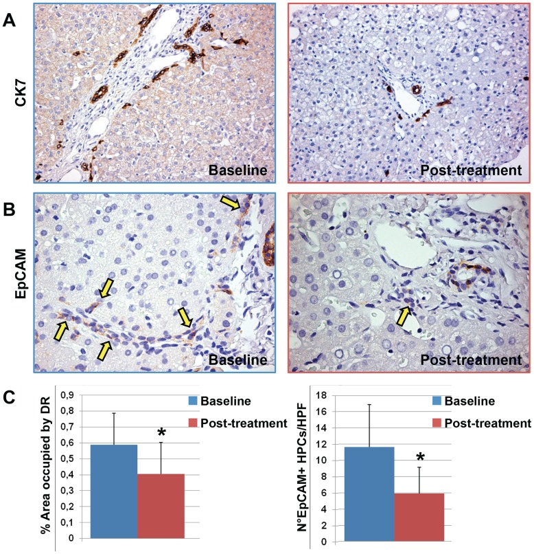
Patients and methods: 20 children with untreated NAFLD were included. Children were treated with DHA for 18 months. Liver biopsies before and after the treatment were analyzed. Hepatic progenitor cell activation, macrophage pool and GPR120 expression were evaluated and correlated with clinical and histo-pathological parameters.

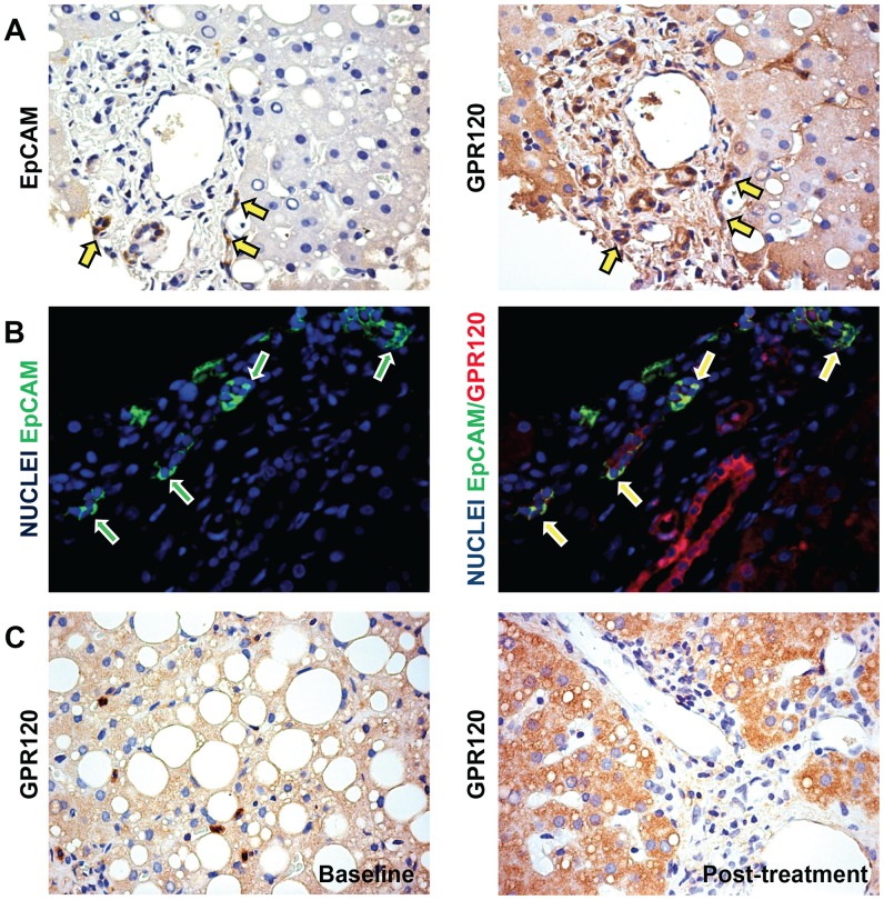
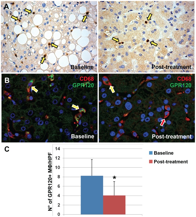
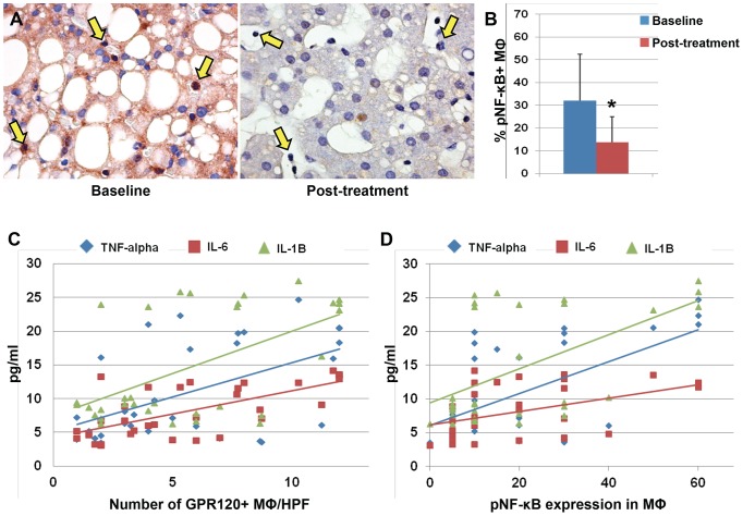
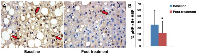
Results: GPR120 was expressed by hepatocytes, liver macrophages, and hepatic progenitor cells. After DHA treatment, the following modifications were present: i) the improvement of histo-pathological parameters such as NAFLD activity score, ballooning, and steatosis; ii) the reduction of hepatic progenitor cell activation in correlation with histo-pathological parameters; iii) the reduction of the number of inflammatory macrophages; iv) the increase of GPR120 expression in hepatocytes; v) the reduction of serine-311-phosphorylated nuclear factor kappa B (NF-κB) nuclear translocation in hepatocytes and macrophages in correlation with serum inflammatory cytokines.

Conclusions: DHA could modulate hepatic progenitor cell activation, hepatocyte survival and macrophage polarization through the interaction with GPR120 and NF-κB repression. In this scenario, the modulation of GPR120 exploits a novel crucial role in the regulation of the cell-to-cell cross-talk that drives inflammatory response, hepatic progenitor cell activation and hepatocyte survival.


Conflict of interest statement

Competing Interests: The authors have declared that no competing interests exist.

Figures

Figure 1
Figure 1. Immunohistochemistry for Cytokeratin(CK) 7 and EpCAM in liver biopsies of pediatric NAFLD patients.
A) at the beasline, pediatric NAFLD biopsies were characterized by a prominent expansion of hepatic progenitor cell (HPC) pool and the presence of reactive ductules at the periphery of portal spaces. After DHA treatment, a minimal involvement of the HPC compartment was present. Original magnification 20×. B) EpCAM-positive HPCs (arrows) were significant reduced by DHA treatment in comparison with the biopsies at the baseline. Original magnification 40×. C) Histograms show the reduction of HPC expansion (ductular reaction extension) and activation (number of EpCAM+ cells) after DHA treatment. Data are shown as Means ± Standard Deviation. * = p<0.05.
Figure 2
Figure 2. GPR120 expression by HPC and Hepatocytes in pediatric NAFLD patients.
A) Immunohistochemistry for EpCAM and GPR120 in serial sections of pediatric NAFLD biopsies. CK-7-positive HPCs highly express GPR120 (arrows). B) Double immunofluorescence for EpCAM (green) and GPR120 (red) in NAFLD biopsies. Nuclei are shown in blue. Numerous EpCAM-positive progenitor cells co-express GPR120 (yellow cells: yellow arrows) confirming the findings obtained through immunohistochemistry in serial sections. C) Immunohistochemistry for GPR120 in sections of pediatric NAFLD biopsies before and after DHA treatment. A significant increase of GPR120-positive hepatocytes was clearly seen after DHA treatment. Original magnification: 40×.
Figure 3
Figure 3. GPR120 expressions by Macrophages in pediatric NAFLD patients.
A) Immunohistochemistry for GPR120 in sections of pediatric NAFLD biopsies. B) Double immunofluorescence for CD68 (red) and GPR120 (green) in NAFLD biopsies. Nuclei are shown in blue. CD68-positive macrophages express GPR120 (yellow arrows). Original magnification: 40×. C) The histogram shows the number of GPR120-positive macrophages per high power field (HPF) at the baseline and after DHA treatment. A significant reduction of GPR120-positive macrophages was seen after DHA treatment. * = p<0.05.
Figure 4
Figure 4. Nuclear expression of phosphorylated (p) NF-κB by Macrophages in pediatric NAFLD patients.
A) Immunohistochemistry for serine-311-phosphorylated NF-κB (pNF-κB) in pediatric NAFLD biopsies before and after DHA treatment. In parallel with GPR120 expression, the DHA treatment determined a reduction of pNF-κB expression and its nuclear translocation in macrophages (A, yellow arrows). Original magnification: 40×. B) Histogram shows the significant reduction of pNF-κB nuclear translocation in macrophages after DHA treatment. Data are shown as Means ± Standard Deviation. * = p<0.05. C–D) The number of GPR120-positive macrophages (C) and the pNF-κB nuclear expression in macrophages. (D) were correlated with serum level of inflammatory cytokines.
Figure 5
Figure 5. Nuclear expression of phosphorylated (p) NF-κB by hepatocytes in pediatric NAFLD patients.
A) Immunohistochemistry for pNF-κB in pediatric NAFLD biopsies demonstrates the reduction of pNF-κB in hepatocyte nuclei (red arrows) after DHA treatment. B) DHA treatment determined a significant reduction of pNF-κB expression and its nuclear translocation in hepatocytes. Data are shown as Means ± Standard Deviation. * = p<0.05.

References

    1. Alisi A, Feldstein AE, Villani A, Raponi M, Nobili V (2012) Pediatric nonalcoholic fatty liver disease: a multidisciplinary approach. Nat Rev Gastroenterol Hepatol 9: 152–161. - PubMed
    1. Carpino G, Renzi A, Onori P, Gaudio E (2013) Role of Hepatic Progenitor Cells in Nonalcoholic Fatty Liver Disease Development: Cellular Cross-Talks and Molecular Networks. Int J Mol Sci 14: 20112–20130. - PMC - PubMed
    1. Nobili V, Carpino G, Alisi A, Franchitto A, Alpini G, et al. (2012) Hepatic progenitor cells activation, fibrosis and adipokines production in pediatric nonalcoholic fatty liver disease. Hepatology 56: 2142–2153. - PubMed
    1. Masterton GS, Plevris JN, Hayes PC (2010) Review article: omega-3 fatty acids - a promising novel therapy for non-alcoholic fatty liver disease. Aliment Pharmacol Ther 31: 679–692. - PubMed
    1. Parker HM, Johnson NA, Burdon CA, Cohn JS, O'Connor HT, et al. (2012) Omega-3 supplementation and non-alcoholic fatty liver disease: a systematic review and meta-analysis. J Hepatol 56: 944–951. - PubMed

Publication types

MeSH terms