. 2014 Feb 5;9(2):e88276.
doi: 10.1371/journal.pone.0088276. eCollection 2014.

Identification and glycerol-induced correction of misfolding mutations in the X-linked mental retardation gene CASK

Affiliations

Identification and glycerol-induced correction of misfolding mutations in the X-linked mental retardation gene CASK

Leslie E W LaConte et al. PLoS One. .

Abstract

The overwhelming amount of available genomic sequence variation information demands a streamlined approach to examine known pathogenic mutations of any given protein. Here we seek to outline a strategy to easily classify pathogenic missense mutations that cause protein misfolding and are thus good candidates for chaperone-based therapeutic strategies, using previously identified mutations in the gene CASK. We applied a battery of bioinformatics algorithms designed to predict potential impact on protein structure to five pathogenic missense mutations in the protein CASK that have been shown to underlie pathologies ranging from X-linked mental retardation to autism spectrum disorder. A successful classification of the mutations as damaging was not consistently achieved despite the known pathogenicity. In addition to the bioinformatics analyses, we performed molecular modeling and phylogenetic comparisons. Finally, we developed a simple high-throughput imaging assay to measure the misfolding propensity of the CASK mutants in situ. Our data suggests that a phylogenetic analysis may be a robust method for predicting structurally damaging mutations in CASK. Mutations in two evolutionarily invariant residues (Y728C and W919R) exhibited a strong propensity to misfold and form visible aggregates in the cytosolic milieu. The remaining mutations (R28L, Y268H, and P396S) showed no evidence of aggregation and maintained their interactions with known CASK binding partners liprin-α3 Mint-1, and Veli, indicating an intact structure. Intriguingly, the protein aggregation caused by the Y728C and W919R mutations was reversed by treating the cells with a chemical chaperone (glycerol), providing a possible therapeutic strategy for treating structural mutations in CASK in the future.


Conflict of interest statement

Competing Interests: The authors have declared that no competing interests exist.

Figures

Figure 1
Figure 1. Domain location and conservation of five CASK mutations.
A. Five CASK XLMR mutations are shown in reference to CASK’s domain structure. B. A comparison of the five mutation sites in CASK orthologs from nine species. Conserved residues, white. Residues identical to the mutation, black. Residues that differ from the wild-type and mutant hCASK sequence are gradiently shaded to indicate their similarity to the native hCASK residue.
Figure 2
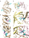
Figure 2. Structural modeling of four CASK mutations.
Dotted lines indicate contacts. A. CAMK domain of CASK (3c0i.pdb) showing R28 and Y268. B. Native (Arg, cyan) and mutant (Leu, magenta) side-chains at position 28. C. Native (Tyr, cyan) and mutant (His, magenta) side-chains at position 268. D. SH3-GuK domain homology model showing Y728 and W919. SH3 region, yellow. GuK region, pink. E. Native (Tyr, cyan) and mutant (Cys, magenta) side-chains at position 728. F. Native (Trp, cyan) and mutant (Arg, magenta) side-chains at position 919.
Figure 3
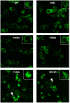
Figure 3. Subcellular localization of GFP-hCASK and GFP-hCASK mutants in HEK-393 cells.
Images were obtained with a 63X Plan-apochromat 1.4 N.A oil lens. White arrows indicate representative intracellular aggregates. Insert shows higher magnification.
Figure 4
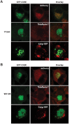
Figure 4. Characterization of aggregates.
Images of HEK cells transfected with A) GFP-CASK-Y728C or B) GFP-CASK-W919R were obtained with a 63X Plan-apochromat 1.4 N.A oil lens. First column shows aggregated GFP-CASK protein. Panels labeled “mCherry” show cells that were co-transfected with GFP-CASK and mCherry, which remains cytosolic. Panels labeled “Thioflavin T” represent coverslips that were fixed and then stained with Thioflavin T, which shows enhanced fluorescence in the presence of amyloid fibrils. Panels labeled “Golgi-RFP” represent coverslips that were treated with CellLight® Golgi-RFP which labels the Golgi network. Third column shows an overlay, demonstrating that aggregates are cytosolic (mCherry, Golgi-RFP) but not amyloid in nature (Thioflavin T).
Figure 5
Figure 5. Functional CASK XLMR mutations (R28L, Y268H and P396S) do not disrupt interactions with liprin-α, Mint-1, or Veli.
Lysates from HEK-293 cells co-transfected with GFP-CASK (wild-type or mutants R28L, Y268H, or P396S) and either liprin-α3 or FLAG-tagged Mint-1 were incubated with anti-GFP beads to pull down GFP-CASK and binding partners. To assess Veli interaction, no co-transfection was performed; native Veli was pulled down after incubation of lysates from GFP-CASK-transfected HEK-293 cells with anti-GFP beads to pull down GFP-CASK. Western blots of samples containing whole cell lysate (Input) or immunoprecipitates (Co-IP) were probed with anti-liprin-α3, anti-Veli, or anti-FLAG primary antibodies.
Figure 6
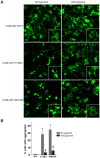
Figure 6. Glycerol treatment eliminates intracellular aggregates.
Six hours after transfection, media was exchanged for either fresh media alone or containing 10% glycerol. A. Images, 40X. Insert shows higher magnification. B. Using five representative 20X images (Figure S7) for each condition, individual cells were classified as free of or containing aggregates in Image J. Bars and error bars represent the average and standard deviation of three independent analyses. * and # indicate statistically significant differences from the wild-type images.

References

    1. Hata Y, Butz S, Sudhof TC (1996) CASK: a novel dlg/PSD95 homolog with an N-terminal calmodulin-dependent protein kinase domain identified by interaction with neurexins. The Journal of neuroscience : the official journal of the Society for Neuroscience 16: 2488–2494. - PMC - PubMed
    1. Hsueh YP (2006) The role of the MAGUK protein CASK in neural development and synaptic function. Current medicinal chemistry 13: 1915–1927. - PubMed
    1. LaConte LEW, Mukherjee K (in press) Structural constraints and functional divergences in CASK evolution. Biochemical Society Transactions. - PubMed
    1. Mukherjee K, Sharma M, Jahn R, Wahl MC, Sudhof TC (2010) Evolution of CASK into a Mg2+-sensitive kinase. Science signaling 3: ra33. - PMC - PubMed
    1. Su AI, Wiltshire T, Batalov S, Lapp H, Ching KA, et al. (2004) A gene atlas of the mouse and human protein-encoding transcriptomes. Proceedings of the National Academy of Sciences of the United States of America 101: 6062–6067. - PMC - PubMed

LinkOut - more resources