. 2014 Apr;155(4):1222-34.
doi: 10.1210/en.2013-1646. Epub 2014 Feb 7.

The PTTG1-binding factor (PBF/PTTG1IP) regulates p53 activity in thyroid cells

Affiliations

The PTTG1-binding factor (PBF/PTTG1IP) regulates p53 activity in thyroid cells

Martin L Read et al. Endocrinology. 2014 Apr.

Abstract

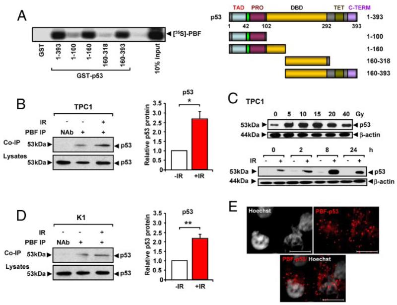
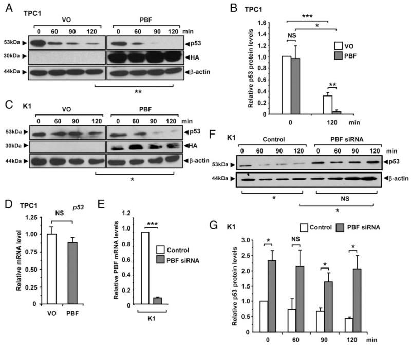
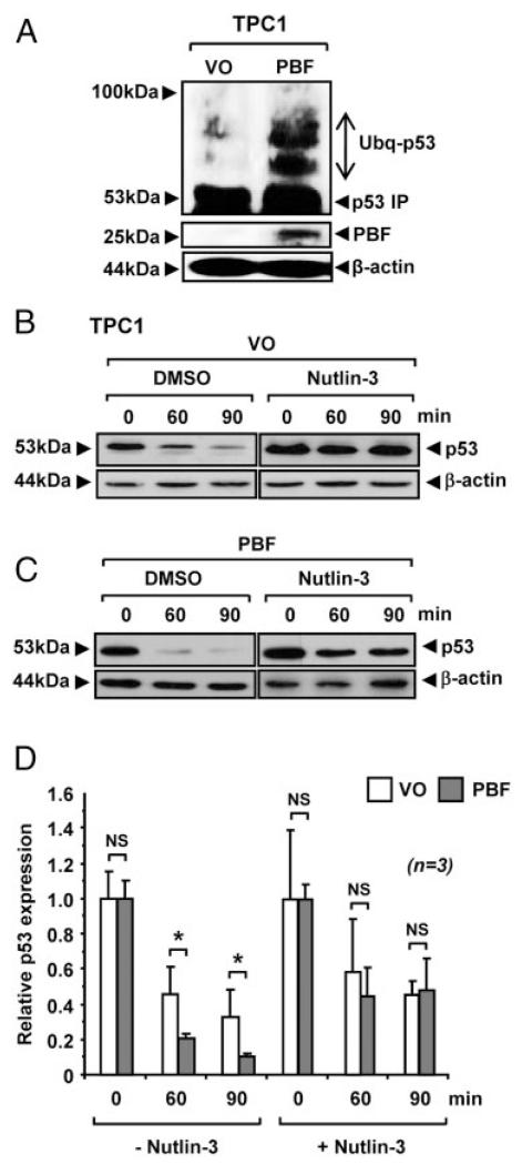
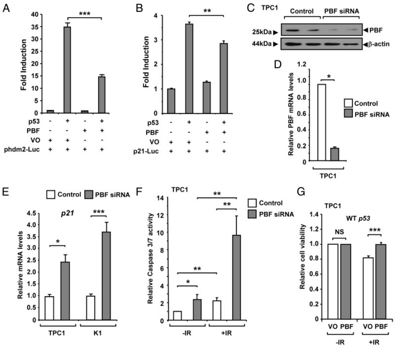
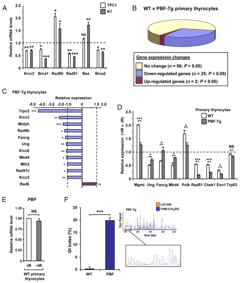
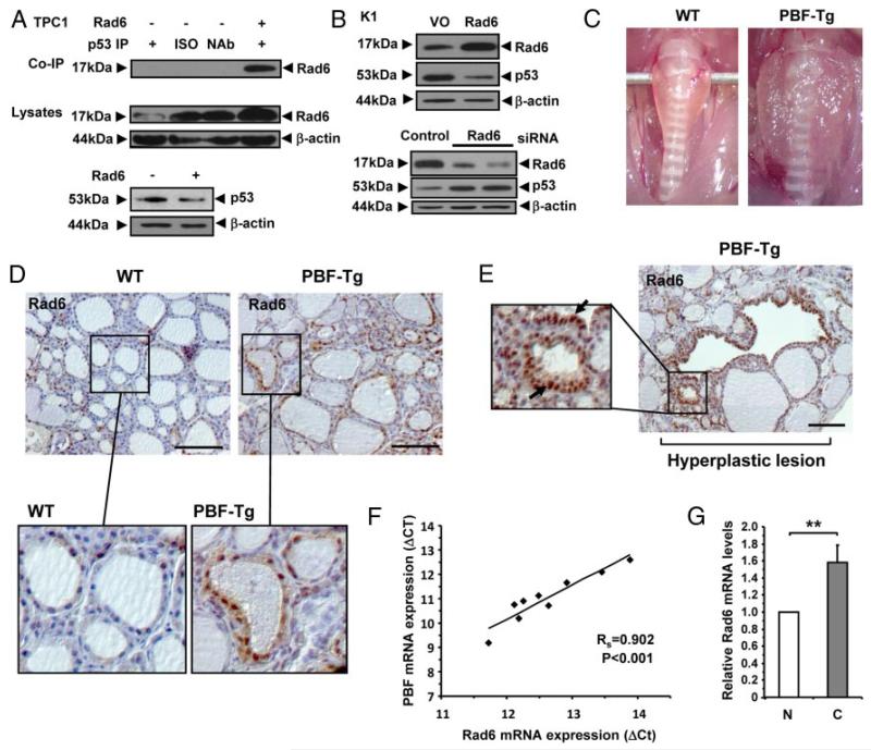
The PTTG1-binding factor (PBF/PTTG1IP) has an emerging repertoire of roles, especially in thyroid biology, and functions as a protooncogene. High PBF expression is independently associated with poor prognosis and lower disease-specific survival in human thyroid cancer. However, the precise role of PBF in thyroid tumorigenesis is unclear. Here, we present extensive evidence demonstrating that PBF is a novel regulator of p53, a tumor suppressor protein with a key role in maintaining genetic stability, which is infrequently mutated in differentiated thyroid cancer. By coimmunoprecipitation and proximity-ligation assays, we show that PBF binds specifically to p53 in thyroid cells and significantly represses transactivation of responsive promoters. Further, we identify that PBF decreases p53 stability by enhancing ubiquitination, which appears dependent on the E3 ligase activity of Mdm2. Impaired p53 function was evident in a transgenic mouse model with thyroid-specific PBF overexpression (transgenic PBF mice), which had significantly increased genetic instability as indicated by fluorescent inter simple sequence repeat-PCR analysis. Consistent with this, approximately 40% of all DNA repair genes examined were repressed in transgenic PBF primary cultures, including genes with critical roles in maintaining genomic integrity such as Mgmt, Rad51, and Xrcc3. Our data also revealed that PBF induction resulted in up-regulation of the E2 enzyme Rad6 in murine thyrocytes and was associated with Rad6 expression in human thyroid tumors. Overall, this work provides novel insights into the role of the protooncogene PBF as a negative regulator of p53 function in thyroid tumorigenesis, in which PBF is generally overexpressed and p53 mutations are rare compared with other tumor types.


Figures

Figure 1
Figure 1
Interaction between PBF and p53. A, Binding of [35S]-PBF to GST-p53 (1–393) and GST-p53 deletion mutants as indicated vs a GST-only control. Schematic of domain structure of p53 and deletion mutants showing a transcriptional activation domain (TAD), proline rich domain (PRO), DNA-binding domain (DBD), tetramerization domain (TET) and C-terminal (C-TERM). B, Co-IP of p53 with PBF in TPC1 cells either untreated (−IR) or irradiated with 15 Gy for 8h (+IR). NAb=no antibody control. Graph shows quantified levels of p53 ± SE from 3 independent experiments. C, Western blot analysis of p53 in TPC1 cells irradiated with 0 to 40 Gy dose as indicated for 8 hours (upper), or irradiated (+) with 15 Gy dose and p53 protein levels monitored at 0, 2, 8 or 24 hours post-treatment compared to untreated (−) controls (lower). D, Co-IP of p53 with PBF in K1 cells either untreated (−IR) or irradiated with 15 Gy for 8h (+IR). NAb=no antibody control. Graph shows quantified levels of p53 ± SE from 3 independent experiments. E, PLA assay to demonstrate specific PBF and p53 interaction (red spots) in TPC1 cells transiently transfected for 24 hours with plasmid expression vectors for p53 and HA-tagged PBF. Scale bars: 10 μM. *, P < .05; **, P < .01.
Figure 2
Figure 2
PBF decreases p53 intracellular stability. Representative Western blot analysis of p53 in (A) TPC1 and (C) K1 cells transfected with either vector only (VO) or PBF and then lysed at indicated times post-treatment with 100 μM anisomycin. Detection of HA epitope was used to monitor transfection. Mean p53 protein levels relative to β-actin in TPC1 cells are shown in (B) from 3 independent experiments. D, Relative mRNA levels of p53 in TPC1 cells transfected with either VO or PBF. E, Relative mRNA levels of PBF in K1 cells transfected with either PBF-specific or control siRNA for 72 hours at a concentration of 100 nM. F, Representative Western blot analysis of p53 in K1 cells transfected with either PBF-specific or control siRNA and then lysed at indicated times post-treatment with 100 μM anisomycin. G, Quantification of p53 protein levels relative to β-actin from p53 half-life experiments in K1 cells transfected with either PBF-specific or control siRNA. Data presented as mean p53 levels ± SE from 3 independent experiments. *, P < .05; ***, P < .001; NS = not significant.
Figure 3
Figure 3
Effect of PBF on p53 ubiquitination. A, Detection of high mwt p53 conjugates by Western blot analysis in TPC1 cells transfected with either VO or PBF and then treated with 10 μM MG132. B-C, Western blot analysis of p53 in TPC1 cells transfected with either VO or PBF and then incubated with 50 μM nutlin-3 prior to 100 μM anisomycin treatment. DMSO was used as vehicle. D, Mean p53 protein levels relative to β-actin quantified from 3 independent experiments are shown. Data presented as mean ± SE. *, P < .05; NS = not significant.
Figure 4
Figure 4
PBF inhibits p53 transcriptional activity. H1299 cells were transfected with p53 luciferase reporter plasmids for either (A) hdm2 (phdm2-Luc) or (B) p21 (p21-Luc), as well as p53 and PBF expression vectors, or vector only (VO) as indicated. Luciferase activity was measured 24 hours post-transfection. C-D, TPC1 cells were transfected with either PBF-specific or control siRNA at a final concentration of 100 nM. PBF expression was assessed by Western blotting and qRT-PCR analysis as shown. E, Relative mRNA levels of p21 mRNA in TPC1 and K1 cells transfected with either PBF-specific or control siRNA for 48 hours at a concentration of 100 nM. F, Analysis of caspase-3/7 activity in TPC1 cells transfected with either PBF-specific or control siRNA for 48 hours and then irradiated with a 15 Gy dose (+IR) or untreated (−IR). Normalized mean caspase-3/7 values ± SE are shown from 4 independent experiments. For each experiment caspase-3/7 activity was determined from n = 5 per condition at 24 hours postirradiation. G, TPC1 cells were transfected with either VO or PBF for 24 hours and either untreated (−IR) or irradiated (+IR) with a 15 Gy dose. Cells were then replated and viability measured after 24 hours. Data presented as mean ± SE from four independent experiments. *, P < .05; **, P < .01; ***, P < .001; NS = not significant.
Figure 5
Figure 5
PBF dysregulates DNA repair gene expression and promotes genetic instability. A, Relative mRNA expression of indicated genes in TPC1 and K1 cells transfected with either PBF-specific or control siRNA for 48 hours at a concentration of 100 nM. B, Pie chart summarizes expression changes of DNA repair genes between PBF-Tg and WT thyrocytes (n = 3 arrays). C, Relative mRNA expression levels of 12 genes (≥ 1.5-fold; P < .05) in PBF-Tg thyrocytes compared to WT. D, Relative fold changes in mRNA expression of indicated 9 genes following irradiation of either WT or PBF-Tg thyrocytes compared to nonirradiated controls as indicated. Data presented as mean ± SE from at least 3 independent experiments. E, Relative mRNA levels of PBF in wild-type (WT) primary thyrocytes either untreated (−IR) or irradiated with a 15 Gy dose (+IR). F, Quantification of genetic instability in murine PBF-Tg thyrocytes compared to WT (n = 5). Data presented as mean GI index ± SE. A representative FISSR-PCR trace amplified from PBF-Tg thyrocyte gDNA is shown plotted against a LIZ1200 size standard. *, P < .05; **, P < .01; ***, P < .001; NS = not significant.
Figure 6
Figure 6
Association of Rad6 and PBF in vivo. A, Western blot analysis (upper) of co-IP of Rad6 with p53 in TPC1 cells transfected for 24 hours with either VO (−) or pCMV6-Ube2a (+). ISO = isotype control antibody. NAb = no antibody control. Western blot analysis of Rad6 (middle) and p53 (lower) in cell lysates from transfected TPC1 cells are also shown. B, Western blot analysis of Rad6 and p53 following either overexpression (upper) or depletion (lower) of Rad6 in K1 cells. Control lanes are indicated. C, Representative enlarged thyroids dissected from aged PBF-Tg mice compared to WT. Thyroids were typically ~3–4 fold heavier in PBF-Tg mice. D-E, Representative images of Rad6 staining in normal regions (D) and hyperplastic lesions (E) in WT (n = 6) and PBF-Tg (n = 9) thyroids are shown. Arrows highlight elevated nuclear expression of Rad6 (E). F, Correlation of PBF and Rad6 mRNA expression in human thyroid tumors (n = 11). Statistics analyzed using Spearman rank correlation. G, Graph shows quantification of Rad6 mRNA expression in thyroid tumors relative to normal thyroid. Scale bars: 100 μm. Data presented as mean ± SE. **, P < .01.

References

    1. Chen AY, Jemal A, Ward EM. Increasing Incidence of Differentiated Thyroid Cancer in the United States, 1988–2005. Cancer. 2009;115:3801–3807. - PubMed
    1. Bhaijee F, Nikiforov YE. Molecular Analysis of Thyroid Tumors. Endocr Pathol. 2011;22:126–133. - PubMed
    1. Mazzaferri EL, Jhiang SM. Long-Term Impact of Initial Surgical and Medical Therapy on Papillary and Follicular Thyroid-Cancer. Am J Med. 1994;97:418–428. - PubMed
    1. Nixon IJ, Ganly I, Patel SG, Palmer FL, Di Lorenzo MM, Grewal RK, Larson SM, Tuttle RM, Shaha A, Shah JP. The Results of Selective Use of Radioactive Iodine on Survival and on Recurrence in the Management of Papillary Thyroid Cancer, Based on Memorial Sloan-Kettering Cancer Center Risk Group Stratification. Thyroid. 2013;23:683–694. - PubMed
    1. Xing MZ, Alzahrani AS, Carson KA, Viola D, Elisei R, Bendlova B, Yip L, Mian C, Vianello F, Tuttle RM, Robenshtok E, Fagin JA, Puxeddu E, Fugazzola L, Czarniecka A, Jarzab B, O’Neill CJ, Sywak MS, Lam AK, Riesco-Eizaguirre G, Santisteban P, Nakayama H, Tufano RP, Pai SI, Zeiger MA, Westra WH, Clark DP, Clifton-Bligh R, Sidransky D, Ladenson PW, Sykorova V. Association Between BRAF V600E Mutation and Mortality in Patients With Papillary Thyroid Cancer. Jama-J Am Med Assoc. 2013;309:1493–1501. - PMC - PubMed

Publication types

MeSH terms