Review
. 2014 Feb 18;53(6):957-78.
doi: 10.1021/bi4016617. Epub 2014 Feb 7.

Designing hydrolytic zinc metalloenzymes

Affiliations
Review

Designing hydrolytic zinc metalloenzymes

Melissa L Zastrow et al. Biochemistry. .

Abstract

Zinc is an essential element required for the function of more than 300 enzymes spanning all classes. Despite years of dedicated study, questions regarding the connections between primary and secondary metal ligands and protein structure and function remain unanswered, despite numerous mechanistic, structural, biochemical, and synthetic model studies. Protein design is a powerful strategy for reproducing native metal sites that may be applied to answering some of these questions and subsequently generating novel zinc enzymes. From examination of the earliest design studies introducing simple Zn(II)-binding sites into de novo and natural protein scaffolds to current studies involving the preparation of efficient hydrolytic zinc sites, it is increasingly likely that protein design will achieve reaction rates previously thought possible only for native enzymes. This Current Topic will review the design and redesign of Zn(II)-binding sites in de novo-designed proteins and native protein scaffolds toward the preparation of catalytic hydrolytic sites. After discussing the preparation of Zn(II)-binding sites in various scaffolds, we will describe relevant examples for reengineering existing zinc sites to generate new or altered catalytic activities. Then, we will describe our work on the preparation of a de novo-designed hydrolytic zinc site in detail and present comparisons to related designed zinc sites. Collectively, these studies demonstrate the significant progress being made toward building zinc metalloenzymes from the bottom up.


Figures
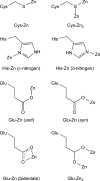
Figure 1
Figure 1
Zinc(II)–amino acid side chain binding modes as described in the text. Most zinc ligands in proteins are the side chains of cysteine (S donor), histidine (N donor), and glutamate or aspartate (O donor).
Figure 2
Figure 2
ZnHis3 sites in various proteins., (a) Insulin (PDB entry 1AIO), in which Zn(II) organizes the hexamer with His ligands originating from three different subunits. (b) Carbonic anhydrase II (PDB entry 2CBA), in which Zn(II) forms a hydrolytic active site and each of the three His ligands is on a β-sheet. (c) Matrix metalloproteinase adamalysin II (PDB entry 1AIG), in which Zn(II) forms a hydrolytic active site with two His ligands on an α-helix and the third from a loop. (d) Serine protease tonin (PDB entry 1TON), in which Zn(II) binding inhibits activity. (e) Zinc transporter ZnuA (PDB entry 1PQ4), in which the structure mediates Zn(II) mobility for transport.
Figure 3
Figure 3
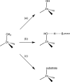
General mechanisms for mononuclear Zn(II) enzymes. (a) Ionization to form a Zn(II)–hydroxide nucleophile. (b) Polarization with the assistance of a general base to generate a nucleophile. (c) Displacement by the substrate that can be subsequently activated to generate a nucleophile.
Figure 4
Figure 4
(a) Modeled structure for the design of the minibody and its predicted metal-binding site (His3). Panel a was reprinted from ref (59). Copyright 1993 Nature Publishing Group. (b) Model of the redesigned scorpion toxin charybdotoxin (orthogonal views) with a His3 metal-binding site. Disulfide bonds are colored yellow. Metal-binding ligands are colored red. Panel b was reproduced from ref (67). Copyright 1995 National Academy of Sciences.
Figure 5
Figure 5
(a) Model of Zα4. The side chains (clockwise from top right) are Cys21, His25, Cys47, and His51. Panel a was reproduced from ref (70). Copyright 1995 American Chemical Society. (b) Model of the metal-binding site in the B1 domain of streptococcal protein G. The ribbon diagram is specifically of Zβ1M. The side chains around the metal-binding site are His16, His18, His30, and Cys33. Panel b was reproduced from ref (76). Copyright 1995 Nature Publishing Group.
Figure 6
Figure 6
Distribution of the designed Cys2His2 sites in thioredoxin (letters identify each design). This figure was reproduced ref (84). Copyright 1998 American Chemical Society.
Figure 7
Figure 7
Molecular model of the Zn(II)–GGG complex with a Cys4 site rendered using Biosystem Insight II. This figure was reproduced from ref (86). Copyright 2007 American Chemical Society.
Figure 8
Figure 8
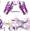
(a) Crystal structure of the Zn–MBPPhen2 dimer (PDB entry 3MNK). (b) Superposition of Ni–MBPPhen2 (yellow) and Zn–MBPPhen2 (magenta) metal centers. (c) Close-up showing the proximity between the coordinatively unsaturated metal centers in the asymmetric unit of the Zn–MBPPhen22 structure. The 2FoFc electronic density map is contoured at 1.2σ. The dimer is formed in solution and in the solid state. This figure was reproduced from ref (92). Copyright 2011 Elsevier.
Figure 9
Figure 9
Overall structure and example of a Zn(II) site in a self-assembling protein cryptand templated by disulfide bonds. (a) Overlay of protein backbones of the apo and Zn(II)-bound forms of C81/C96RIDCl4. (b) One of four ZnHis3(H2O) sites in A74/C81/C96RIDCl4 with the 2FoFc electron density map contoured at 1.4σ (cyan) and 7σ (magenta). This figure was reproduced from ref (102). Copyright 2013 American Chemical Society.
Figure 10
Figure 10
Schematic of the active site structure of carbonic anhydrase II displaying the extended active site around the Zn(II) center.
Figure 11
Figure 11
(a) Amino acid sequence of the zinc finger parent peptide. Panel a was reproduced from ref (105). Copyright 2004 American Chemical Society. (b) Natural zinc finger fold (αββ structure) and (c) zinc finger αββ fold modified as a metallohydrolase. Panels b and c were reproduced from ref (151). Copyright 2010 Elsevier.
Figure 12
Figure 12
Comparison of the size of the active site cavities of (a) the modeled His3 site using the structure of [As(III)]S(CSL9C)3 (PDB entry 2JGO) and (b) the actual structure containing the His3 site, [Hg(II)]S[Zn(II)(H2O/OH)]N(CSL9PenL23H)3n+ (PDB entry 3PBJ). (c) Overlay of the two sites with the model colored gray and the actual structure colored cyan. This figure was reproduced from ref (47). Copyright 2013 Elsevier.
Figure 13
Figure 13
Ribbon diagrams of the [Hg(II)]S[Zn(II)(H2O/OH)]N(CSL9PenL23H)3n+ parallel 3SCC (one of two different three-helix bundles present in the asymmetric unit) at pH 8.5. Shown are the main chain atoms represented as helical ribbons (cyan) and the Pen and His side chains in stick form (yellow for sulfur, blue for nitrogen, and red for oxygen). (a) One of two trimers found in the asymmetric unit of the crystal structure. (b) Top-down view of the structural trigonal thiolate site, HgS3, confirming the proposed structure of Hg(II) in Cys-containing TRI peptides. This metal site should mimic well the structural site in the metalloregulatory protein MerR. (c) Side view of the tetrahedral catalytic site, ZnN3O, which closely mimics carbonic anhydrase and matrix metalloproteinase active sites. All figures are shown with 2FoFc electron density contoured at 1.5σ overlaid. This figure was reproduced from ref (152). Copyright 2012 Nature Publishing Group.
Figure 14
Figure 14
Overlay of the ZnN3O site in [Hg(II)]S[Zn(II)(H2O/OH)]N(CSL9PenL23H)3n+ with the active site of human CAII and the matrix metalloproteinase (MMP) adamalysin II. [Hg(II)]S[Zn(II)(H2O/OH)]N(CSL9PenL23H)3n+ is colored cyan (PDB entry 3PBJ), CAII tan (PDB entry 2CBA), and adamalysin II gray (PDB entry 1IAG). (a) Top-down view of the overlay with CAII. The solvent molecule associated with [Hg(II)]S[Zn(II)(H2O/OH)]N(CSL9PenL23H)3n+ is colored red, and that associated with CAII lies underneath. (b) Side-on view of the overlay with CAII. The model displays an excellent structural overlay for the first coordination-sphere atoms with CAII; however, the orientation of the imidazoles differs between the two proteins. Another subtle difference is that the present structure has three ε-amino nitrogens bound to the Zn(II) ion whereas CAII has a mixed two-ε and one-δ coordination sphere. (c) Top-down view of the overlay with adamalysin II. The solvent molecule associated with adamalysin II is colored gray. (d) Side-on view of the overlay with adamalysin II. While the position of the His rings is close between the model and adamalysin II, the locations of the solvent molecules differ noticeably. Unlike for CAII, three ε-amino nitrogens bind to Zn(II) in adamalysin II. The overlay was performed manually in PyMOL. This figure was adapted from ref (152).
Figure 15
Figure 15
Comparison of the X-ray crystal structure of [Hg(II)]S[Zn(II)(H2O/OH)]N(CSL9PenL23H)3n+ (left, PDB entry 3PBJ) with a PyMOL model of [Zn(II)(H2O/OH)]N(TRIL2WL23H)3n+ lacking the HgS3 structural site (right, based on PDB entry 3PBJ). This figure was reproduced from ref (168). Copyright 2013 American Chemical Society.
Figure 16
Figure 16
X-ray crystal structure of MID1-zinc, a designed protein with a metal-mediated protein interface. The red mesh represents the active site cleft above the open coordination site of the ZnHis3 metal site. This figure was reproduced from ref (96). Copyright 2012 American Chemical Society.
Figure 17
Figure 17
Comparison of the X-ray crystal structure of (a) [Hg(II)]S[Zn(II)(H2O/OH)]N(CSL9PenL23H)3n+ (PDB entry 3PBJ) with PyMOL models of (b) [Zn(II)(H2O/OH)]N[Hg(II)]S(TRIL9HL23C)3n+ based on the coordinates of PDB entry 2JGO(194) and (c) [Hg(II)]S[Zn(II)(H2O/OH)]N(TRIL9CL19H)3n+ based on the coordinates of PDB entry 3PBJ. Models were prepared in PyMOL using the mutagenesis option and PyMOL’s rotamer library. This figure was adapted from ref (168).

References

    1. Andreini C.; Banci L.; Bertini I.; Rosato A. (2006) Counting the Zinc-Proteins Encoded in the Human Genome. J. Proteome Res. 5, 196–201. - PubMed
    1. Sousa S. F.; Lopes A. B.; Fernandes P. A.; Ramos M. J. (2009) The Zinc Proteome: A Tale of Stability and Functionality. Dalton Trans. 7946–7956. - PubMed
    1. Keilin D.; Mann T. (1939) Carbonic Anhydrase. Nature 144, 442–443.
    1. Vallee B. L.; Neurath H. (1954) Carboxypeptidase, a Zinc Metalloprotein. J. Am. Chem. Soc. 76, 5006–5007.
    1. Vallee B. L. (1959) Biochemistry, Physiology and Pathology of Zinc. Physiol. Rev. 39, 443–490. - PubMed

Publication types

LinkOut - more resources