. 2014 Feb 6;14(2):228-36.
doi: 10.1016/j.stem.2014.01.006.

Small molecules facilitate the reprogramming of mouse fibroblasts into pancreatic lineages

Affiliations

Small molecules facilitate the reprogramming of mouse fibroblasts into pancreatic lineages

Ke Li et al. Cell Stem Cell. .

Abstract

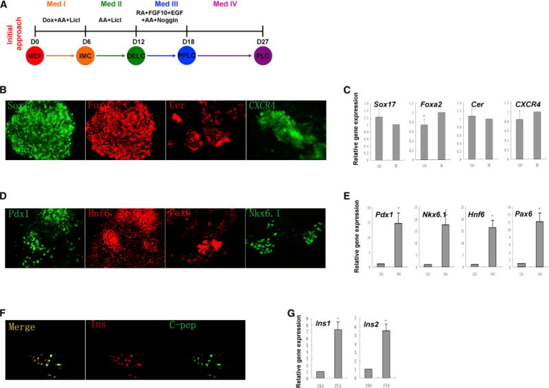
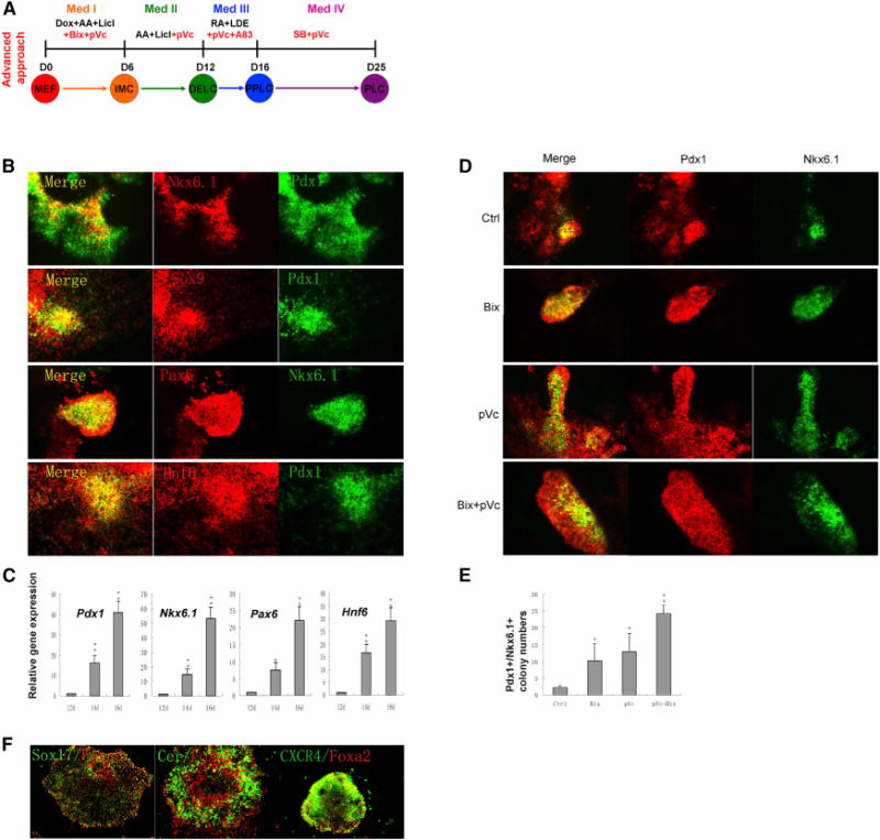
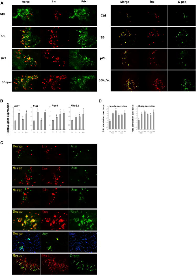
Pancreatic β cells are of great interest for the treatment of type 1 diabetes. A number of strategies already exist for the generation of β cells, but a general approach for reprogramming nonendodermal cells into β cells could provide an attractive alternative in a variety of contexts. Here, we describe a stepwise method in which pluripotency reprogramming factors were transiently expressed in fibroblasts in conjunction with a unique combination of soluble molecules to generate definitive endoderm-like cells that did not pass through a pluripotent state. These endoderm-like cells were then directed toward pancreatic lineages using further combinations of small molecules in vitro. The resulting pancreatic progenitor-like cells could mature into cells of all three pancreatic lineages in vivo, including functional, insulin-secreting β-like cells that help to ameliorate hyperglycemia. Our findings may therefore provide a useful approach for generating large numbers of functional β cells for disease modeling and, ultimately, cell-based therapy.


Figures

Figure 1
Figure 1. Initial Approach for Reprogramming MEFs into Pancreatic β-like Cells
(A) Scheme of the initial approach. Med-IV contains laminin, nicotinamide, and B27 etc., as described (Schroeder et al., 2006). (B) Immunostaining and (C) gene expression of definitive endoderm markers Sox17, CXCR4, Foxa2, and Cerberus 1 on day 12. NGFP1 iPSC-derived definitive endoderm-like cells (DE) were used as a positive control. (D) Immunostaining and (E) gene expression of pancreatic progenitor markers Pdx1, Hnf6, Pax6, and Nkx6.1 on day 18. (F) Immunostaining of pancreatic β cell markers insulin (Ins) and C-peptide (C-pep) on day 27. (G) Gene expression analysis of Ins1 and Ins2 from day 18 to day 27. Results in (C), (E), and (G) are the average of at least three independent experiments. *p < 0.05, **p < 0.01. See also Figure S1.
Figure 2
Figure 2. Identification of Small Molecule Conditions that Enhance Generation of PPLCs
(A) Scheme of the advanced approach. Red indicates small molecules that significantly improved induction efficiencies. (B) Immunostaining of pancreatic progenitor markers Pdx1, Hnf6, Sox9, Pax6, and Nkx6.1 on day 16. Cells were treated with the combination of four small molecules (RA, A83-01, LDE225, and pVc) from day 12 to day 16. (C) Gene expression analysis of markers during the pancreatic progenitor induction process by qPCR. Results are the average of three independent experiments. *p < 0.05, **p < 0.01. (D) Immunostaining of Pdx1 and Nkx6.1 on day 16. (E) Pdx1+/Nkx6.1+ colony numbers under indicated conditions on day 16. Four thousand cells were seeded into each well of 24-well plate on day 0. Results are the average of three independent experiments. *p < 0.05. (F) Immunostaining of Sox17, CXCR4, Foxa2, and Cerberus 1 on day 12. In (D), (E), and (F), Bix (Bix-01294) at 1 μM was added from day 0 to day 6 and pVc (2-Phospho-L-ascorbic acid trisodium salt) at 280 μM was added from day 0 to day 12. See also Figures S2, S3, and S4.
Figure 3
Figure 3. PPLCs Can Be Further Induced into PLCs
(A) Immunostaining of Pdx1/insulin and insulin/C-peptide on day 25 after treatment with SB (SB203580) at 5 μM and pVc (2-Phospho-L-ascorbic acid trisodium salt) at 280 μM from day 16 to day 25. (B) Gene expression analysis of Ins1, Ins2, Pdx1, and Nkx6.1 by qPCR. Cells treated with dissolvent were used as control. Results are the average of three independent experiments. *p < 0.05, **p < 0.01. (C) Immunostaining of pancreatic cell markers insulin, C-peptide, Glucagon, Somatostatin, Amylase, Pdx1, and Nkx6.1 on day 25. (D) Insulin and C-peptide release on day 25. Fold stimulation of insulin and C-peptide release over the respective basal condition for glucose and other insulin secretion agonists. Native mouse islets served as a control. Glu, 16.7mM D-glucose; TOL, 100 μM tolbutamide; IBMX, 0.5 mM 3 isobutil-1-methylxanthine. Results are the average of four independent experiments. See also Figures S3 and S4.
Figure 4
Figure 4. In Vivo Characterizations of PPLCs
(A) Blood glucose level of normal and STZ-induced diabetic mice without grafts or transplanted with PPLCs, MEFs, or islets. Under anesthesia, the left kidney of diabetic mice received a renal subcap-sular transplant of 300 native mouse islets in 30 μl Matrigel (Islet, n = 4), 3 × 10⁶ PPLCs reprogrammed from MEFs (MEF-derived PPLC, n = 10), 3 × 10⁶ NGFP1 mouse iPSC-derived PPLCs by our own optimized differentiation conditions (iPSC-derived PPLC1, n = 6), 3 × 10⁶ NGFP1 mouse iPSC-derived PPLCs by published differentiation conditions (iPSC-derived PPLC2, n = 6), or 3 × 10⁶ secondary MEF cells (MEF, n = 6). Untreated normal mice (Norm, n = 4) and mice treated with STZ only (STZ, n = 4) were controls. Eight weeks after transplantation, the left kidneys were surgically removed. (B) Glucose-stimulated insulin secretion (GSIS). Mice were fasted overnight, D-glucose (3 g/kg body weight) was injected intraperitoneally, and serum insulin levels of each group were measured before and 30 min after glucose stimulation. (C) Intraperitoneal glucose tolerance tests (IPGTTs). Mice were treated as in (B) and blood glucose levels were measured at 0, 15, 30, 60, 90, and 120 min after glucose stimulation. (D and E) Immunofluorescence staining of kidneys engrafted with MEF-derived PPLCs. (F) Quantification of endocrine cell numbers of mice from MEF, PPLC, and islet grafts. For each experimental group, endocrine cells of four mice were counted and averaged as described in the Experimental Procedures.

Comment in

References

    1. D’Amour KA, Bang AG, Eliazer S, Kelly OG, Agulnick AD, Smart NG, Moorman MA, Kroon E, Carpenter MK, Baetge EE. Production of pancreatic hormone-expressing endocrine cells from human embryonic stem cells. Nat Biotechnol. 2006;24:1392–1401. - PubMed
    1. Efe JA, Hilcove S, Kim J, Zhou H, Ouyang K, Wang G, Chen J, Ding S. Conversion of mouse fibroblasts into cardiomyocytes using a direct reprogramming strategy. Nat Cell Biol. 2011;13:215–222. - PubMed
    1. Ferber S, Halkin A, Cohen H, Ber I, Einav Y, Goldberg I, Barshack I, Seijffers R, Kopolovic J, Kaiser N, Karasik A. Pancreatic and duodenal homeobox gene 1 induces expression of insulin genes in liver and ameliorates streptozotocin-induced hyperglycemia. Nat Med. 2000;6:568–572. - PubMed
    1. Jiang W, Shi Y, Zhao D, Chen S, Yong J, Zhang J, Qing T, Sun X, Zhang P, Ding M, et al. In vitro derivation of functional insulin-producing cells from human embryonic stem cells. Cell Res. 2007;17:333–344. - PubMed
    1. Kelly OG, Chan MY, Martinson LA, Kadoya K, Ostertag TM, Ross KG, Richardson M, Carpenter MK, D’Amour KA, Kroon E, et al. Cell-surface markers for the isolation of pancreatic cell types derived from human embryonic stem cells. Nat Biotechnol. 2011;29:750–756. - PubMed

Publication types

MeSH terms

Substances