. 2014 Feb 5;81(3):536-543.
doi: 10.1016/j.neuron.2013.12.018.

Axonal transport of TDP-43 mRNA granules is impaired by ALS-causing mutations

Affiliations

Axonal transport of TDP-43 mRNA granules is impaired by ALS-causing mutations

Nael H Alami et al. Neuron. .

Abstract

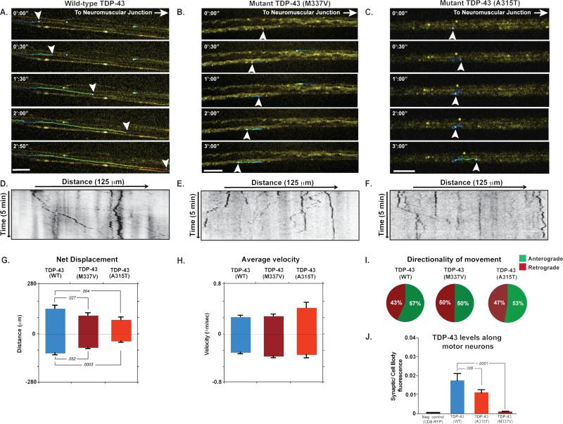
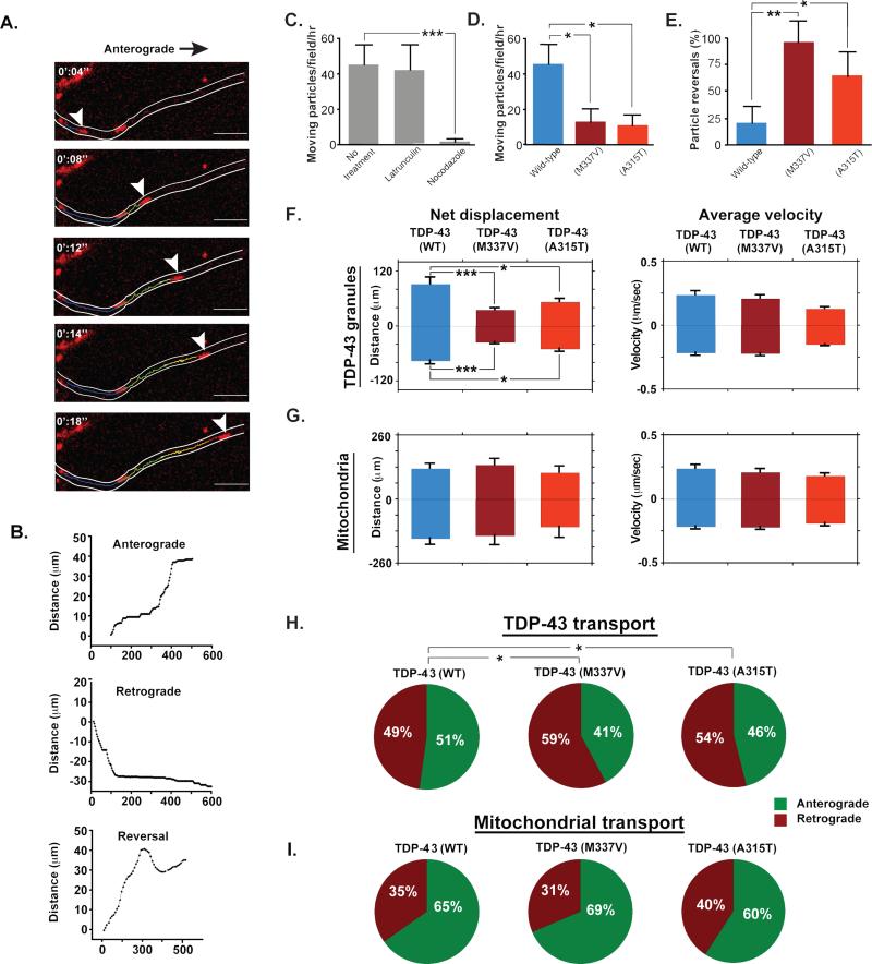
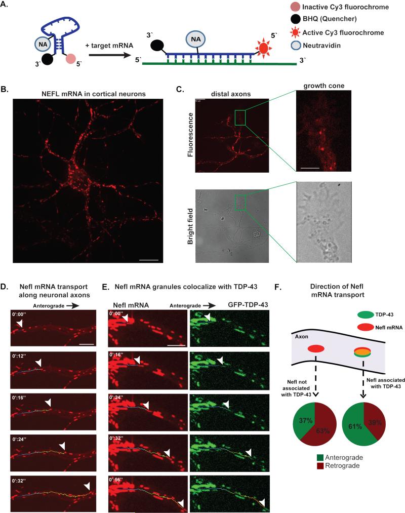
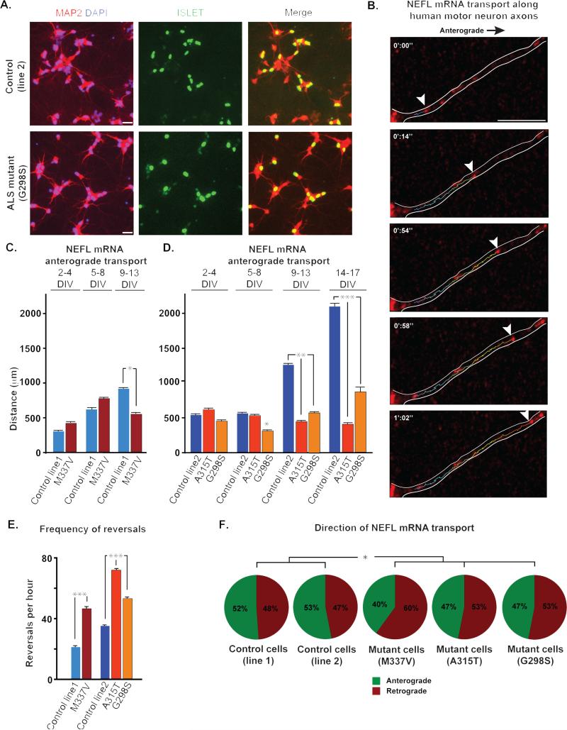
The RNA-binding protein TDP-43 regulates RNA metabolism at multiple levels, including transcription, RNA splicing, and mRNA stability. TDP-43 is a major component of the cytoplasmic inclusions characteristic of amyotrophic lateral sclerosis and some types of frontotemporal lobar degeneration. The importance of TDP-43 in disease is underscored by the fact that dominant missense mutations are sufficient to cause disease, although the role of TDP-43 in pathogenesis is unknown. Here we show that TDP-43 forms cytoplasmic mRNP granules that undergo bidirectional, microtubule-dependent transport in neurons in vitro and in vivo and facilitate delivery of target mRNA to distal neuronal compartments. TDP-43 mutations impair this mRNA transport function in vivo and in vitro, including in stem cell-derived motor neurons from ALS patients bearing any one of three different TDP-43 ALS-causing mutations. Thus, TDP-43 mutations that cause ALS lead to partial loss of a novel cytoplasmic function of TDP-43.


Figures

Fig. 1
Fig. 1. Localization and transport kinetics of TDP-43 granules in Drosophila motor neurons
(A-C) Venus-TDP-43WT, Venus-TDP-43M337V, and Venus-TDP-43A315T granules (arrowheads) displayed dynamic bidirectional movement along motor neuron axons of 3rd instar larvae. Transport of mutant TDP-43 granules showed normal instantaneous velocities, but an increased frequency of reversals and diminished net anterograde movement. Scale bar: 10 μm. (D-F) Kymographs tracing TDP-43 granules in motor neuron axons. (G-E) Quantification of displacement and instantaneous velocities of wild type and mutant granules along Drosophila motor neuron axons. Anterograde and retrograde movements are represented as positive or negative values on the y-axis, respectively. (I) Analysis of movement directionality shows a reduced ratio of anterograde to retrograde movement for mutant TDP-43 granules as compared to TDP-43WT. (J) Quantification of fluorescence intensities in Drosophila motor neuron axons. The ratio of average fluorescence intensity per pixel at the synaptic terminal divided by the fluorescence at the cell body shows that Venus-TDP-43WT signal is higher than that of mutant TDP-43. Error bars are shown as mean ± SEM.
Fig. 2
Fig. 2. TDP-43 transport in primary cortical neurons
(A) A wild-type TDP-43 granule (arrowhead) moves along the axon of mouse cortical neuron in the anterograde direction. Scale bar: 10 μm. (B) 3 representative tracings of TDP-43 granules moving along the axons of cortical neurons illustrating anterograde displacement, retrograde displacement, and reversal of directionality. The x-axis represents time and the y-axis total distance along the axon. (C) The number of moving TDP-43 granules in wild type and mutant-transfected cells were normalized to movements per hour and plotted. Latrunculin treatment had no effect on TDP-43 transport, while nocodazole-treated cells contained dramatically fewer motile TDP-43 granules, demonstrating microtubule-dependent movement. (D) The fraction of motile mutant TDP-43 granules was significantly reduced compared to TDP-43WT granules. (E) Mutant TDP-43 granules reversed direction significantly more frequently than TDP-43WT granules. (F) Kinetics of wild-type and mutant TDP-43 transport show a significant decrease in net displacement of TDP-43M337V (p< 0.001) and TDP-43A315T (p=0.030 for anterograde and 0.045 for retrograde) as compared to TDP-43WT, and no significant change in instantaneous velocities. (G) Kinetics of mitochondrial transport show that there is no significant difference in mitochondrial transport in cells transfected with wild-type or mutant TDP-43. (H) The direction of transport of TDP-43 granules was altered in cells transfected with mutant TDP-43. A significantly higher percentage of TDP-43M337V and TDP-43A315T granules moved in the retrograde direction as compared to TDP-43WT granules (p= 0.037 and 0.042, respectively). (I) The direction of mitochondrial transport was not altered in cells transfected with wild-type or mutant TDP-43. All error bars are shown as mean ± SEM.
Fig. 3
Fig. 3. Nefl mRNA is transported bidirectionally along primary cortical neuron axons and is part of an mRNP granule that contains TDP-43
(A) mRNA beacons are designed to target Nefl mRNA. The beacon emits a fluorescence signal once it linearizes upon hybridization with its target mRNA. (B) Nefl mRNA fluorescence is present in the soma and along primary cortical neuron axons. Scale bar: 10 μm. (C) Nefl mRNA was detected in distal axonal compartments and inside growth cones. Scale bar: axons 10 μm; growth cone 2 μm. (D) Nefl mRNA granule (arrowheads) is transported along the axon of a cortical neuron. Scale bar: 5 μm. (E) TDP-43 is present in the motile mRNA granule in a cortical neuron. Scale bar: 10 μm. (F) Granules containing TDP-43 and Nefl mRNA show predominately anterograde movements and net anterograde displacement, whereas those that contain Nefl without TDP-43 show predominately retrograde movements and net retrograde displacement.
Fig. 4
Fig. 4. NEFL mRNA transport defect along human motor neuron axons
(A) Human motor neurons were derived from iPS cells, as illustrated with immunostaining for motor neuron markers like Islet. For live imaging, we identified cells based on expression of the HB9 responsive GFP construct. (B) Live imaging of stem cell derived human motor neurons shows endogenous NEFL mRNA transport along axons. Scale bar: 5μm. (C) There is no significant difference in net anterograde displacement of NEFL mRNA granules in control vs. patient motor neurons with TDP-43M337V mutation during the first week of observation. The transport defect was apparent 9 days after plating when a significant decrease in net anterograde transport of NEFL mRNA granules is observed. (D) NEFL mRNA anterograde displacement was significantly altered in human motor neurons from iPS cells of ALS patients with TDP-43G298S mutation after 5 days in culture as compared to control. After 9 days in culture, mRNA anterograde transport is significantly altered in motor neurons from all ALS patients as compared to control. This defect becomes more pronounced over time. (E) Frequency of NEFL mRNA granule reversals per hour is significantly higher in neurons with mutant TDP-43 as compared to control, 2 weeks after plating (p<0.001). (F) The percentage of retrogradely moving NEFL mRNA granules is significantly higher in 9 DIV neurons from ALS patients with mutant TDP-43. All error bars are shown as mean ± SEM.

References

    1. Al-Chalabi A, Jones A, Troakes C, King A, Al-Sarraj S, van den Berg LH. The genetics and neuropathology of amyotrophic lateral sclerosis. Acta neuropathologica. 2012;124:339–352. - PubMed
    1. Arnold ES, Ling SC, Huelga SC, Lagier-Tourenne C, Polymenidou M, Ditsworth D, Kordasiewicz HB, McAlonis-Downes M, Platoshyn O, Parone PA, et al. ALS-linked TDP-43 mutations produce aberrant RNA splicing and adult-onset motor neuron disease without aggregation or loss of nuclear TDP-43. Proc Natl Acad Sci U S A. 2013;110:E736–745. - PMC - PubMed
    1. Barbee SA, Estes PS, Cziko AM, Hillebrand J, Luedeman RA, Coller JM, Johnson N, Howlett IC, Geng C, Ueda R, et al. Staufen-and FMRP-containing neuronal RNPs are structurally and functionally related to somatic P bodies. Neuron. 2006;52:997–1009. - PMC - PubMed
    1. Barmada SJ, Skibinski G, Korb E, Rao EJ, Wu JY, Finkbeiner S. Cytoplasmic mislocalization of TDP-43 is toxic to neurons and enhanced by a mutation associated with familial amyotrophic lateral sclerosis. J Neurosci. 2010;30:639–649. - PMC - PubMed
    1. Bilican B, Serio A, Barmada SJ, Nishimura AL, Sullivan GJ, Carrasco M, Phatnani HP, Puddifoot CA, Story D, Fletcher J, et al. Mutant induced pluripotent stem cell lines recapitulate aspects of TDP-43 proteinopathies and reveal cell-specific vulnerability. Proc Natl Acad Sci U S A. 2012;109:5803–5808. - PMC - PubMed

Publication types

MeSH terms