Review
. 2014 Jun;55(6):290-6.
doi: 10.1016/j.ceca.2014.01.001. Epub 2014 Jan 22.

Calcium signaling in lacrimal glands

Affiliations
Review

Calcium signaling in lacrimal glands

James W Putney et al. Cell Calcium. 2014 Jun.

Abstract

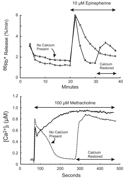
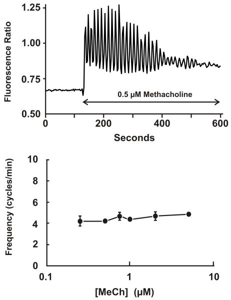
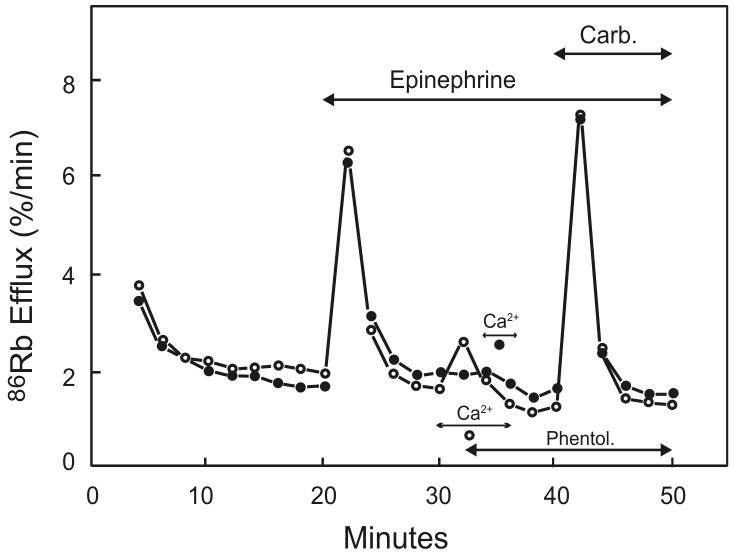
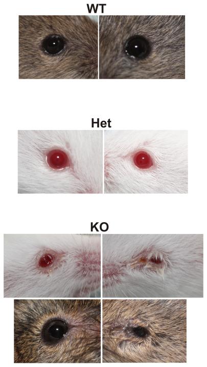
Lacrimal glands provide the important function of lubricating and protecting the ocular surface. Failure of proper lacrimal gland function results in a number of debilitating dry eye diseases. Lacrimal glands secrete lipids, mucins, proteins, salts and water and these secretions are at least partially regulated by neurotransmitter-mediated cell signaling. The predominant signaling mechanism for lacrimal secretion involves activation of phospholipase C, generation of the Ca(2+)-mobilizing messenger, IP3, and release of Ca(2+) stored in the endoplasmic reticulum. The loss of Ca(2+) from the endoplasmic reticulum then triggers a process known as store-operated Ca(2+) entry, involving a Ca(2+) sensor in the endoplasmic reticulum, STIM1, which activates plasma membrane store-operated channels comprised of Orai subunits. Recent studies with deletions of the channel subunit, Orai1, confirm the important role of SOCE in both fluid and protein secretion in lacrimal glands, both in vivo and in vitro.

Keywords: Calcium entry; Calcium oscillations; Calcium release; Calcium signaling; Fluid secretion; IP3; Lacrimal; Orai; Protein secretion; STIM.


Figures

Figure 1
Figure 1. Two phases of Ca2+ signaling in in vitro lacrimal gland preparations
Top: Changes in intracellular Ca2+ in slices of rat lacrimal gland are inferred from the efflux rate of 86Rb+. Redrawn from data originally presented in [11]. Bottom: Changes in intracellular Ca2+ in mouse lacrimal acinar cells are measured with the Ca2+ indicator, Fura-2. In both cases, in the absence of extracellular Ca2+, the response is transient, and subsequently restored by addition of Ca2+.
Figure 2
Figure 2. Muscarinic receptor-induced sinusoidal Ca2+ oscillations in mouse lacrimal cells
Top: A single, fura-2-loaded mouse lacrimal acinar cell was exposed to 0.5 μM methacholine (MeCh) inducing sinusoidal Ca2+ oscillations on an elevated basal level of Ca2+. MeCh-induced oscillations occur over a narrow concentration range, and with frequencies that are relatively constant at different agonist concentrations (Bottom figure). These findings were originally published in [46].
Figure 3
Figure 3. Evidence for the independence of Ca2+ influx from receptor activation
In both experiments, Ca2+ stores were discharged by addition of epinephrine in the absence of extracellular Ca2+, the α-adrenergic receptors were then blocked by phentolamine (Phentol.), and the status of Ca2+ stores assessed by addition of carbachol (Carb.). In the experiment with open circles, Ca2+ was added before phentolamine, so Ca2+ could flow into the cell through presumed receptor activated channels. In the experiment with closed circles, phentolamine was added before Ca2+ such that receptor-activated channels would presumably be closed. Nonetheless, the stores were refilled with similar efficiency in both cases, indicating that it is not receptor activation per se that is responsible for Ca2+ entry and refilling of intracellular stores. Redrawn from data originally presented in [25].
Figure 4
Figure 4. Appearance of eyes from wild type (WT), heterozygous (Het) and homozygous Orai1 knockout (KO) mice
The eyes from wild type and heterozygous mice appeared normal, while in many (but not all) cases the eyes from knockout animals showed signs of inflammation.

References

    1. Dartt DA. Neural regulation of lacrimal gland secretory processes: relevance in dry eye diseases. Prog Retin Eye Res. 2009;28:155–177. - PMC - PubMed
    1. Dartt DA. Dysfunctional neural regulation of lacrimal gland secretion and its role in the pathogenesis of dry eye syndromes. Ocul Surf. 2004;2:76–91. - PubMed
    1. Bothelo SY. Tears and the lacrimal gland. Scientific American. 1964;211:78–86. - PubMed
    1. Bothelo SY, Hisada M, Fuenmayor N. Functional innervation of the lacrimal gland in the cat. Origin of secretomotor fibers in the lacrimal nerve. Arch Opthalmol. 1966;76:581–588. - PubMed
    1. Herzog V, Sies H, Miller F. Exocytosis in secretory cells of rat lacrimal gland. Peroxidase release from lobules and isolated cells upon cholinergic stimulation. The Journal of Cell Biology. 1976;70:692–706. - PMC - PubMed

Publication types

MeSH terms