

Pontine malformation, undecussated pyramidal tracts, and regional polymicrogyria: a new syndrome
- PMID: 24507697
- PMCID: PMC3959267
- DOI: 10.1016/j.pediatrneurol.2013.12.013
Pontine malformation, undecussated pyramidal tracts, and regional polymicrogyria: a new syndrome
Abstract
Background: Horizontal gaze palsy and progressive scoliosis is caused by mutations in the ROBO3 gene, which plays a role in axonal guidance during brain development. Horizontal gaze palsy and progressive scoliosis is characterized by the congenital absence of conjugate lateral eye movements with preserved vertical gaze and progressive scoliosis as well as dysgenesis of brainstem structures and ipsilateral projection of the pyramidal tract.
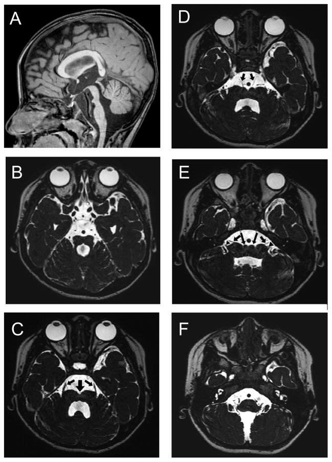
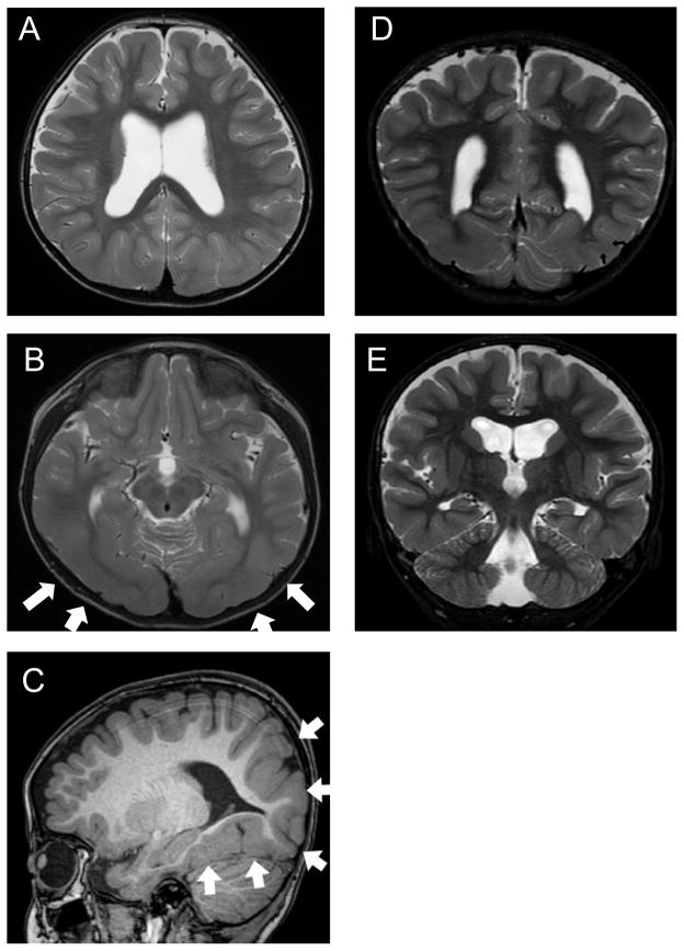
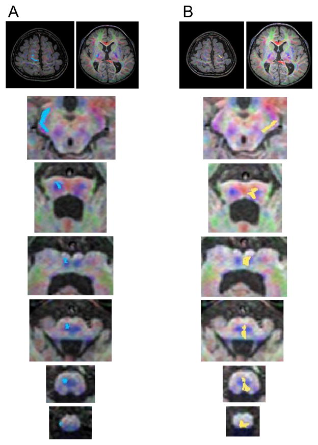
Patient: A 4-year, 11-month, girl presented with psychomotor retardation and autistic traits. Magnetic resonance imaging revealed hypoplasia and malformation of the ventral portion of the pons and medulla oblongata. Diffusion tensor imaging revealed the absence of decussation of the bilateral pyramidal tracts. These findings were similar to the typical findings for horizontal gaze palsy and progressive scoliosis. However, restriction of horizontal eye movement was minimal, and bilateral polymicrogyria were also noted in the occipitotemporal cortex in the present patient. These findings have not been previously reported in patients with horizontal gaze palsy and progressive scoliosis. No mutations in the ROBO3, SLIT1, SLIT2, NTN1, SEMA3 A, or SEMA3 F genes were identified.
Conclusion: This child may have a disorder caused by an unidentified factor, other than a mutation in the genes analyzed, involved in corticogenesis, axonal guidance, and brainstem morphogenesis.
Keywords: axonal guidance; brainstem hypoplasia; decussation of the pyramidal tract; horizontal gaze palsy; polymicrogyria; pontine malformation; progressive scoliosis.
Copyright © 2014 Elsevier Inc. All rights reserved.
Figures
References
-
- Dretakis EK, Kondoyannis PN. Congenital scoliosis associated with encephalopathy in five children of two families. J Bone Joint Surg Am. 1974;56(8):1747–1750. - PubMed
-
- Bosley TM, Salih MA, Jen JC, et al. Neurologic features of horizontal gaze palsy and progressive scoliosis with mutations in ROBO3. Neurology. 2005;64(7):1196–1203. - PubMed
-
- Sicotte NL, Salamon G, Shattuck DW, et al. Diffusion tensor MRI shows abnormal brainstem crossing fibers associated with ROBO3 mutations. Neurology. 2006;67(3):519–521. - PubMed
Publication types
MeSH terms
Grants and funding
LinkOut - more resources
Full Text Sources
Other Literature Sources
Molecular Biology Databases
