Review
. 2014 Apr;25(4):178-87.
doi: 10.1016/j.tem.2014.01.002. Epub 2014 Feb 4.

Emerging roles of hematopoietic cells in the pathobiology of diabetic complications

Affiliations
Review

Emerging roles of hematopoietic cells in the pathobiology of diabetic complications

Hideto Kojima et al. Trends Endocrinol Metab. 2014 Apr.

Abstract

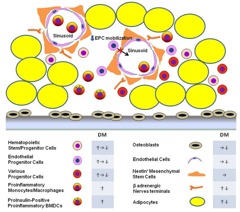
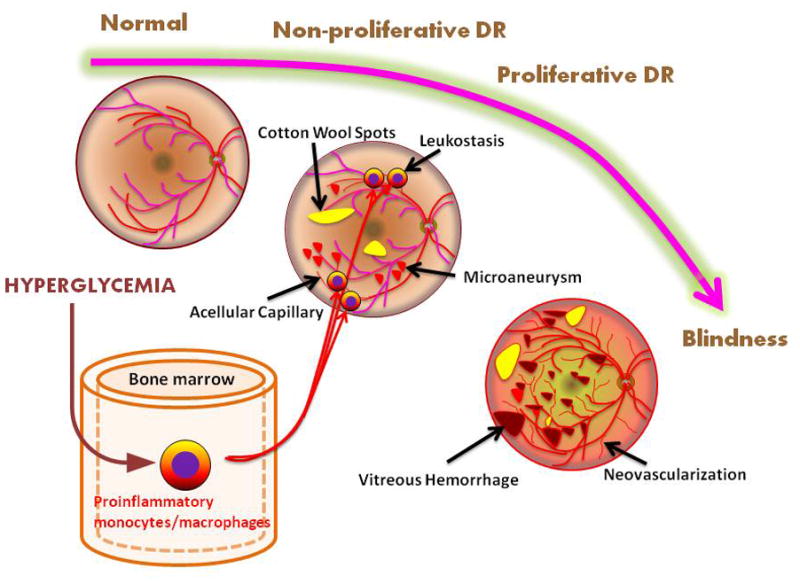
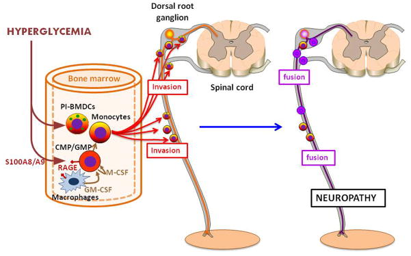
Diabetic complications encompass macrovascular events, mainly the result of accelerated atherosclerosis, and microvascular events that strike the eye (retinopathy), kidney (nephropathy), and nervous system (neuropathy). The traditional view is that hyperglycemia-induced dysregulated biochemical pathways cause injury and death of cells intrinsic to the organs affected. There is emerging evidence that diabetes compromises the function of the bone marrow (BM), producing a stem cell niche-dependent defect in hematopoietic stem cell mobilization. Furthermore, dysfunctional BM-derived hematopoietic cells contribute to diabetic complications. Thus, BM cells are not only a victim but also an accomplice in diabetes and diabetic complications. Understanding the underlying molecular mechanisms may lead to the development of new therapies to prevent and/or treat diabetic complications by specifically targeting these perpetrators.

Keywords: bone marrow; complications; diabetes mellitus; diabetic nephropathy; diabetic neuropathy; diabetic retinopathy; hematopoietic cells.


Figures

Figure 1
Figure 1. Diabetic BM microangiopathy and niche dysfunction
Hyperglycemia induces structural and functional changes in the cellular composition of the BM, including the ones depicted in this figure. These include: unchanged capillary [10] or capillary rarefaction (↓endothelial cells) [7]; decreased [7, 10, 11], unchanged [36] or increased [9] fractions of hematopoietic and other progenitor cells; unchanged [10] or decreased [9] osteoblasts; decreased [11] or increased [7, 37] fat accumulation; and decreased [37] or increased [9] sympathetic nerve fibers. Functional defects, i.e. niche dysfunction, accompany the structural changes. Different laboratories have observed impaired mobilization of EPCs in response to ischemic injury [5] and of HSPCs in response to G-CSF [9] associated with β adrenergic dysfunction. There is down-regulated proliferative potential of multipotent progenitor cells [7], but increased production of pro-inflammatory monocytes [11, 38]. In addition, PI-BMDCs appear in the diabetic BM [42]. PI-BMDCs share many of the characteristics of monocytes/macrophages. They are proinflammatory in nature and are actively involved in the pathogenesis of diabetic complications (see Box 2) Abbreviations: DM, abnormalities in diabetic mellitus BM; CSF, colony stimulating factor; EPC, endothelial progenitor cell; HSPC, hematopoietic stem and progenitor cell.
Figure 2
Figure 2. Role of hematopoietic cells in diabetic retinopathy
Vasoregression is an early event in diabetic retinopathy associated with hyperglycemia-induced leukostasis, causing loss of capillary endothelial cells and pericytes and thus leading to acellular capillaries without perfusion [16]. Ischemia secondary to vasoregression manifests as cotton wool spots (nerve fiber layer infarcts) and microaneurysms (aborted attempts at neovascularization). Progressive ischemia results in fragile neovascularization that is prone to preretinal and vitreous hemorrhage. Fibrotic changes from hemorrhages and fibrovascular proliferation can eventually lead to retinal detachment and loss of vision. Macular edema can occur at any stage of diabetic retinopathy and is another cause of loss of vision.
Figure 3
Figure 3. Role of hematopoietic cells, especially PI-BMDCs, in diabetic neuropathy
Hyperglycemia increases myelopoiesis through the activation of RAGE on the macrophages and common myeloid progenitors (CMPs) in the bone marrow [38]. Stimulated macrophages and CMPs secrete granulocyte-macrophage colony stimulating factor (GM-CSF) and macrophage-colony stimulating factor (M-CSF) leading to proliferation of granulocyte macrophage progenitors (GMPs) and CMPs/GMPs, respectively. RAGE+ BM-derived cells exhibit impaired axon regeneration after injury in diabetic mice [84]. Hyperglycemia also induces the emergence of proinsulin positive and TNF-α producing inflammatory cell in the BM [42]. These PI-BMDCs invade the peripheral nervous system, where they fuse with neurons and Schwann cells in the peripheral nerves, e.g., sciatic nerve, and with neurons in the dorsal root ganglia (DRG). The fusion damages the neurons and causes premature apoptosis. Furthermore, the PI-BMDC-neuron fusion cells continue to be a source of TNF-α, which amplifies the ill effects of PI-BMDCs [40, 43] (see Box 2).

References

    1. Brownlee M. The pathobiology of diabetic complications a unifying mechanism. Diabetes. 2005;54:1615–1625. - PubMed
    1. Manigrasso MB, et al. Unlocking the biology of RAGE in diabetic microvascular complications. Trends Endocrinol Metab. 2013 http://dx.doi.org/10.1016/j.tem.2013.08.002. - DOI - PMC - PubMed
    1. Rask-Madsen C, King GL. Vascular complications of diabetes: mechanisms of injury and protective factors. Cell Metab. 2013;17:20–33. - PMC - PubMed
    1. Fadini GP, et al. Circulating endothelial progenitor cells are reduced in peripheral vascular complications of type 2 diabetes mellitus. J Am Coll Cardiol. 2005;45:1449–1457. - PubMed
    1. Fadini GP, et al. Diabetes impairs progenitor cell mobilisation after hindlimb ischaemia-reperfusion injury in rats. Diabetologia. 2006;49:3075–3084. - PubMed

Publication types

MeSH terms