. 2014 May;8(3):565-80.
doi: 10.1016/j.molonc.2014.01.004. Epub 2014 Jan 15.

Honokiol inhibits epithelial-mesenchymal transition in breast cancer cells by targeting signal transducer and activator of transcription 3/Zeb1/E-cadherin axis

Affiliations

Honokiol inhibits epithelial-mesenchymal transition in breast cancer cells by targeting signal transducer and activator of transcription 3/Zeb1/E-cadherin axis

Dimiter B Avtanski et al. Mol Oncol. 2014 May.

Abstract

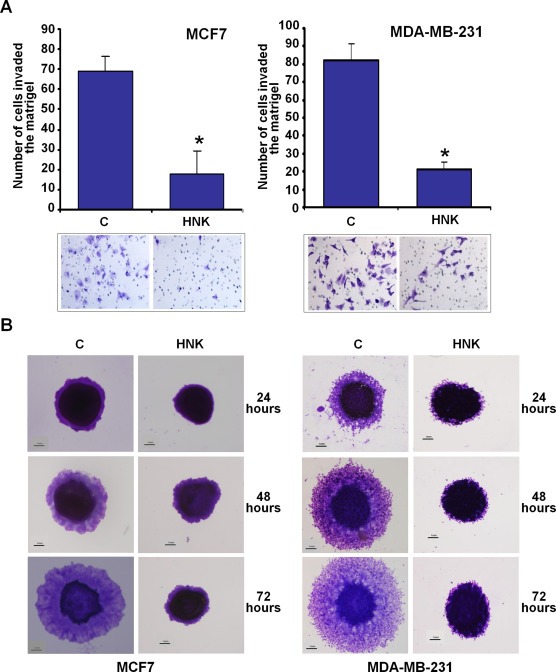
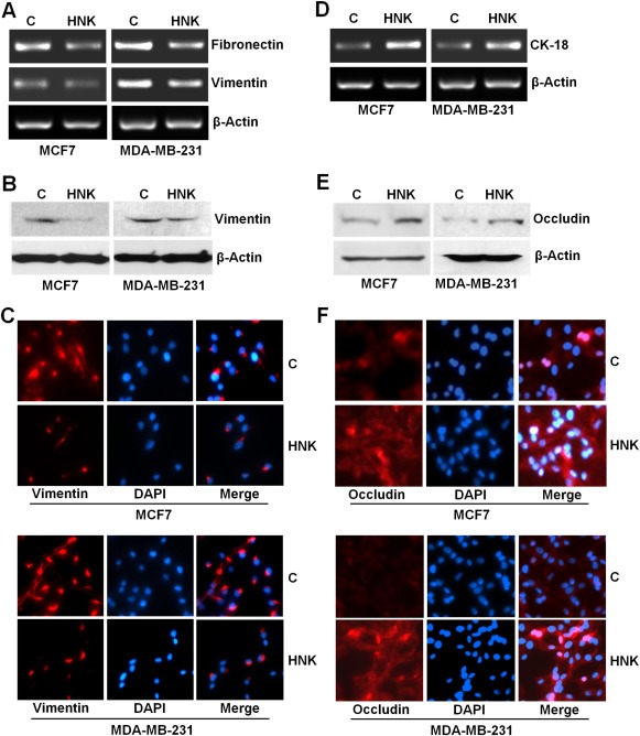
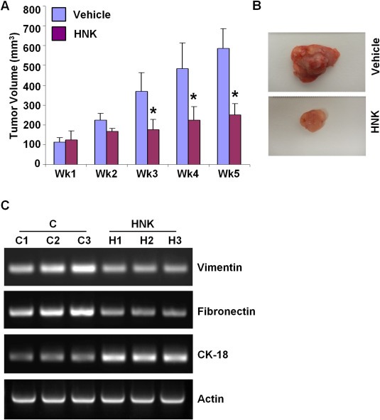
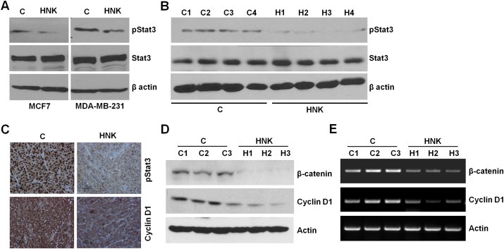
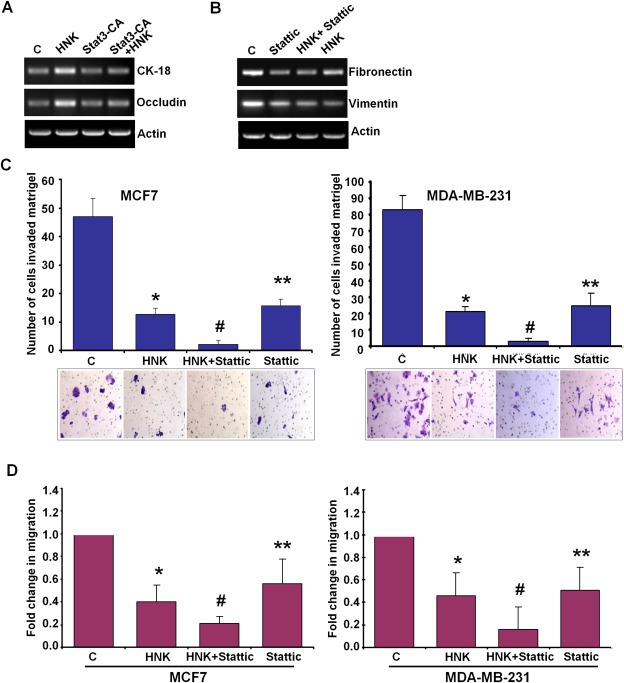
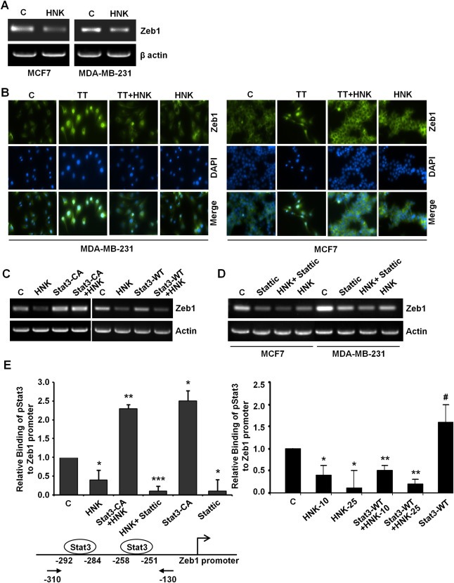
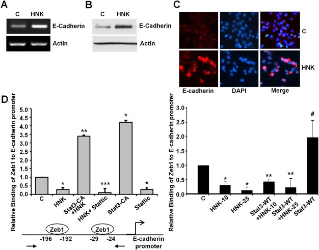
Epithelial-mesenchymal transition (EMT), a critical step in the acquisition of metastatic state, is an attractive target for therapeutic interventions directed against tumor metastasis. Honokiol (HNK) is a natural phenolic compound isolated from an extract of seed cones from Magnolia grandiflora. Recent studies from our lab show that HNK impedes breast carcinogenesis. Here, we provide molecular evidence that HNK inhibits EMT in breast cancer cells resulting in significant downregulation of mesenchymal marker proteins and concurrent upregulation of epithelial markers. Experimental EMT induced by exposure to TGFβ and TNFα in spontaneously immortalized nontumorigenic human mammary epithelial cells is also completely reversed by HNK as evidenced by morphological as well as molecular changes. Investigating the downstream mediator(s) that may direct EMT inhibition by HNK, we found functional interactions between HNK, Stat3, and EMT-signaling components. In vitro and in vivo analyses show that HNK inhibits Stat3 activation in breast cancer cells and tumors. Constitutive activation of Stat3 abrogates HNK-mediated activation of epithelial markers whereas inhibition of Stat3 using small molecule inhibitor, Stattic, potentiates HNK-mediated inhibition of EMT markers, invasion and migration of breast cancer cells. Mechanistically, HNK inhibits recruitment of Stat3 on mesenchymal transcription factor Zeb1 promoter resulting in decreased Zeb1 expression and nuclear translocation. We also discover that HNK increases E-cadherin expression via Stat3-mediated release of Zeb1 from E-cadherin promoter. Collectively, this study reports that HNK effectively inhibits EMT in breast cancer cells and provide evidence for a previously unrecognized cross-talk between HNK and Stat3/Zeb1/E-cadherin axis.

Keywords: Breast cancer; E-cadherin; EMT; Honokiol; Stat3; Zeb1.


Figures

Figure 1
Figure 1
Honokiol inhibits invasion and migration of breast cancer cells. A, MCF7 and MDA‐MB‐231 cells were cultured in matrigel‐invasion chambers followed by treatment with 5 μM honokiol (HNK) for 24 h as indicated. C represents vehicle controls. The number of cells that invaded through the matrigel was counted in five different regions. *P < 0.005, compared with untreated controls. B, MCF7 and MDA‐MB‐231 cells were subjected to spheroid‐migration assay. Culture media were replaced with media containing honokiol (5 μM) or vehicle control media (C). The spheroids were photographed at 24 h, 48 h and 72 h‐post treatment. The results shown are representative of three independent experiments performed in triplicates.
Figure 2
Figure 2
Honokiol inhibits the expression of mesenchymal genes and induces the expression of epithelial markers in breast cancer cells. A, D, MCF7 and MDA‐MB‐231 cells were treated with vehicle (C) or 5 μM honokiol (HNK). Total RNA was isolated and examined for the expression of fibronectin, vimentin and CK‐18 using specific primers. B, E, MCF7 and MDA‐MB‐231 cells were treated with vehicle (C) or 5 μM honokiol (HNK). Total lysates were immunoblotted for vimentin and occludin expression levels. C, F, MCF7 and MDA‐MB‐231 cells were treated with vehicle (C) or 5 μM honokiol (HNK) and subjected to immunofluorescence analysis of vimentin and occludin.
Figure 3
Figure 3
Honokiol abrogates TGFβ/TNFα‐induced epithelial–mesenchymal transition in mammary epithelial cells. A, MCF10A cells were treated with vehicle control (control), TGFβ + TNFα (10 ng/ml of each), 5 μM honokiol (HNK) or TGFβ + TNFα +HNK for 24 h. Morphological changes associated with EMT are shown in phase‐contrast images. The presence of spindle‐shaped cells, increased intercellular separation and pseudopodia were noted in TGFβ + TNFα‐treated cells but not in HNK‐treated or TGFβ + TNFα + HNK‐treated cells. B, MCF10A cells were treated as in A and total lysates were immunoblotted for vimentin and occludin expression levels. Actin was used as control. C, MCF10A cells were treated as in A, total RNA was isolated and expression of fibronectin and CK‐18 was analyzed. Actin was included as control. D, MCF10A cells were treated as in A, and subjected to immunofluorescence analysis of E‐Cadherin, occludin and vimentin.
Figure 4
Figure 4
Honokiol treatment inhibits breast tumor growth in nude mice. MDA‐MB‐231 cells derived tumors were developed in nude mice and treated with vehicle or honokiol (HNK). At the end of five weeks of treatment, tumors were collected, measured, weighed and photographed. A, Tumor growth was monitored by measuring the tumor volume for 5 weeks (n = 8 mice per group), *P < 0.001, compared with vehicle‐treated controls. B, Representative tumor images are shown here. C, Total RNA was isolated from tumor samples and subjected to RT‐PCR analysis. Expression of epithelial and mesenchymal markers (vimentin, fibronectin and CK‐18) was analyzed.
Figure 5
Figure 5
Evidence for honokiol‐mediated inhibition of signal transducer and activator of transcription 3. A, MCF7 and MDA‐MB‐231 cells were treated with vehicle (C) or 5 μM honokiol (HNK). Total lysates were immunoblotted for phosphorylated Stat3 (pStat3) and total Stat3 expression levels. B, MDA‐MB‐231 cells derived tumors were developed in nude mice, treated with vehicle or honokiol (HNK) for five weeks. Tumors were collected at the end of five weeks and subjected to western blot analysis for phosphorylated Stat3 (pStat3) and total Stat3 expression levels. C, MDA‐MB‐231 cells derived tumors, treated with vehicle or honokiol (HNK) were subjected to immunohistochemical analysis using phosphorylated Stat3 and cyclinD1 antibodies. D, Tumor lysates (from three different tumors from each set) were subjected to immunoblot analysis using β‐catenin and cyclinD1 antibodies. Actin antibody was used as control. E, Total RNA was isolated from three different tumors from each set were subjected to RT‐PCR analysis using β‐catenin and cyclinD1 primers. Actin primers were used as control.
Figure 6
Figure 6
Stat3‐inhibition plays an important role in honokiol‐mediated modulation of EMT markers, and inhibition of invasion and migration of breast cancer cells. A, MCF7 cells were treated with vehicle (C) or 5 μM honokiol (HNK), transfected with constitutively active Stat3 (Stat3‐CA) and treated with Honokiol (Stat3‐CA + HNK). Total RNA was isolated and subjected to RT‐PCR analysis using CK‐18 and occludin primers. Actin was included as control. B, MDA‐MB‐231 cells were treated with vehicle (C), 10 μM Stattic, 5 μM honokiol (HNK) alone, or in combination (HNK + Stattic), total RNA was isolated and subjected to RT‐PCR analysis using fibronectin and vimentin primers. Actin was included as control. C, MCF7 and MDA‐MB‐231 cells were cultured in matrigel‐invasion chambers followed by treatment with 10 μM Stattic, 5 μM honokiol (HNK) alone, or in combination (HNK + Stattic) for 24 h as indicated. C represents vehicle controls. The number of cells that invaded through the matrigel was counted in five different regions. *P < 0.005, compared with vehicle‐treated controls; **P < 0.001, compared with vehicle‐treated controls; #P < 0.005, compared with HNK‐treated cells. D, MCF7 and MDA‐MB‐231 cells were subjected to spheroid‐migration assay. Culture media were replaced with media containing 10 μM Stattic, 5 μM honokiol (HNK) alone, in combination (HNK + Stattic) or vehicle control (C). The spheroids were photographed 48 h‐post treatment. The results are shown as fold‐change in migration of breast cancer cells in response to HNK and Stattic treatments. These are representative of three independent experiments performed in triplicates. *P < 0.01, compared with vehicle‐treated controls; **P < 0.05, compared with vehicle‐treated controls; #P < 0.05, compared with HNK‐treated cells.
Figure 7
Figure 7
Honokiol inhibits Zeb1 expression, nuclear translocation and releases Stat3 from Zeb1 promoter. A, MCF7 and MDA‐MB‐231 cells were treated with vehicle (C) or 5 μM honokiol (HNK). Total RNA was isolated and examined for the expression of Zeb1 using specific primers. B, MCF7 and MDA‐MB‐231 cells were treated with vehicle control (C), TGFβ and TNFα (10 ng/ml of each) (TT), 5 μM honokiol (HNK) or TT + HNK and subjected to immunofluorescence analysis of Zeb1. TT induces nuclear translocation of Zeb1 which is abrogated by HNK treatment. C, MCF7 cells were treated with vehicle (C) or 5 μM honokiol (HNK), transfected with constitutively active Stat3 (Stat3‐CA) and treated with Honokiol (Stat3‐CA + HNK). In another set, MCF7 cells were treated with vehicle (C) or 5 μM honokiol (HNK), transfected with wild‐type Stat3 (Stat3‐WT) and treated with Honokiol (Stat3‐WT + HNK). Total RNA was isolated and subjected to RT‐PCR analysis using Zeb1 primers. Actin was included as control. D, MCF7 and MDA‐MB‐231 cells were treated with vehicle (C), 10 μM Stattic, 5 μM honokiol (HNK) alone, or in combination (HNK + Stattic), total RNA was isolated and subjected to RT‐PCR analysis using Zeb1 primers. Actin was included as control. E, Soluble chromatin was prepared from MCF7 cells treated with vehicle (C), 5 μM honokiol (HNK), transfected with constitutively active Stat3 (Stat3‐CA) alone or in combination with honokiol (Stat3‐CA + HNK), 10 μM Stattic alone or in combination (HNK + Stattic) and subjected to chromatin immunoprecipitation assay using pStat3 antibody. In another set, soluble chromatin was prepared from MCF7 cells treated with vehicle (C), 10 μM and 25 μM honokiol (HNK), transfected with wild‐type Stat3 (Stat3‐WT) alone or in combination with honokiol (Stat3‐WT + HNK‐10 and Stat3‐WT + HNK‐25) and subjected to chromatin immunoprecipitation assay using pStat3 antibody. The purified DNA was analyzed by real‐time quantitative PCR using primers spanning the Stat3‐binding sites at Zeb1 promoter.
Figure 8
Figure 8
Honokiol increases E‐cadherin expression and inhibits the recruitment of Zeb1 on E‐cadherin promoter in a Stat3‐dependent manner. A, MCF7 cells were treated with vehicle (C) or 5 μM honokiol (HNK). Total RNA was isolated and examined for the expression of E‐cadherin using specific primers. B, MCF7 cells were treated as in A, total lysates were immunoblotted for E‐cadherin expression levels. C, MCF7 cells were treated as in A and subjected to immunofluorescence analysis of E‐cadherin. D, Soluble chromatin was prepared from MCF7 cells treated with vehicle (C), 5 μM honokiol (HNK), transfected with constitutively active Stat3 (Stat3‐CA) alone or in combination with honokiol (Stat3‐CA + HNK), 10 μM Stattic alone or in combination (HNK + Stattic) and subjected to chromatin immunoprecipitation assay using Zeb1 antibody. In another set, soluble chromatin was prepared from MCF7 cells treated with vehicle (C), 10 μM and 25 μM honokiol (HNK), transfected with wild‐type Stat3 (Stat3‐WT) alone or in combination with honokiol (Stat3‐WT + HNK‐10 and Stat3‐WT + HNK‐25) and subjected to chromatin immunoprecipitation assay using Zeb1 antibody. The purified DNA was analyzed by real‐time quantitative PCR using primers spanning the Zeb1‐binding sites at E‐cadherin promoter.

References

    1. Ahn, K.S. , Sethi, G. , Shishodia, S. , Sung, B. , Arbiser, J.L. , Aggarwal, B.B. , 2006. Honokiol potentiates apoptosis, suppresses osteoclastogenesis, and inhibits invasion through modulation of nuclear factor-kappaB activation pathway. Mol. Cancer Res. MCR. 4, 621–633. - PubMed
    1. Arora, S. , Bhardwaj, A. , Srivastava, S.K. , Singh, S. , McClellan, S. , Wang, B. , Singh, A.P. , 2011. Honokiol arrests cell cycle, induces apoptosis, and potentiates the cytotoxic effect of gemcitabine in human pancreatic cancer cells. PLoS One. 6, e21573 - PMC - PubMed
    1. Arora, S. , Singh, S. , Piazza, G.A. , Contreras, C.M. , Panyam, J. , Singh, A.P. , 2012. Honokiol: a novel natural agent for cancer prevention and therapy. Curr. Mol. Med.. 12, 1244–1252. - PMC - PubMed
    1. Bai, X. , Cerimele, F. , Ushio-Fukai, M. , Waqas, M. , Campbell, P.M. , Govindarajan, B. , Der, C.J. , Battle, T. , Frank, D.A. , Ye, K. , Murad, E. , Dubiel, W. , Soff, G. , Arbiser, J.L. , 2003. Honokiol, a small molecular weight natural product, inhibits angiogenesis in vitro and tumor growth in vivo. J. Biol. Chem.. 278, 35501–35507. - PubMed
    1. Bar-Sela, G. , Epelbaum, R. , Schaffer, M. , 2010. Curcumin as an anti-cancer agent: review of the gap between basic and clinical applications. Curr. Med. Chem.. 17, 190–197. - PubMed

Publication types

MeSH terms