. 2014 Feb 20;40(2):262-73.
doi: 10.1016/j.immuni.2014.01.003. Epub 2014 Feb 6.

The cytokine IL-22 promotes pathogen colonization by suppressing related commensal bacteria

Affiliations

The cytokine IL-22 promotes pathogen colonization by suppressing related commensal bacteria

Judith Behnsen et al. Immunity. .

Abstract

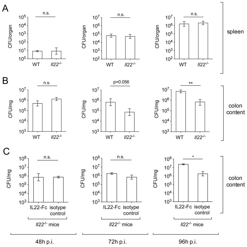
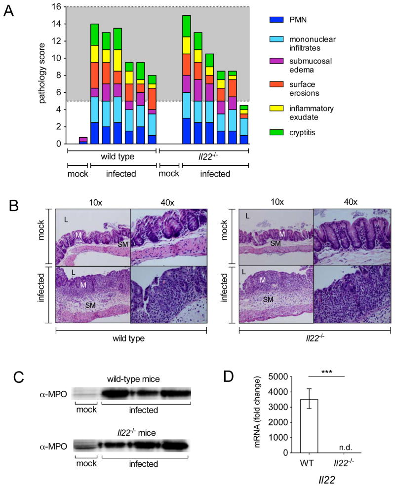
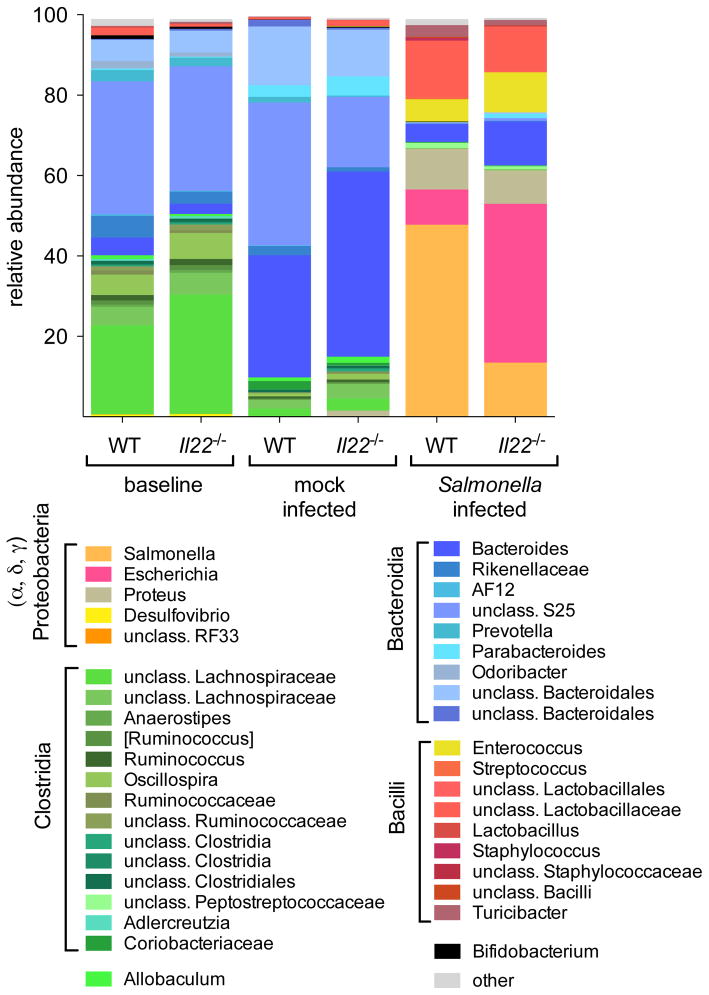
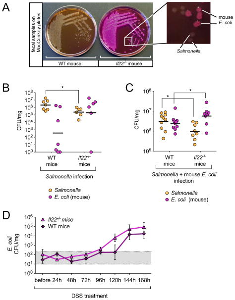
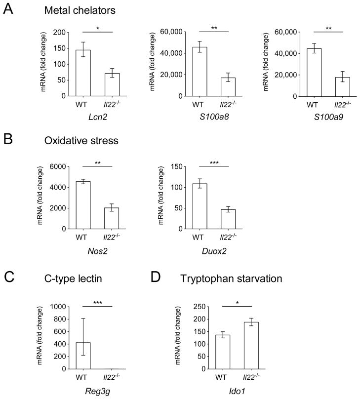
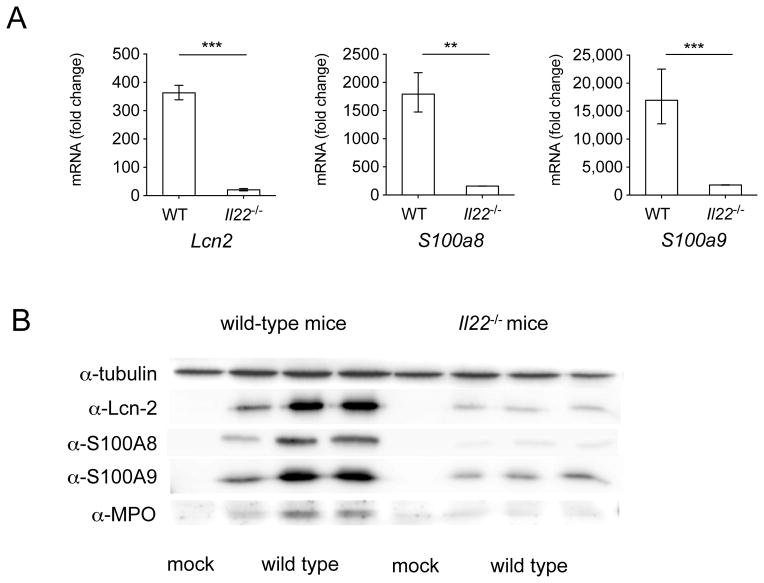
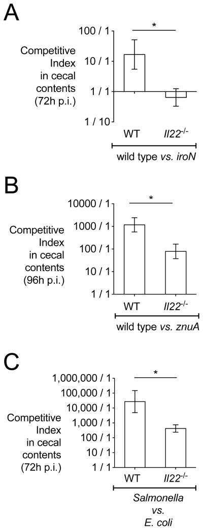
Interleukin-22 (IL-22) is highly induced in response to infections with a variety of pathogens, and its main functions are considered to be tissue repair and host defense at mucosal surfaces. Here we showed that IL-22 has a unique role during infection in that its expression suppressed the intestinal microbiota and enhanced the colonization of a pathogen. IL-22 induced the expression of antimicrobial proteins, including lipocalin-2 and calprotectin, which sequester essential metal ions from microbes. Because Salmonella enterica ser. Typhimurium can overcome metal ion starvation mediated by lipocalin-2 and calprotectin via alternative pathways, IL-22 boosted its colonization of the inflamed intestine by suppressing commensal Enterobacteriaceae, which are susceptible to the antimicrobial proteins. Thus, IL-22 tipped the balance between pathogenic and commensal bacteria in favor of a pathogen. Taken together, IL-22 induction can be exploited by pathogens to suppress the growth of their closest competitors, thereby enhancing pathogen colonization of mucosal surfaces.


Figures

Figure 1
Figure 1. Colonization of S. Typhimurium in WT and Il22−/− mice
WT and Il22−/− mice were infected with S. Typhimurium and colony forming units (CFUs) in spleen (A) and colon content (B) were determined at 48h (WT n=10, Il22−/− n=10), 72h (WT n=11, Il22−/− n=9) and 96h (WT n=11, Il22−/− n=10) after infection. (C) Il22−/− mice were administered IL22-Fc or an isotype control antibody (n=3/group) and fecal samples were collected at 48h, 72h and 96h after infection. Data shown represent CFUs per mg of fecal material for colon content and CFUs per organ for spleen. Data represent the geometric mean ± standard error. n.s.= not significant. A significant decrease over WT control is indicated by ** (P value ≤ 0.01). (See also Fig. S1 and Table S1).
Figure 2
Figure 2. Histopathology of WT and Il22−/− mice after infection with S. Typhimurium
(A) Blinded histopathology score indicating the score of individual mice 96h after either mock infection (treated with streptomycin but not infected) or infection with S. Typhimurium. The grey region includes scores indicative of moderate to severe inflammation. (B) H&E stained cecal sections from representative animals in each group. An image at lower magnification (10x) and one at higher magnification (40x) from the same section are shown. L=lumen; M=mucosa; SM=submucosa. Note marked edema in the submucosa and inflammation in mice infected with S. Typhimurium. (C) Myeloperoxidase (MPO) was detected 72h post infection by immunoblot in protein samples prepared from the cecum of mice that were mock infected or infected with S. Typhimurium. (D) Il22 was detected by quantitative real time PCR in the cecum of WT mice (n=6) and Il22−/− mice (n=6) 96h after infection with WT S. Typhimurium. A significant increase over mock control is indicated by *** (P value ≤ 0.001), n.d. = not detected. (See also Fig. S2 and Table S2).
Figure 3
Figure 3. Analysis of colonic microbiota in WT and Il22−/− mice by sequencing
Fecal samples were collected from mice before streptomycin treatment (n=9/group) and mock-infected animals (n=4/group) or S. Typhimurium-infected animals (n=5/group) at 96h post infection. The colonic microbiota was analyzed by sequencing using an Illumina MiSeq system. Graphed is the average relative abundance of each bacterial genus (See also Fig. S3 and Tables S3 and S4).
Figure 4
Figure 4. Competition of S. Typhimurium with E. coli
(A) Colon content samples collected from WT and Il22−/− mice at 96h post infection were resuspended in PBS and streaked on MacConkey agar plates for single colonies. Magnification shows the presence of lactose fermenting (pink) and lactose non-fermenting colonies in a sample from an Il22−/− mouse. (B+C) WT and Il22−/− mice were either (B) infected with S. Typhimurium (WT n=7, Il22−/− n=6) or (C) infected with S. Typhimurium and 24h later given 109 CFUs of mouse E. coli strain JB2 (WT n=10, Il22−/− n=8). Four days after infection with S. Typhimurium, colon content samples were collected and MacConkey plates were used to enumerate lactose fermenting (E. coli) and non lactose-fermenting (S. Typhimurium) colonies. Horizontal lines represent the geometric mean. (D) Fecal samples were collected from WT and Il22−/− mice before and every 24h after mice were given DSS in water. Samples were resuspended in PBS and plated on MacConkey agar plates to enumerate CFUs of E. coli (WT n=4, Il22−/− n=4). Gray area (101 to 3×10²) indicates basal E. coli level in mice (See also Fig. S4).
Figure 5
Figure 5. Expression of antimicrobial peptide genes
(A) Lcn2, S100a8, S100a9, (B) Duox2, Nos2, (C) Reg3g, and (D) Ido1 were detected by quantitative real time PCR in the cecum of WT mice and Il22−/− mice 72h after infection with WT S. Typhimurium. Infected WT n=6, infected Il22−/− n=6, mock n=4. Data are expressed as fold increase over mock-infected WT mice. Data represent the geometric mean ± standard error. A significant increase over mock control is indicated by * (P value ≤ 0.05), ** (P value ≤ 0.01) and *** (P value ≤ 0.001). (See also Fig. S5).
Figure 6
Figure 6. Expression of antimicrobial peptide genes in colonic crypts
(A) Lcn2, S100a8 and S100a9 were detected by quantitative real time PCR in isolated colonic crypts of WT mice and Il22−/− mice 48h after infection with WT S. Typhimurium. Infected WT n=6, infected Il22−/− n=6, mock n=2. Data are expressed as fold increase over mock-infected WT mice. Data represent the geometric mean ± standard error (for some conditions error marks are not visible due to small error). A significant increase over mock control is indicated by ** (P value ≤ 0.01) and *** (P value ≤ 0.001). (B) Lcn-2, S100A8, S100A9, myeloperoxidase (MPO), and tubulin were detected 48h post infection by immunoblot in isolated crypts of WT and Il22−/− mice that were mock-infected or infected with S. Typhimurium.
Figure 7
Figure 7. Competition in wild-type and Il22−/− mice of S. Typhimurium WT with strains of known sensitivity to antimicrobial proteins
Colon content samples were collected from mice three or four days after infection with S. Typhimurium. Competitive index was calculated by dividing the output CFU ratio (WT/mutant or E. coli) by the input CFU ratio (WT/mutant or E. coli). Competitive indices of S. Typhimurium strains in the colon contents of WT and Il22−/− mice (n=5/group) infected with (A) an equal mixture of WT S. Typhimurium and the iroN mutant, (B) WT S. Typhimurium and the znuA mutant or (C) WT S. Typhimurium and E. coli. Data represent the geometric mean ± standard error. A significant decrease over the competitive index in WT mice is indicated by * (P value ≤ 0.05).

Comment in

  • The battle in the gut.
    Monack DM. Monack DM. Immunity. 2014 Feb 20;40(2):173-5. doi: 10.1016/j.immuni.2014.01.007. Immunity. 2014. PMID: 24560194

References

    1. Aujla SJ, Chan YR, Zheng M, Fei M, Askew DJ, Pociask DA, Reinhart TA, McAllister F, Edeal J, Gaus K, et al. IL-22 mediates mucosal host defense against Gram-negative bacterial pneumonia. Nat Med. 2008;14:275–281. - PMC - PubMed
    1. Awoniyi M, Miller SI, Wilson CB, Hajjar AM, Smith KD. Homeostatic regulation of Salmonella-induced mucosal inflammation and injury by IL-23. PLoS One. 2012;7:e37311. - PMC - PubMed
    1. Barman M, Unold D, Shifley K, Amir E, Hung K, Bos N, Salzman N. Enteric salmonellosis disrupts the microbial ecology of the murine gastrointestinal tract. Infection and immunity. 2008;76:907–915. - PMC - PubMed
    1. Barthel M, Hapfelmeier S, Quintanilla-Martinez L, Kremer M, Rohde M, Hogardt M, Pfeffer K, Russmann H, Hardt WD. Pretreatment of mice with streptomycin provides a Salmonella enterica serovar Typhimurium colitis model that allows analysis of both pathogen and host. Infection and immunity. 2003;71:2839–2858. - PMC - PubMed
    1. Berger T, Togawa A, Duncan GS, Elia AJ, You-Ten A, Wakeham A, Fong HE, Cheung CC, Mak TW. Lipocalin 2-deficient mice exhibit increased sensitivity to Escherichia coli infection but not to ischemia-reperfusion injury. Proc Natl Acad Sci U S A. 2006;103:1834–1839. - PMC - PubMed

Publication types