

Morphological and physiological development of auditory synapses
- PMID: 24508369
- PMCID: PMC4122648
- DOI: 10.1016/j.heares.2014.01.007
Morphological and physiological development of auditory synapses
Abstract
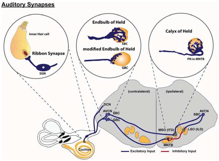
Acoustic communication requires gathering, transforming, and interpreting diverse sound cues. To achieve this, all the spatial and temporal features of complex sound stimuli must be captured in the firing patterns of the primary sensory neurons and then accurately transmitted along auditory pathways for additional processing. The mammalian auditory system relies on several synapses with unique properties in order to meet this task: the auditory ribbon synapses, the endbulb of Held, and the calyx of Held. Each of these synapses develops morphological and electrophysiological characteristics that enable the remarkably precise signal transmission necessary for conveying the miniscule differences in timing that underly sound localization. In this article, we review the current knowledge of how these synapses develop and mature to acquire the specialized features necessary for the sense of hearing.
Copyright © 2014 Elsevier B.V. All rights reserved.
Figures
Similar articles
-
Molecularly and structurally distinct synapses mediate reliable encoding and processing of auditory information.Hear Res. 2015 Dec;330(Pt B):178-90. doi: 10.1016/j.heares.2015.07.008. Epub 2015 Jul 15. Hear Res. 2015. PMID: 26188105 Review.
-
The ion channels and synapses responsible for the physiological diversity of mammalian lower brainstem auditory neurons.Hear Res. 2019 May;376:33-46. doi: 10.1016/j.heares.2018.12.011. Epub 2018 Dec 26. Hear Res. 2019. PMID: 30606624 Review.
-
Transmission of auditory sensory information decreases in rate and temporal precision at the endbulb of Held synapse during age-related hearing loss.J Neurophysiol. 2016 Dec 1;116(6):2695-2705. doi: 10.1152/jn.00472.2016. Epub 2016 Sep 28. J Neurophysiol. 2016. PMID: 27683884 Free PMC article.
-
Remodelling at the calyx of Held-MNTB synapse in mice developing with unilateral conductive hearing loss.J Physiol. 2014 Apr 1;592(7):1581-600. doi: 10.1113/jphysiol.2013.268839. Epub 2014 Jan 27. J Physiol. 2014. PMID: 24469075 Free PMC article.
-
Synaptic transmission at the calyx of Held under in vivo like activity levels.J Neurophysiol. 2007 Aug;98(2):807-20. doi: 10.1152/jn.00355.2007. Epub 2007 May 16. J Neurophysiol. 2007. PMID: 17507501
Cited by
-
Cellular and synaptic specializations for sub-millisecond precision in the mammalian auditory brainstem.Front Cell Neurosci. 2025 May 19;19:1568506. doi: 10.3389/fncel.2025.1568506. eCollection 2025. Front Cell Neurosci. 2025. PMID: 40458470 Free PMC article. Review.
-
Ribbon synapses in zebrafish hair cells.Hear Res. 2015 Dec;330(Pt B):170-7. doi: 10.1016/j.heares.2015.04.003. Epub 2015 Apr 25. Hear Res. 2015. PMID: 25916266 Free PMC article. Review.
-
The Purinergic Receptor P2rx3 is Required for Spiral Ganglion Neuron Branch Refinement during Development.eNeuro. 2020 Aug 10;7(4):ENEURO.0179-20.2020. doi: 10.1523/ENEURO.0179-20.2020. Print 2020 Jul/Aug. eNeuro. 2020. PMID: 32675174 Free PMC article.
-
Developmental plasticity shapes synaptic phenotypes of autism-associated neuroligin-3 mutations in the calyx of Held.Mol Psychiatry. 2017 Oct;22(10):1483-1491. doi: 10.1038/mp.2016.157. Epub 2016 Oct 11. Mol Psychiatry. 2017. PMID: 27725662 Free PMC article.
-
Visualization of Exo- and Endocytosis of AMPA Receptors During Hippocampal Synaptic Plasticity Around Postsynaptic-Like Membrane Formed on Glass Surface.Front Cell Neurosci. 2018 Nov 21;12:442. doi: 10.3389/fncel.2018.00442. eCollection 2018. Front Cell Neurosci. 2018. PMID: 30519162 Free PMC article.
References
-
- Ashmore JF, Kolston PJ. Hair cell based amplification in the cochlea. Current opinion in neurobiology. 1994;4:503–8. - PubMed
Publication types
MeSH terms
Grants and funding
LinkOut - more resources
Full Text Sources
Other Literature Sources
