. 2014 May:50:87-98.
doi: 10.1016/j.jaut.2014.01.002. Epub 2014 Feb 7.

Ly108 expression distinguishes subsets of invariant NKT cells that help autoantibody production and secrete IL-21 from those that secrete IL-17 in lupus prone NZB/W mice

Affiliations

Ly108 expression distinguishes subsets of invariant NKT cells that help autoantibody production and secrete IL-21 from those that secrete IL-17 in lupus prone NZB/W mice

Xiaobin Tang et al. J Autoimmun. 2014 May.

Abstract

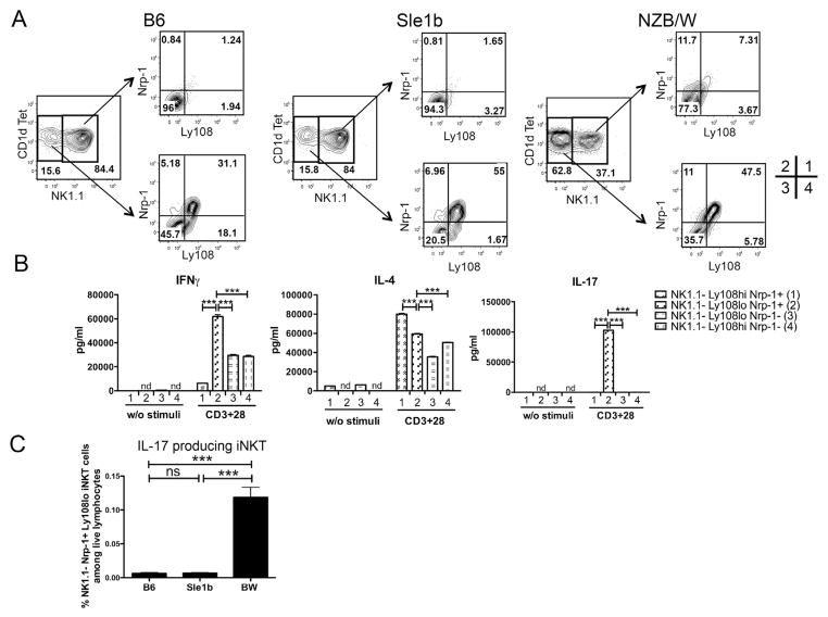
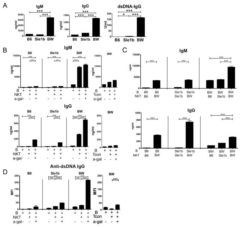
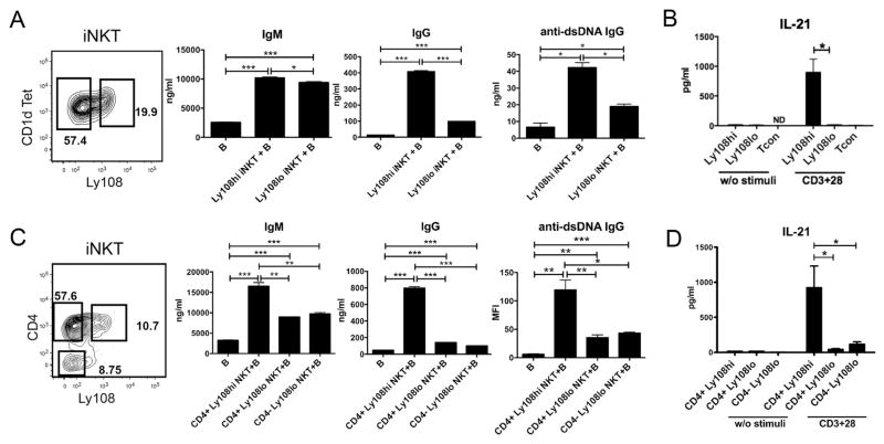
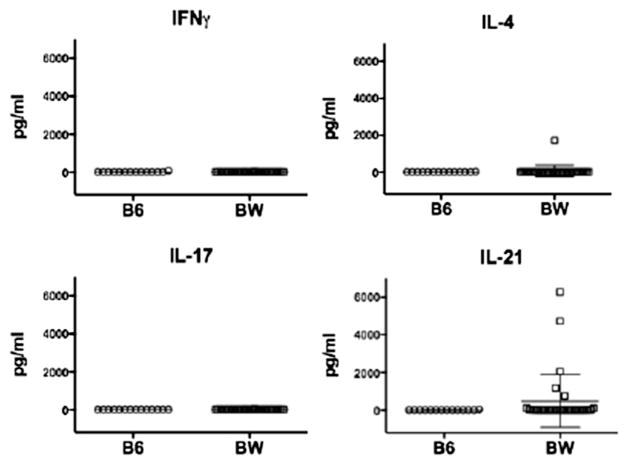
Lupus is a systemic autoimmune disease characterized by anti-nuclear antibodies in humans and genetically susceptible NZB/W mice that can cause immune complex glomerulonephritis. T cells contribute to lupus pathogenesis by secreting pro-inflammatory cytokines such as IL-17, and by interacting with B cells and secreting helper factors such as IL-21 that promote production of IgG autoantibodies. In the current study, we determined whether purified NKT cells or far more numerous conventional non-NKT cells in the spleen of NZB/W female mice secrete IL-17 and/or IL-21 after TCR activation in vitro, and provide help for spontaneous IgG autoantibody production by purified splenic CD19(+) B cells. Whereas invariant NKT cells secreted large amounts of IL-17 and IL-21, and helped B cells, non-NKT cells did not. The subset of IL-17 secreting NZB/W NKT cells expressed the Ly108(lo)CD4(-)NK1.1(-) phenotype, whereas the IL-21 secreting subset expressed the Ly108(hi)CD4(+)NK1.1(-) phenotype and helped B cells secrete a variety of IgG anti-nuclear antibodies. α-galactocylceramide enhanced the helper activity of NZB/W and B6.Sle1b NKT cells for IgG autoantibody secretion by syngeneic B cells. In conclusion, different subsets of iNKT cells from mice with genetic susceptibility to lupus can contribute to pathogenesis by secreting pro-inflammatory cytokines and helping autoantibody production.

Keywords: Autoantibodies; IL-17; IL-21; Natural killer T cells; Systemic lupus erythematosus.


Figures

Figure 1
Figure 1
Flow cytometric analyses of Ly108 expression on iNKT and Tcon cells from B6, Sle1b and BW splenocytes. Single cell suspensions were obtained from young mice that range 8 to 16 weeks old (mean 14 weeks) and analyzed by flow cytometry. (A) iNKT (TCR+CD1dtet+) and Tcon (TCR+CD1dtet) populations of B6, Sle1b and BW mice are enclosed in boxes, and percentage within each box is shown; mean frequencies (percentage among gated lymphocytes) of iNKT and Tcon cells are shown on the right. (B) Percentage of Ly108hi cells among iNKT and Tcon cells from B6, Sle1b and BW mice are enclosed in boxes; mean percentages for Ly108hi population in iNKT and Tcon gates are shown on the right. (C) NK1.1 staining on iNKT cells from B6, Sle1b and BW mice, and boxes show NK1.1+ and NK1.1 cells; mean percentages for NK1.1 population in iNKT cells are shown on the right. (D) Ly108hi cells among NK1.1+ and NK1.1 iNKT populations from BW mice shown on the left; mean percentages for Ly108hi population in NK1.1+ (closed square) and NK1.1 (open circle) iNKT cells from B6, Sle1b and BW mice are shown on the right. Each symbol in graphs represents an individual mouse. Data are presented as the mean ± s.e.m. Group differences with P>0.05 were not considered statistically significant (ns). *, P<0.05; **, P<0.01; ***, P<0.001 (two-tailed Student’s t-test).
Figure 2
Figure 2
BW iNKT cells, produce significantly higher amounts of IL-4 and IL-17 than Tcon cells and Ly108lo iNKT cells are predominant IL-17 producing iNKT cells. Cells were sorted from three pooled spleens of young mice and cultured (5X10⁵/ml) for 3 days with or without plate bound anti-CD3 and CD28 mAbs, culture supernatants were then collected for IFNγ, IL-4 and IL-17 measurement by ELISA or Luminex. (A) IFNγ, IL-4 and IL-17 concentrations in supernatants of stimulated iNKT and Tcon cells from BW mice. Cytokines measurements are shown on the right. (B) IFNγ, IL-4 and IL-17 production by BW CD4+ Ly108hi, CD4+ Ly108lo and CD4 Ly108lo iNKT cells. Sorting gates are shown on the left, and cytokines measurements were shown on the right. (C) IFNγ, IL-4 and IL-17 production by BW NK1.1+ and NK1.1 iNKT cells. FACS profile is shown for the sorting gates on the left, and cytokines measurements are shown on the right. (D) IFNγ, IL-4 and IL-17 production by BW NK1.1Ly108hi and NK1.1Ly108lo iNKT cells. FACS profile is shown for the sort gates on the left, and cytokines measurements are shown on the right. Group differences with P>0.05 was not considered statistically significant (ns). *, P<0.05; **, P<0.01; ***, P<0.001; ****, P<0.0001 (two-tailed Student’s t-test). Data (mean ± s.e.m.) are calculated from four independent experiments (A, B) or derived from triplicate cultures collected from one representative experiment of three with similar results (C, D). Cytokine concentrations without anti-CD3 and CD28 mAbs were less than 100pg/ml.
Figure 3
Figure 3
NK1.1Ly108loNrp-1+ iNKT cells are IL-17 producing iNKT cells. (A) Ly108 and Nrp-1 expression on gated NK1.1+ and NK1.1 iNKT cells from young B6, Sle1b and BW mice. (B) IFNγ, IL-4 and IL-17 production by sorted BW NK1.1Ly108hiNrp-1+ (indicated as population 1 as shown in A), NK1.1Ly108lo Nrp-1+ (population 2), NK1.1Ly108loNrp-1 (population 3), NK1.1Ly108hiNrp-1 (population 4) iNKT cells. Cells were sorted from three to five pooled spleens from young BW mice and cultured (5X10⁵/ml) for 3 days with anti-CD3 and CD28 mAbs. (C) Percentages of IL-17 producing iNKT cells, defined as NK1.1Ly108loNrp-1+TCR+CD1dTet+ population, among live lymphocytes of B6, Sle1b and BW mice. nd, not detected. Group differences with P>0.05 was not considered statistically significant (ns); ***, P<0.001 (two-tailed Student’s t-test). Bar graphs show mean± s.e.m. Results are representative of four independent experiments (A), or two independent experiments (B), or four independent experiments (C). Independent experiments showed similar p values for group comparisons.
Figure 4
Figure 4
BW iNKT cells provide help for spontaneous IgG secretion of B cells from B6, Sle1b and NZB/W mice, and α-GalCer augments production of IgG and IgG autoantibodies. Splenocytes or sorted CD19+ B cells with or without sorted iNKT cells were cultured for 10 days, and supernatants were collected for determination of IgM, IgG and IgG anti-dsDNA antibody concentrations. (A) IgM, IgG and IgG anti-dsDNA antibody concentrations from 10-day culture supernatants of 6 month old B6, Sle1b and NZB/W whole splenocytes. (B, C) IgM and IgG production from 6 month old mice, with B cells cultured with or without iNKT or Tcon cells from same strain (B) or different strains (C), in the presence of α-GalCer or not. (D) IgG anti-dsDNA antibody production from cell cultures of (B) detected by proteomic microarray. *, P<0.05; **, P<0.01; ***, P<0.001 (two-tailed Student’s t-test). Results (mean ± s.e.m.) are calculated from triplicate cultures from one representative experiment of two or three experiments with similar results.
Figure 5
Figure 5
CD4+Ly108hi iNKT cells provide help for spontaneous antibody secretion of B cells. Sorted B cells with or without sorted iNKT cells, were cultured for 10 days, then supernatant were collected for determination of IgM, IgG and IgG anti-dsDNA antibody concentrations. For IL-21 production assays, cells were sorted and cultured (5X10⁵/ml) for 3 days with anti-CD3 and CD28 mAbs. Different populations of iNKT cells were sorted from three pooled spleens of BW mice. (A) IgM, IgG and IgG anti-dsDNA antibody production from cultures of B cells, with or without sorted Ly108hi or Ly108lo iNKT cells from 6 month old BW mice. (B) IL-21 production by anti-CD3/CD28 stimulated iNKT and Tcon cells of young BW mice. (C) IgM, IgG and IgG anti-dsDNA antibody production from B cells cultures, with or without sorted CD4+Ly108hi, CD4+Ly108lo or CD4Ly108lo iNKT cells from 6 month old BW mice. (D) IL-21 production by sorted CD4+Ly108hi, CD4+Ly108lo or CD4- Ly108lo iNKT from young BW mice. ND, Not Detected. *, P<0.05; **, P<0.01; ***, P<0.001 (two-tailed Student’s t-test). Data (mean ± s.e.m.) shown are derived from three or four independent experiments (B, D) or from triplicate collected from one representative experiment of two to five with similar results (A, C)
Figure 6
Figure 6
IL-21 is increased in the serum of young BW mice. Serum IFNγ, IL-4, IL-17 and IL-21 concentrations in young B6 (n=14) and BW (n=32) mice were determined by Lumenix assays. Bar graphs show mean ± s.e.m.
Figure 7
Figure 7
Phenotype of infiltrating BW kidney T cells and production of IL-17. (A, B) Representative flow cytometric analyses of 6 month old BW kidney mononuclear cells (KMC) (A) and spleen cells (B). (C) Mean percentages of B, total T, iNKT and Tcon cells among gated lymphocytes. (D) Mean percentages of different subsets of iNKT cells among iNKT cells. (E) Mean percentages of CD4+CD8, CD4CD8+ and DN cells among BW kidney Tcon (left) and iNKT cells (middle), and percentages of total T, Tcon and iNKT DN cells among BW live kidney lymphocytes (right). (F) IFNγ and IL-17 production by BW KMC cells (5X10⁵/ml) upon stimulation with anti-CD3 and CD28 mAbs. 5X10⁵/ml KMC cells prepared from one or two kidneys from 6 month old BW mice were cultured in 96 well plates for 3 days. (G) IFNγ and IL-17 production by BW KMC cells with stimulation by α-GalCer. KMC (1X10⁵) cells obtained from two 6 month old BW mice were cultured for 4 days with or without DCs (5X10³), in the presence of α-GalCer (100 ng/mL) or not. Culture supernatants were collected for IFNγ and IL-17 measurements by Lumenix. ***, P<0.001 (two-tailed Student’s t-test). Bar graphs show mean ± s.e.m. Data are derived from six mice from three independent experiments (A-E), or from three independent experiments (F), or from two independent experiments (G).

Similar articles

Cited by

References

    1. Kotzin BL. Systemic lupus erythematosus. Cell. 1996;85:303–6. - PubMed
    1. Theofilopoulos AN, Kofler R, Singer PA, Dixon FJ. Molecular genetics of murine lupus models. Adv Immunol. 1989;46:61–109. - PubMed
    1. Tsokos GC. Systemic lupus erythematosus. N Engl J Med. 2011;365:2110–21. - PubMed
    1. Ando DG, Sercarz EE, Hahn BH. Mechanisms of T and B cell collaboration in the in vitro production of anti-DNA antibodies in the NZB/NZW F1 murine SLE model. J Immunol. 1987;138:3185–90. - PubMed
    1. Borchers A, Ansari AA, Hsu T, Kono DH, Gershwin ME. The pathogenesis of autoimmunity in New Zealand mice. Semin Arthritis Rheum. 2000;29:385–99. - PubMed

Publication types

MeSH terms