. 2014 Feb 27;6(4):657-69.
doi: 10.1016/j.celrep.2014.01.013. Epub 2014 Feb 6.

Destruction of full-length androgen receptor by wild-type SPOP, but not prostate-cancer-associated mutants

Affiliations

Destruction of full-length androgen receptor by wild-type SPOP, but not prostate-cancer-associated mutants

Jian An et al. Cell Rep. .

Abstract

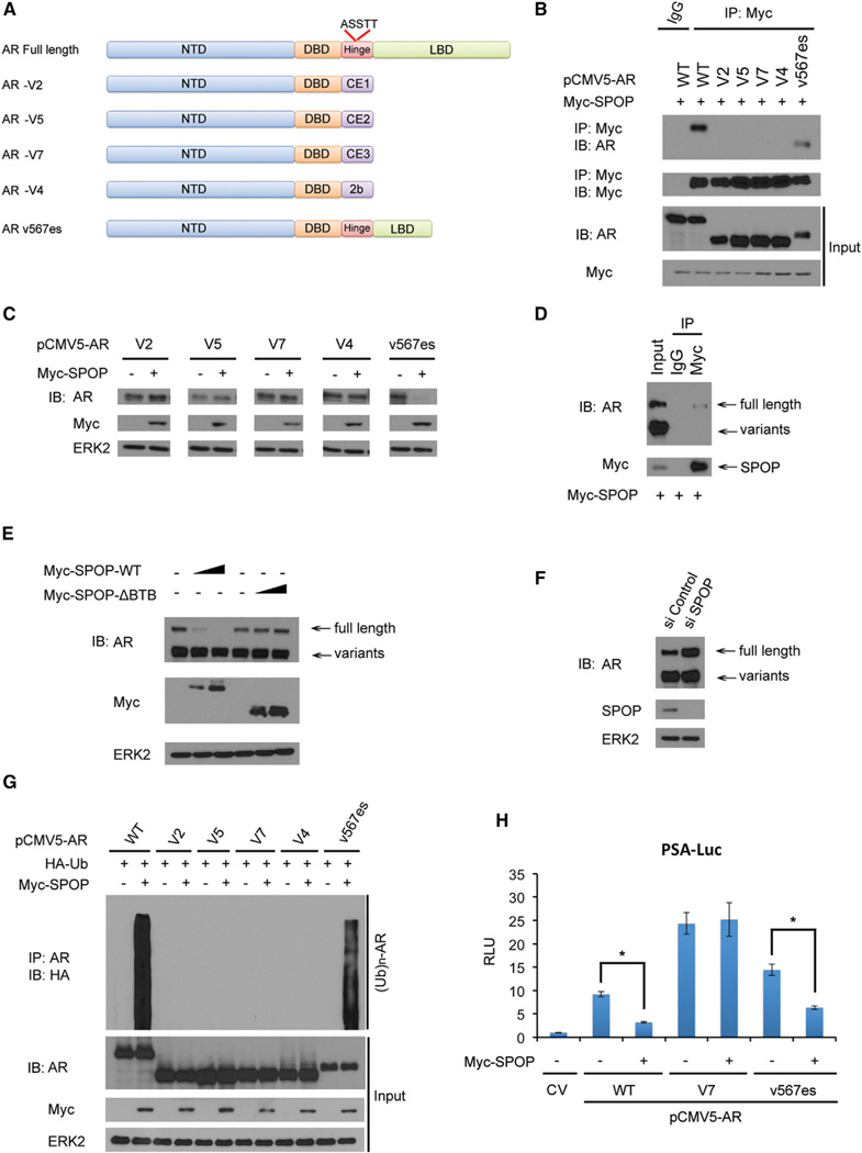
The SPOP E3 ubiquitin ligase gene is frequently mutated in human prostate cancers. Here, we demonstrate that SPOP recognizes a Ser/Thr-rich degron in the hinge domain of androgen receptor (AR) and induces degradation of full-length AR and inhibition of AR-mediated gene transcription and prostate cancer cell growth. AR splicing variants, most of which lack the hinge domain, escape SPOP-mediated degradation. Prostate-cancer-associated mutants of SPOP cannot bind to and promote AR destruction. Furthermore, androgens antagonize SPOP-mediated degradation of AR, whereas antiandrogens promote this process. This study identifies AR as a bona fide substrate of SPOP and elucidates a role of SPOP mutations in prostate cancer, thus implying the importance of this pathway in resistance to antiandrogen therapy of prostate cancer.


Figures

Figure 1
Figure 1. The SPOP-CUL3-RBX1 Complex Targets AR for Ubiquitination and Degradation
(A) Comparison of putative SPOP binding sites in AR with the SPOP-binding consensus motif defined in the known SPOP substrates. (B) Ectopically expressed SPOP promotes exogenous AR protein degradation. 293T cells were transfected with indicated constructs for 24 hr followed by western blot (WB). ERK2, a loading control. (C) SPOP regulates AR protein levels through the proteasome pathway. 293T cells were transfected with 4 µg pCMV5-AR and 0, 2, or 4 µg Myc-SPOP plasmids. After 16 hr, cells were treated with 20 µM MG132 for 8 hr and harvested for WB. (D) SPOP regulates endogenous AR protein levels. LNCaP cells were transfected with 0, 2,or4 µgHA-SPOP plasmid for 24 hr before cells were harvested for WB. (E) Knockdown of SPOP increases endogenous AR protein levels. LNCaP and C4-2 cells were transfected with control or two independent SPOP-specific siRNA. After 48 hr, cells were harvested for WB. (F and G) SPOP knockdown prolongs AR protein half-life. LNCaP cells were transfected with control or SPOP-specific siRNA. After 48 hr, cells were treated with 50 µg/µl cycloheximide (CHX). At different time points, cells were harvested for WB (F). At each time point, the intensity of AR was normalized to the intensity of ERK2 (loading control) first and then to the value at the 0 hr time point (G). A similar result was obtained in two independent experiments. (H) The BTB domain in SPOP is essential for SPOP-mediated degradation of AR. We transfected 4 µg pCMV5-AR and 0, 2, or 6 µg Myc-SPOP-WT or Myc-SPOP-ΔBTB plasmids into 293T cells for 24 hr, followed by WB. (I) Knockdown of RBX1 or CUL3 increases endogenous AR protein levels. LNCaP cells were transfected with control siRNA or siRNAs for RBX1 or CUL3 for 48 hr followed by WB. (J) SPOP promotes AR polyubiquitination in vivo. 293T cells were transfected with indicated plasmids for 16 hr followed by treatment with 20 µM MG132 for 8 hr. Immunoprecipitated AR proteins were analyzed by WB for ubiquitination. (K) Knockdown of SPOP decreases ubiquitination of endogenous AR. LNCaP cells were transfected with indicated plasmids and siRNAs for 40 hr followed by treatment with 20 µM MG132 for 8 hr, and then the same procedure was performed as shown in (J). (L) Quantitative RT-PCR measurement of the mRNA level of SPOP, AR, and two AR target genes (PSA and TMPRSS2) in SPOP-knockdown LNCaP cells. The mRNA level of GAPDH was used for normalization. All data shown are mean values ± SD (error bar) from three independent experiments. p < 0.01. (M and N) SPOP inhibits prostate cancer cell growth via regulation of AR. C4-2 cells were infected with lentivirus expressing control or SPOP and/or AR-specific small hairpin RNAs. Forty-eight hours after infection, cells were harvested for WB (M) or cultured in androgen-depleted medium for MTS assay (N). All data shown are mean values ± SD (error bar) from six replicates. p < 0.01.
Figure 2
Figure 2. SPOP Interacts with AR In Vitro and In Vivo
(A and B) Ectopically expressed SPOP and AR proteins interact with each other. 293T cells were transfected with indicated plasmids for 16 hr followed by treatment with 20 µM MG132 for 8 hr and coIP with anti-HA (A) or anti-AR (B) and WB. (C) Endogenous SPOP and AR proteins interact with each other in LNCaP cells. After treatment with 20 µM MG 132 for8 hr, cell lysates were prepared for coIP with AR antibody and WB. SKP2 was included as a negative control. (D) Schematic diagram of three GST-AR recombinant proteins. NTD, NH2-terminal domain; DBD, DNA binding domain; LBD, ligand binding domain. (E) SPOP binds to the central part of AR including DBD and the hinge domain. Bacterially expressed GST fusion proteins of NTD, LBD, and DBD plus the hinge domain were incubated with cell lysates of 293T cells transfected with HA-SPOP and subjected to GST pull-down assay. Bound HA-SPOP was detected by WB with HA antibody, and GST fusion proteins were detected by GelCode blue staining.
Figure 3
Figure 3. The 645ASSTT649 Motif in AR Is a Degron Recognized by SPOP
(A) Diagram showing two putative SPOP-binding motifs located in AR NTD and the hinge domain, respectively. (B) The 645ASSTT649 motif is required for AR binding to SPOP. 293T cells were transfected with plasmids for Myc-SPOP and wild-type (WT) and three deletion mutants of AR(ΔEGSSS, ΔASSTT, and double deletion 2Δ). After16hr, cells were treated with 20 µM MG132 for8 hr followed by coIP with Myc antibody and WB. (C) The 645ASSTT649 motif is required for AR degradation by SPOP. 293T cells were transfected with indicated plasmids for 24 hr followed by WB. (D) S647 and T648 residues in the 645ASSTT649 motif are critical for SPOP-induced degradation of AR. 293T cells were transfected with the indicated plasmids for 24 hr followed by WB. The density of AR was determined by normalizing to ERK2 (loading control) first and then to the normalized value in Myc-SPOP-untransfected cells. (E) The 645ASSTT649 motif is essential for SPOP-induced AR polyubiquitination. 293T cells were transfected with the indicated plasmids for 16 hr followed by treatment with 20 µM MG132 for 8 hr, IP, and WB. (F and G) Deletion of the 645ASSTT649 motif prolongs the half-life of AR protein. AR WT or AR ΔASSTT mutant was transfected into 293T cells for 24 hr followed by treatment with 50 µg/µl cycloheximide (CHX). At various time points, cells were harvested for WB (F). At each time point, the intensity of AR was normalized to the intensity of ERK2 first and then to the value at the 0 hr time point (G). A similar result was obtained in two independent experiments.
Figure 4
Figure 4. Hinge Domain Null AR Splicing Variants Are Resistant to SPOP-Mediated Degradation
(A) Schematic diagram of full-length AR and five AR splicing variants (V2, V5, V7, V4, and v567es). Only full-length AR and v567es contain the 645ASSTT649 motif. (B) Hinge domain-deficient AR splicing variants lose the capacity of binding to SPOP. 293T cells were transfected with the indicated plasmids for 16 hr followed by treatment with 20 µM MG132 for 8 hr, coIP, and WB. (C) Hinge domain null AR splicing variants are resistant to SPOP-promoted degradation. 293T cells were transfected with the indicated plasmids for24 hr followed by WB. (D) SPOP binds to endogenous full-length AR but not the variants in 22Rv1 cells. Cells were transfected with Myc-SPOP. After 16 hr, cells were treated with MG132 for 8 hr. Cell lysates were subjected to coP and WB. (E) Ectopically expressed SPOP differentially targets endogenous full-length AR and variants for degradation in 22Rv1 cells. Cells were transfected with 0, 2, or 4 µg plasmid for Myc-SPOP-WT or Myc-SPOP-ΔBTB. After 24 hr, cell lysates were prepared for WB. (F) Knockdown of endogenous SPOP increases protein levels of endogenous full-length AR but not variants in 22Rv1 cells. Cells were transfected with control or SPOP-specific siRNAs for 48 hr followed by WB. (G) SPOP cannot induce polyubiquitination of hinge domain null AR splicing variants. 293T cells were transfected with the indicated plasmids for 16 hr followed by treatment with 20 µM MG132 for 8 hr, IP, and WB. (H) Differential effects of SPOP on the transcriptional activity of V7 hinge domain null and v567es variants of AR. 293T cells were transfected with PSA-Luc firefly luciferase reporter, renilla luciferase reporter, and the indicated plasmids. After 24 hr, cells were harvested for measurement of luciferase activities. Relative luciferase units (RLU) were determined by first normalizing the firefly units with the renilla activity and then normalized to the value of cells transfected with control vector (CV). All data shown are mean values ± SD (error bar) from three independent experiments. p < 0.01.
Figure 5
Figure 5. Prostate-Cancer-Associated Mutants of SPOP Fail to Promote AR Degradation
(A) Computer modeling of the SPOP MATH domain indicating the positions of the residues mutated in prostate cancer. The side chains of mutated residues are shown as sticks in red; the substrate is shown as a tube in green; the MATH domain is shown as transparent surface in light gray. The figure is made using software vmd-1.9 (http://www.ks.uiuc.edu/Research/vmd/). (B) Prostate-cancer-associated mutants of SPOP cannot interact with AR. 293T cells were transfected with the indicated plasmids for 16 hr followed by treatment with 20 µM MG132 for 8 hr, coIP, and WB. (C) Prostate-cancer-associated mutants of SPOP fail to induce degradation of endogenous AR. C4-2 cells were transfected with wild-type (WT) or mutated SPOP as indicated for 24 hr followed by WB. The density of AR was determined by normalizing to ERK2 (loading control) first and then to the normalized value in Myc-SPOP-untransfected cells. (D) Prostate-cancer-associated mutants of SPOP cannot promote AR ubiquitination. 293T cells were transfected with the indicated plasmids. After 16 hr, cells were treated with MG132 for 8 hr, and cell lysates were prepared for IP and WB. (E and F) Prostate-cancer-associated mutants of SPOP have little or no effect on protein turnover of endogenous AR. LNCaP cells were transfected with control vector (CV), wild-type, or mutated SPOP. After 24 hr, cells were treated with 50 µg/ml cycloheximide (CHX), and at various time points cells were harvested for WB (E). At each time point, the intensity of AR was normalized to the intensity of ERK2 first and then to the value at the 0 hr time point (F). A similar result was obtained in two independent experiments.
Figure 6
Figure 6. Androgens Attenuate SPOP-Mediated Degradation of AR
(A) Androgens attenuate the SPOP-AR interaction, and this effect is blocked by the antiandrogen enzalutamide. C4-2 cells were cultured in androgen-depleted medium and treated with the vehicle ethanol (EtOH), 1 nM mibolerone (Mib), and/or 10 µM enzalutamide for 24 hr followed by MG132 treatment for 8 hr, coIP, and WB. (B) The antiandrogen enzalutamide enhances SPOP-mediated degradation of endogenous AR. C4-2 cells were transfected with the indicated plasmids and cultured in regular (androgen-containing) medium for 24 hr followed by treatment with mibolerone and/or enzalutamide for 24 hr and WB. (C) Enzalutamide induces degradation of endogenous full-length AR but not variants in 22Rv1 cells. Cells grown in regular (androgen-containing) medium were pretreated with 10 µg/µl cycloheximide (CHX) for 4 hr and then treated with different doses of enzalutamide for 20 hr followed by WB. (D) Knockdown of SPOP diminishes androgen-induced increase in AR protein levels. LNCaP cells were transfected with control or SPOP-specific siRNA and cultured in androgen-depleted medium for 24 hr. Cells were then treated with or without 1 nM mibolerone (Mib) for 24 hr followed by WB. The density of AR was determined by normalizing to ERK2 first and then to the normalized value in mock-treated cells. (E) Differential effects of androgens on the protein level of wild-type AR and the SPOP degradation-resistant mutant. 293T cells were transfected with the indicated plasmids and cultured in androgen-depleted medium for 24 hr. Cells were then treated with or without 1 nM mibolerone (Mib) for 24 hr followed by WB. The density of AR was determined by normalizing to ERK2 first and then to the normalized value in mock-treated cells. (F) Androgens attenuate SPOP-induced polyubiquitination of AR. 293T cells were transfected with the indicated plasmids (4 µg pCMV5-AR, 0, 2, or 4 µg Myc-SPOP) and treated with or without 1 nM mibolerone for 24 hr followed by treatment with MG132 for 8 hr, IP, and WB.
Figure 7
Figure 7. Models Depicting SPOP-Mediated Degradation of AR in Physiological and Pathological Conditions in Prostate Cancer
(A) Unmutated SPOP promotes degradation of full-length wild-type AR (AR-WT). (B) Prostate-cancer-associated SPOP mutants lose the capacity to promote AR degradation. (C) Prostate-cancer-derived hinge domain-deficient AR splice variants escape from SPOP-mediated degradation. (D) Androgens attenuate SPOP-mediated degradation of AR, whereas the antiandrogen enzalutamide accelerates this process.

Comment in

References

    1. Barbieri CE, Baca SC, Lawrence MS, Demichelis F, Blattner M, Theur-illat JP, White TA, Stojanov P, Van Allen E, Stransky N, et al. Exome sequencing identifies recurrent SPOP, FOXA1 and MED12 mutations in prostate cancer. Nat. Genet. 2012;44:685–689. - PMC - PubMed
    1. Berger MF, Lawrence MS, Demichelis F, Drier Y, Cibulskis K, Siva-chenko AY, Sboner A, Esgueva R, Pflueger D, Sougnez C, et al. The genomic complexity of primary human prostate cancer. Nature. 2011;470:214–220. - PMC - PubMed
    1. Bohrer LR, Liu P, Zhong J, Pan Y, Angstman J, Brand LJ, Dehm SM, Huang H. FOXO1 binds to the TAU5 motif and inhibits constitu-tively active androgen receptor splice variants. Prostate. 2013;73:1017–1027. - PMC - PubMed
    1. Cai C, Chen S, Ng P, Bubley GJ, Nelson PS, Mostaghel EA, Marck B, Matsumoto AM, Simon NI, Wang H, et al. Intratumoral de novo steroid synthesis activates androgen receptor in castration-resistant prostate cancer and is upregulated by treatment with CYP17A1 inhibitors. Cancer Res. 2011;71:6503–6513. - PMC - PubMed
    1. Chen CD, Welsbie DS, Tran C, Baek SH, Chen R, Vessella R, Rosenfeld MG, Sawyers CL. Molecular determinants of resistance to antiandrogen therapy. Nat. Med. 2004;10:33–39. - PubMed

Publication types

MeSH terms