. 2014 Apr:69:88-96.
doi: 10.1016/j.yjmcc.2014.01.016. Epub 2014 Feb 4.

Embryonic domains of the aorta derived from diverse origins exhibit distinct properties that converge into a common phenotype in the adult

Affiliations

Embryonic domains of the aorta derived from diverse origins exhibit distinct properties that converge into a common phenotype in the adult

Elise R Pfaltzgraff et al. J Mol Cell Cardiol. 2014 Apr.

Abstract

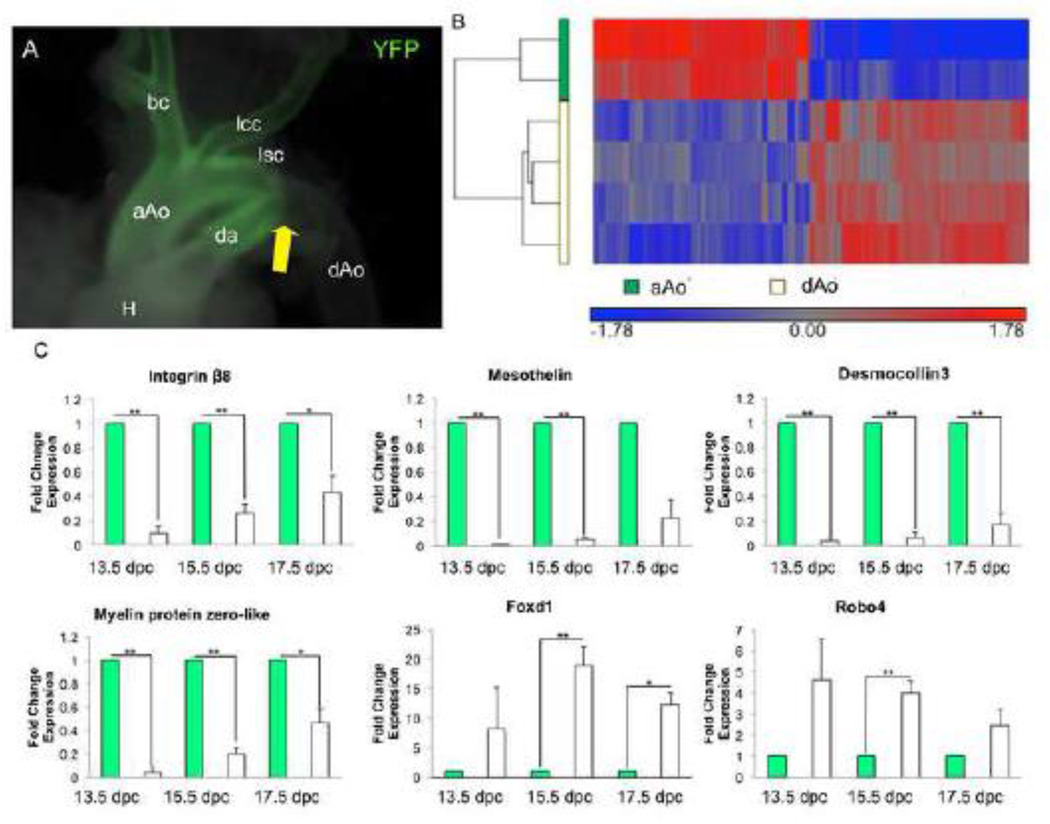
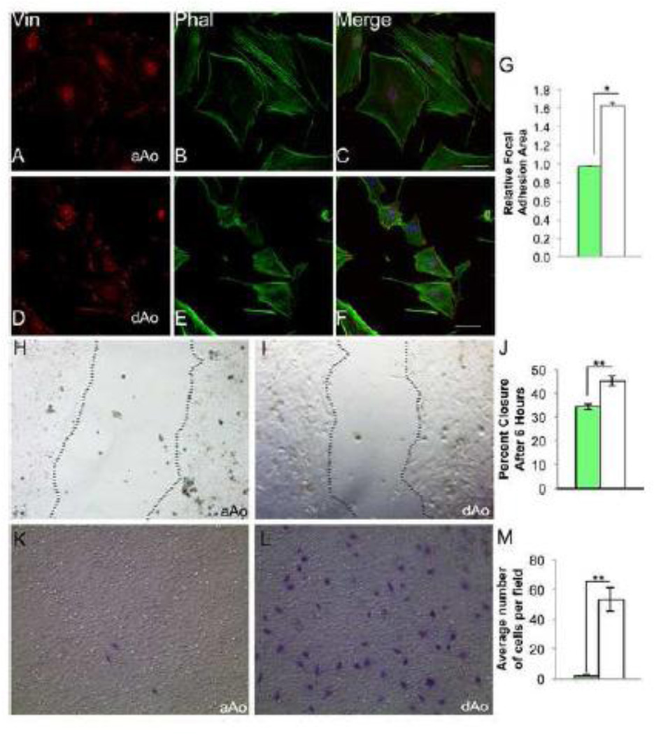
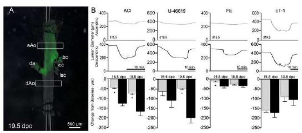
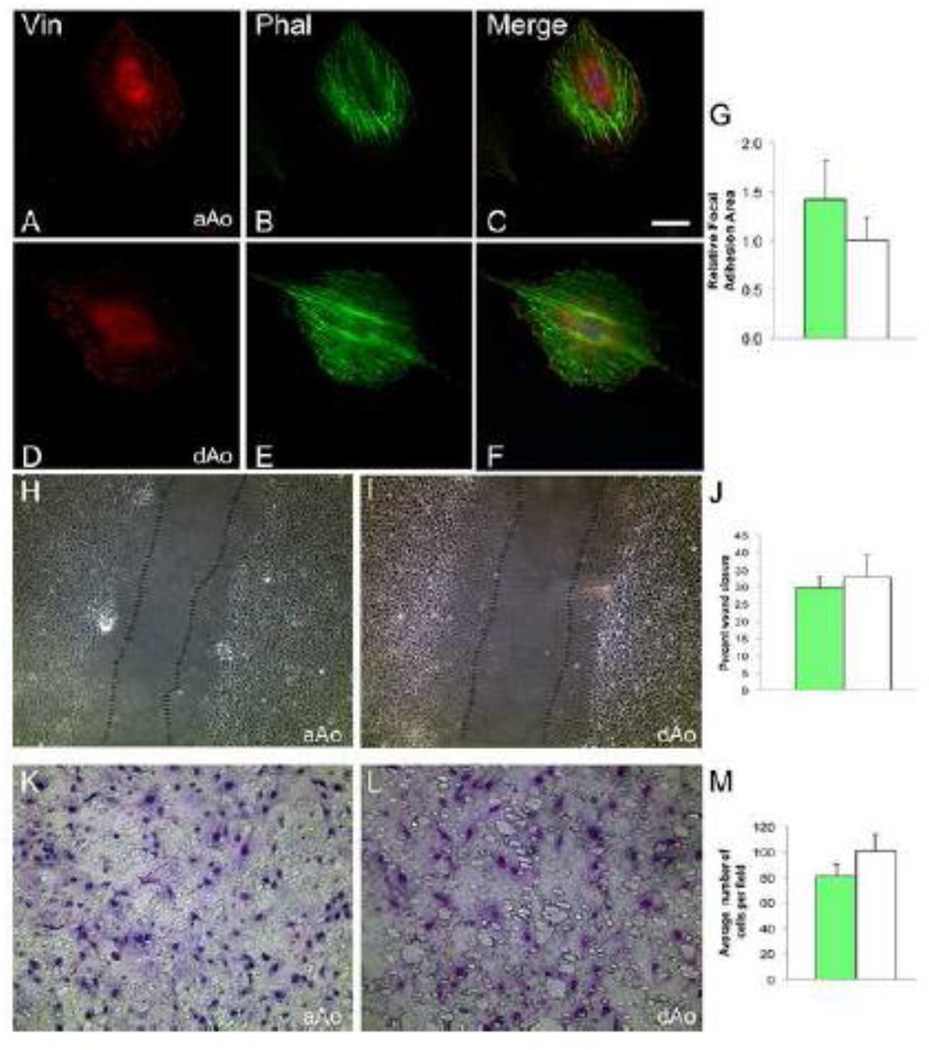
Vascular smooth muscle cells (VSMCs) are derived from distinct embryonic origins. Vessels originating from differing smooth muscle cell populations have distinct vascular and pathological properties involving calcification, atherosclerosis, and structural defects such as aneurysm and coarctation. We hypothesized that domains within a single vessel, such as the aorta, vary in phenotype based on embryonic origin. Gene profiling and myographic analyses demonstrated that embryonic ascending and descending aortic domains exhibited distinct phenotypes. In vitro analyses demonstrated that VSMCs from each region were dissimilar in terms of cytoskeletal and migratory properties, and retention of different gene expression patterns. Using the same analysis, we found that these same two domains are indistinguishable in the adult vessel. Our data demonstrate that VSMCs from different embryonic origins are functionally distinct in the embryonic mouse, but converge to assume a common phenotype in the aorta of healthy adults. These findings have fundamental implications for aortic development, function and disease progression.

Keywords: Mesoderm; Neural crest; Smooth muscle; Vasculature.


Figures

Figure 1
Figure 1
Embryonic regions of the aorta have distinct gene expression profiles. A) Wnt1-Cre; R26RYFP lineage labeled aorta demonstrating the border between the aAo and dAo. White arrow points to the border between the NC- and somitic-derived VSMCs. B) Hierarchical clustering of 1,475 probes detected as significantly different (at least 1.5-fold, B-H p value < 0.05) between control aAo and dAo samples. Values shown are log base 2, and bright red, bright blue, and gray indicate the highest, lowest, and median normalized signal values, respectively. Vertical dendrograms represent the individual samples, of which there are two to four replicates for each sample type. C) qRT-PCR on RNA taken from aorta samples throughout development. Abbreviations: aAo, ascending aorta; bc, brachiocephalic artery; da, ductus arteriosus; dAo, descending aorta; H, heart; lcc, left common carotid; lsc, left subclavian. Error bars represent SEM. *, p≤0.05; **, p≤0.01
Figure 2
Figure 2
Localization of cytoskeletal elements and migratory characteristics differ between cells derived from embryonic aAo versus dAo. A–C) Embryonic aAo VSMCs labeled with anti-vinculin antibody and phalloidin. D–F) Cells derived from the embryonic dAo labeled with anti-vinculin antibodies and phalloidin. DNA labeled with DAPI. G) Quantification of relative focal adhesion area. H, I) Scratch assay using embryonic aAo and dAo VSMC lines, 6 hours after scratch was applied. J) Quantification of percent wound closure in embryonic VSMC lines. K, L) Boyden chamber assay on embryonic VSMCs t=24 hours. Cells stained with Giemsa demonstrate the number of cell that invaded the membrane. M) Quantification of number of cells migrated after 24 hours. Scale bars represent 50µm. Abbreviations: Vin, vinculin; Phal, phalloidin. Error bars represent SEM. *, p≤0.05; **, p≤0.01.
Figure 3
Figure 3
Region-specific contractile response of the embryonic aorta. A) Lineage labeled 19.5 dpc aorta mounted on cannula for experimental analysis. Boxes represent regions where lumen diameters are measured. B) Example traces from aAo (gray) and dAo (black) with the addition of different vasoconstrictors, PE (phenylephrine), ET-1 (endothelin-1). Abbreviations: aAo, ascending aorta; bc, brachiocephalic artery; da, ductus arteriosus; dAo, descending aorta; lcc, left common carotid; lsc, left subclavian. Error bars represent SEM. *, p≤0.05; **, p≤0.01.
Figure 4
Figure 4
Adult vessels have different gene expression patterns, however the regions of the aorta have similar profiles. A) Principal components analysis of four different blood vessel tissues: ascending aorta (red), descending aorta (green), coronary artery (blue), and superior mesenteric artery (purple) is shown. The colors represent the different sample replicates, as indicated in the legend, and circles mark the ellipsoids (+/− standard a deviation of 2) for each group. The X-axis, Y-axis, and Z-axis components represent 17.7%, 14.8%, and 12.1%, respectively, of the total variability between experimental replicates. B) Hierarchical clustering of 1,496 probes detected as significantly different (at least 1.5-fold, B-H p value < 0.05) between ascending aorta, descending aorta, coronary, and superior mesenteric arteries. Values shown are log base 2, and bright red, bright blue, and gray indicate the highest, lowest, and median normalized signal values, respectively. Vertical dendrograms represent the individual samples, of which there are three replicates for each sample type (two replicates for ascending aortic tissue).
Figure 5
Figure 5
VSMC lines from adult aAo and dAo have similar localization of cytoskeletal elements and migratory characteristics. A–C) Representative adult aAo VSMCs labeled with anti-vinculin antibody and phalloidin. D–F) Representative adult dAo VSMC lines labeled with anti-vinculin antibodies and phalloidin. DNA labeled with DAPI. G) Quantification of relative focal adhesion area. H, I) Scratch assay of adult aAo and dAo VSMC lines after 6 hours. J) Quantification of percent wound closure in adult VSMC cultures. K, L) Boyden chamber assay on adult VSMC lines t=24 hours. M) Quantification of number of cells migrated after 24 hours. Scale bars represent 50µm. Abbreviations: aAo, ascending aorta; dAo, descending aorta; Vin, vinculin; Phal, phalloidin. Error bars represent SEM. *, p≤0.05; **, p≤0.01.
Figure 6
Figure 6
Regions of the adult aorta have similar contractile responses to vasoconstrictors. A) Lineage labeled 8 week old aorta on cannula. Boxes represent regions where lumen diameters are measured. B) Example traces from aAo (gray) and dAo (black) with the addition of different vasoconstrictors, PE (phenylephrine), ET-1 (endothelin-1). Abbreviations: aAo, ascending aorta; bc, brachiocephalic artery; da, ductus arteriosus; dAo, descending aorta; H, heart; lcc, left common carotid; lsc, left subclavian. Error bars represent SEM. *, p≤0.05.

References

    1. Katora ME, Hollis TM. Regional variation in rat aortic endothelial surface morphology: relationship to regional aortic permeability. Exp Mol Pathol. 1976;24:23–34. - PubMed
    1. Gadson PF, Jr, Rossignol C, McCoy J, Rosenquist TH. Expression of elastin, smooth muscle alpha-actin, and c-jun as a function of the embryonic lineage of vascular smooth muscle cells. In Vitro Cell Dev Biol Anim. 1993;29A:773–781. - PubMed
    1. Ruckman JL, Luvalle PA, Hill KE, Giro MG, Davidson JM. Phenotypic stability and variation in cells of the porcine aorta: collagen and elastin production. Matrix Biol. 1994;14:135–145. - PubMed
    1. van Meurs-van Woezik H, Klein HW, Markus-Silvis L, Krediet P. Comparison of the growth of the tunica media of the ascending aorta, aortic isthmus and descending aorta in infants and children. J Anat. 1983;136:273–281. - PMC - PubMed
    1. Stenmark KR, Mecham RP. Cellular and molecular mechanisms of pulmonary vascular remodeling. Annu Rev Physiol. 1997;59:89–144. - PubMed

Publication types

MeSH terms