. 2014 Jun:81:134-41.
doi: 10.1016/j.neuropharm.2014.01.044. Epub 2014 Feb 6.

The angiotensin type 2 receptor agonist Compound 21 elicits cerebroprotection in endothelin-1 induced ischemic stroke

Affiliations

The angiotensin type 2 receptor agonist Compound 21 elicits cerebroprotection in endothelin-1 induced ischemic stroke

Jason P Joseph et al. Neuropharmacology. 2014 Jun.

Abstract

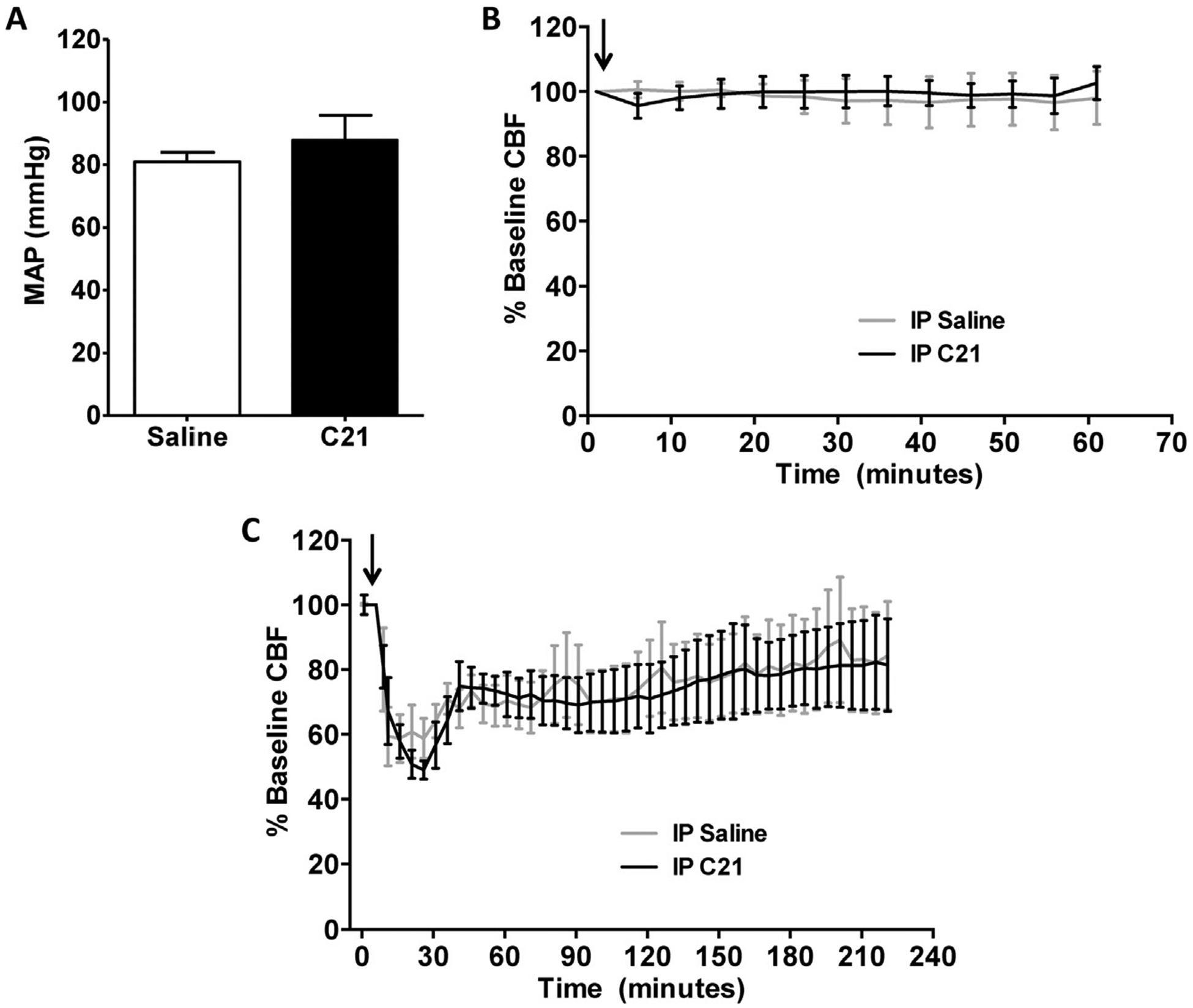
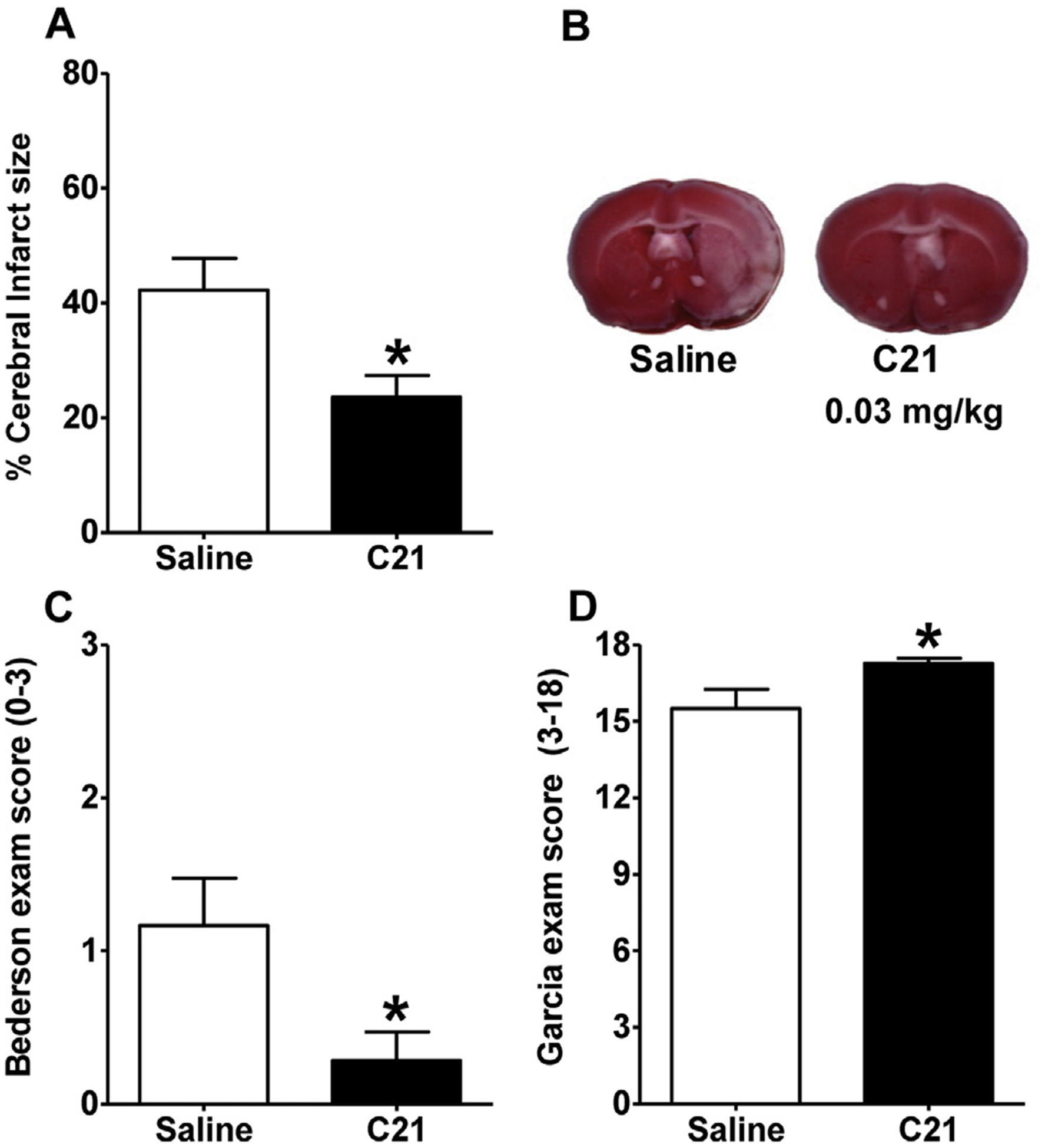
Evidence indicates that angiotensin II type 2 receptors (AT2R) exert cerebroprotective actions during stroke. A selective non-peptide AT2R agonist, Compound 21 (C21), has been shown to exert beneficial effects in models of cardiac and renal disease, as well as hemorrhagic stroke. Here, we hypothesize that C21 may exert beneficial effects against cerebral damage and neurological deficits produced by ischemic stroke. We determined the effects of central and peripheral administration of C21 on the cerebral damage and neurological deficits in rats elicited by endothelin-1 induced middle cerebral artery occlusion (MCAO), a model of cerebral ischemia. Rats infused centrally (intracerebroventricular) with C21 before endothelin-1 induced MCAO exhibited significant reductions in cerebral infarct size and the neurological deficits produced by cerebral ischemia. Similar cerebroprotection was obtained in rats injected systemically (intraperitoneal) with C21 either before or after endothelin-1 induced MCAO. The protective effects of C21 were reversed by central administration of an AT2R inhibitor, PD123319. While C21 did not alter cerebral blood flow at the doses used here, peripheral post-stroke administration of this agent significantly attenuated the MCAO-induced increases in inducible nitric oxide synthase, chemokine (C-C) motif ligand 2 and C-C chemokine receptor type 2 mRNAs in the cerebral cortex, indicating that the cerebroprotective action is associated with an anti-inflammatory effect. These results strengthen the view that AT2R agonists may have potential therapeutic value in ischemic stroke, and provide the first evidence of cerebroprotection induced by systemic post stroke administration of a selective AT2R agonist.

Keywords: Angiotensin type 2 receptor; Chemokine; Compound 21; Endothelin-1; Ischemia; Stroke.


Figures

Fig. 1.
Fig. 1.
Central pre-treatment with C21 is cerebroprotective. Rats were pre-treated with C21 (0.0075 μg/μl/h) or 1 μl of aCSF via ICV infusion for 7 days prior to ET-1 induced MCAO. (A) Bar graphs show % cerebral infarct size (white color) and (B) representative brain sections from each treatment condition. Panels (C) and (D) are data from Bederson and Garcia Neurological Exams, respectively. Data are means ± SEM from 21 (C21-treated) and 16 (aCSF-treated) rats. *p < 0.05 vs. saline control (unpaired t-test for panel A and Mann Whitney test for panels C and D).
Fig. 2.
Fig. 2.
Peripheral pre- and post-stroke treatment with C21 is cerebroprotective. Rats were injected IP with C21 (0.03 mg/kg [n = 11] or 0.1 mg/kg [n = 10]) or saline (0.9%, n = 11) 2 h prior to, as well as 4, 24, and 48 h following MCAO induction. (A) Bar graphs showing % cerebral infarct size and (B) representative brain sections from each treatment condition. (C), (D) are data from Bederson and Garcia Neurological Exams, respectively. Data are means ± SEM. *p < 0.05 vs. saline control (one way ANOVA with Bonferroni’s post hoc analysis for panel A, Kruskal–Wallis test with Dunn’s Multiple Comparison analysis for panels C and D).
Fig. 3.
Fig. 3.
Effects of peripherally administered C21 on blood pressure and CBF. (A) Rats received a single IP injection of C21 (0.03 mg/kg, n = 6) or saline (0.9%, n = 6). Mean arterial blood pressure (MAP) was measured 30 min after injection. Data are presented as means ± SEM, and no significant difference was detected (unpaired t-test). (B) Rats were administered a single IP injection of C21 (0.03 mg/kg, n = 4) or 0.9% saline (n = 4), followed by analysis of CBF for 60 min. Arrow indicates C21 or 0.9% saline injection. (C) Rats were injected IP with C21 (0.03 mg/kg, n = 4) or 0.9% saline (n = 6) 2 h prior to MCAO induction. Graphs are 5 min averages of continuous flow recordings normalized to a 5 min pre-stroke baseline. Arrow indicates ET-1 injection. For panels (B) and (C) graphs are 5 min averages of continuous flow recordings normalized toa5 min pre-stroke baseline, and data are presented as means ± SEM. No significant differences exist between C21 and 0.9% saline treatment groups at any time point (two-way repeated-measures ANOVA).
Fig. 4.
Fig. 4.
Peripheral treatment with C21 at 4, 24 and 48 h post-stroke is cerebroprotective. Rats were injected IP with either C21 (0.03 mg/kg, n = 7) or saline (0.9%, n = 6) at 4, 24, and 48 h following MCAO induction. (A) Bar graphs showing % infarcted gray matter and (B) representative brain sections from each treatment condition. (C), (D) are data from Bederson and Garcia Neurological Exams, respectively. Data are means ± SEM. *p < 0.05 vs. saline control (unpaired t-test for panel A or Mann Whitney test for panels C and D).
Fig. 5.
Fig. 5.
Cerebroprotective effect of C21 is abolished by PD123319. Rats were pre-treated with PD123319 (PD; 0.3 μg/μl/h) or aCSF via ICV infusion for 7 days prior to ET-1 induced MCAO. IP injections with either C21 (0.03 mg/kg) or 0.9% saline were administered at 4, 24, and 48 h following MCAO induction in both pre-treatment groups. (A) Bar graphs showing % cerebral infarct size and (B) representative brain sections from each treatment condition. (C), (D) are data from Bederson and Garcia Neurological Exams, respectively. Data are means ± SEM from 14 (aCSF/saline), 17 (aCSF/C21), 8 (PD/saline) and 8 (PD/C21) treated rats. *p < 0.05 vs. saline control (one way ANOVA with Bonferroni’s analysis for panel A, Kruskal–Wallis test with Dunn’s Multiple Comparison analysis for panels C and D).
Fig. 6.
Fig. 6.
Peripheral treatment with C21 at 4 and 12 h post-stroke is cerebroprotective. Rats were injected IP with either C21 (0.03 mg/kg, n = 6) or saline (0.9%, n = 6) at 4 and 12 h following MCAO induction. Twenty-four hours post-MCAO, rats underwent neurological testing via the Bederson and Garcia tests, and then were euthanized. Panels (A), (B) and (C) are the % cerebral infarct size, Bederson and Garcia scores respectively. Data are means ± SEM. *p < 0.05 vs. saline control (unpaired t-test for panel A or Mann Whitney test for panels B and C).
Fig. 7.
Fig. 7.
Peripheral post-stroke administration of C21 reduces CCL2 and CCR2 mRNAs in the cerebral cortex. Rats were injected IP with C21 (0.03 mg/kg) or saline (0.9%) at 4 and 12 h following MCAO induction by ET-1 or sham stroke. One-day post-MCAO, the ipsilateral cerebral cortex was used for qRT-PCR analyses. Shown are the respective levels of CD11b, GFAP, CCL2 and CCR2 mRNAs in the ipsilateral cerebral cortex in rats that underwent MCAO or sham stroke plus IP C21 or saline treatments. Data (normalized against GAPDH) are means ± SEM from 4 to 8 rats per treatment group. *p < 0.05 vs. respective sham stroke control; ψp < 0.05 vs. rats that underwent MCAO and IP saline treatment (one way ANOVA with Newman–Keuls analysis).
Fig. 8.
Fig. 8.
Peripheral post-stroke administration of C21 reduces iNOS mRNA in the cerebral cortex. Rats were injected IP with C21 (0.03 mg/kg) or saline (0.9%) at 4 and 12 h following MCAO induction by ET-1 or sham stroke. One-day post-MCAO, the ipsilateral cerebral cortex was used for qRT-PCR analyses. Shown are the respective levels of iNOS, eNOS, IL-1α and IL-1β mRNAs in the ipsilateral cerebral cortex in rats that underwent MCAO or sham stroke plus IP C21 or saline treatments. Data (normalized against GAPDH) are means ± SEM from 4 to 8 rats per treatment group. *p < 0.05 vs. respective sham stroke control; ψp < 0.05 vs. rats that underwent MCAO and IP saline treatment (one way ANOVA with Newman–Keuls analysis).

References

    1. Bederson JB, Pitts LH, Tsuji M, Nishimura MC, Davis RL, Bartkowski H, 1986. Rat middle cerebral artery occlusion: evaluation of the model and development of a neurologic examination. Stroke 17, 472–476. - PubMed
    1. Bosnyak S, Welungoda IK, Hallberg A, Alterman M, Widdop RE, Jones ES, 2010. Stimulation of angiotensin AT2 receptors by the non-peptide agonist, Compound 21, evokes vasodepressor effects in conscious spontaneously hypertensive rats. Br. J. Pharmacol 159 (3), 709–716. - PMC - PubMed
    1. Bosnyak S, Jones ES, Christopoulos A, Aguilar MI, Thomas WG, Widdop RE, 2011. Relative affinity of angiotensin peptides and novel ligands at AT1 and AT2 receptors. Clin. Sci. (Lond) 121 (7), 297–303. - PubMed
    1. Carey RM, 2013. Newly discovered components and actions of the renin-angiotensin system. Hypertension 62 (5), 818–822. - PubMed
    1. Cote F, Do TH, Laflamme L, Gallo JM, Gallo-Payet N, 1999. Activation of the AT(2) receptor of angiotensin II induces neurite outgrowth and cell migration in microexplant cultures of the cerebellum. J. Biol. Chem 274, 31686–31692. - PubMed

Publication types

MeSH terms