. 2014 Apr 15;306(8):L786-96.
doi: 10.1152/ajplung.00243.2013. Epub 2014 Feb 7.

Activated alveolar epithelial cells initiate fibrosis through autocrine and paracrine secretion of connective tissue growth factor

Affiliations

Activated alveolar epithelial cells initiate fibrosis through autocrine and paracrine secretion of connective tissue growth factor

Jibing Yang et al. Am J Physiol Lung Cell Mol Physiol. .

Abstract

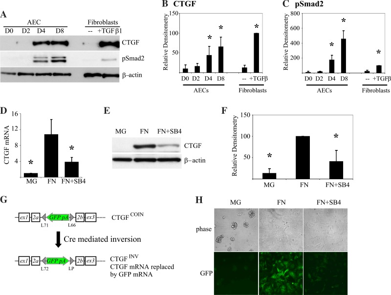
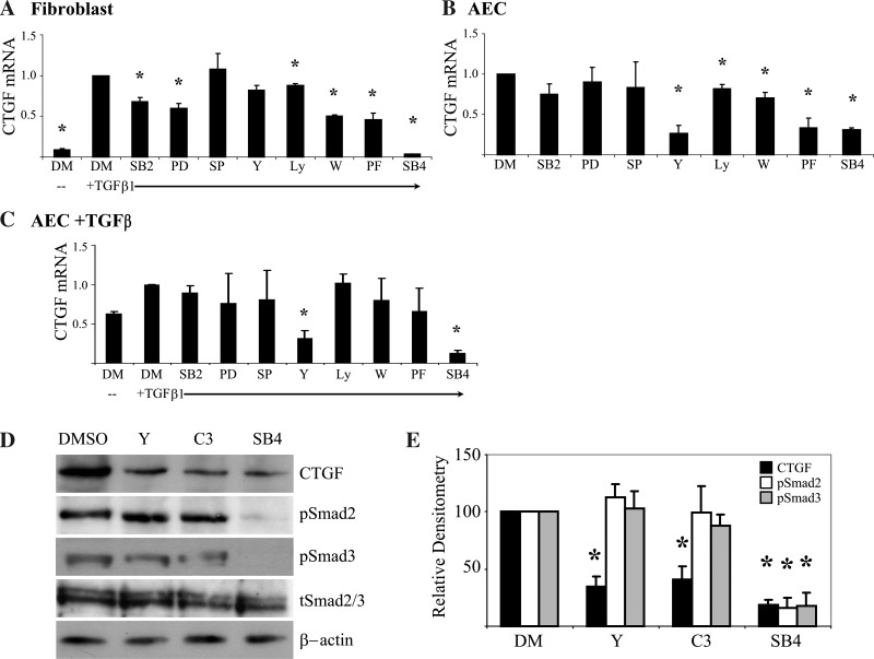
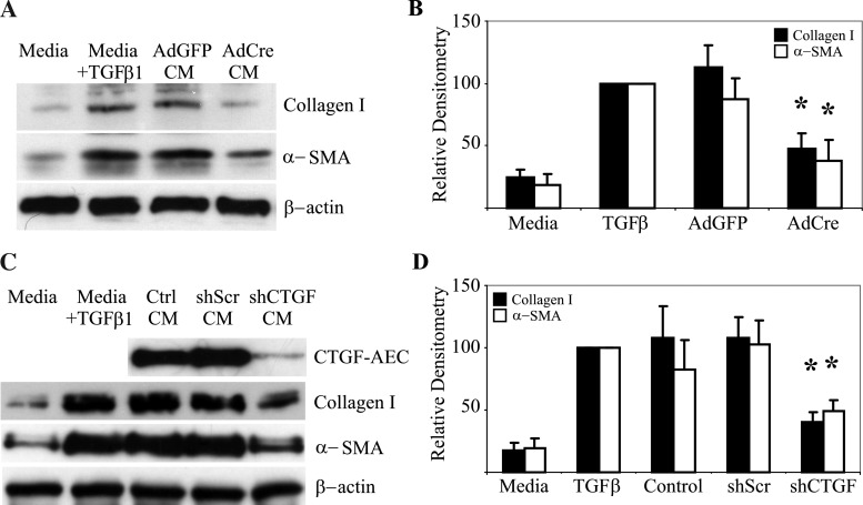
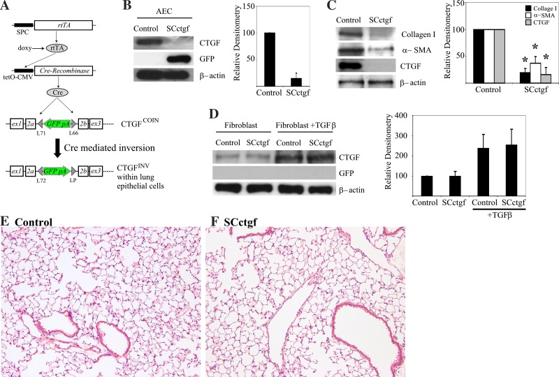
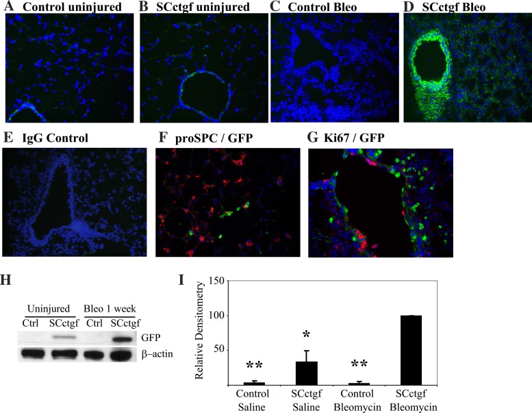
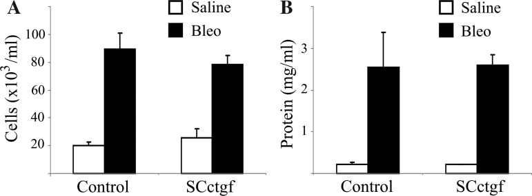
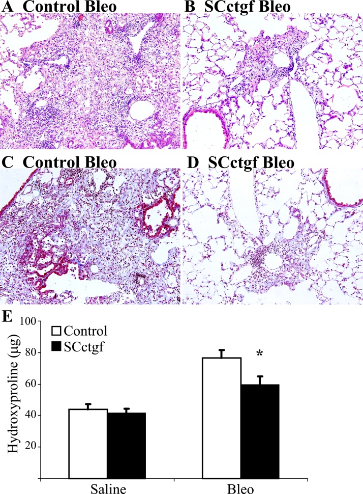
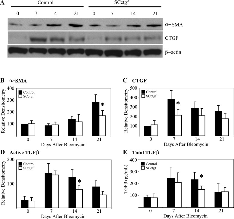
Fibrogenesis involves a pathological accumulation of activated fibroblasts and extensive matrix remodeling. Profibrotic cytokines, such as TGF-β, stimulate fibroblasts to overexpress fibrotic matrix proteins and induce further expression of profibrotic cytokines, resulting in progressive fibrosis. Connective tissue growth factor (CTGF) is a profibrotic cytokine that is indicative of fibroblast activation. Epithelial cells are abundant in the normal lung, but their contribution to fibrogenesis remains poorly defined. Profibrotic cytokines may activate epithelial cells with protein expression and functions that overlap with the functions of active fibroblasts. We found that alveolar epithelial cells undergoing TGF-β-mediated mesenchymal transition in vitro were also capable of activating lung fibroblasts through production of CTGF. Alveolar epithelial cell expression of CTGF was dramatically reduced by inhibition of Rho signaling. CTGF reporter mice demonstrated increased CTGF promoter activity by lung epithelial cells acutely after bleomycin in vivo. Furthermore, mice with lung epithelial cell-specific deletion of CTGF had an attenuated fibrotic response to bleomycin. These studies provide direct evidence that epithelial cell activation initiates a cycle of fibrogenic effector cell activation during progressive fibrosis. Therapy targeted at epithelial cell production of CTGF offers a novel pathway for abrogating this progressive cycle and limiting tissue fibrosis.

Keywords: connective tissue growth factor; epithelial; epithelial-mesenchymal transition; fibrosis; lung.


Figures

Fig. 1.
Fig. 1.
TGF-β-dependent expression of connective tissue growth factor (CTGF) by primary alveolar epithelial cells (AECs). AC: primary AECs cultured on fibronectin (FN) have Smad2 phosphorylation and expression of CTGF similar to primary lung fibroblasts stimulated with exogenous TGF-β1 determined by immunoblot (A) and quantified by densitometry for CTGF (B) and pSmad2 (C). *P < 0.05 compared with day 0 (D0), n = 4. D: RT-PCR of AECs cultured on Matrigel (MG), FN, or FN with 10 μM SB431542 (FN+SB4) for CTGF expression. *P < 0.05 compared with AECs cultured on FN without inhibitor, n = 5. E and F: immunoblot of AECs cultured on MG, FN, or FN+SB4 for CTGF expression (E) and quantification by densitometry (F). *P < 0.05 compared with AECs cultured on FN without inhibitor, n = 3. G: schematic of CTGF conditional-by-inversion (CTGFCOIN) allele. A transmembrane enhanced green fluorescent protein-polyA (GFP pA) sequence is integrated within an intron of the CTGF gene in reverse orientation. The sequence is flanked by lox71 (L71) and lox66 (L66) sequences, enabling Cre recombinase-mediated inversion of the floxed sequence. Cre-mediated inversion yields a CTGFINV allele, in which GFP expression is permanently regulated by the native CTGF promoter, and expression of CTGF itself is permanently blocked by the polyA sequence. H: phase contrast and GFP fluorescence microscopy of AdCre-treated CTGFINV AECs cultured on MG, FN, or FN+SB4, ×100.
Fig. 2.
Fig. 2.
Inhibition of TGF-β-mediated CTGF expression by primary AECs and primary lung fibroblasts. A: CTGF mRNA expression by lung fibroblasts with or without TGF-β1 (4 ng/ml) stimulation and treated with intracellular signaling pathway chemical inhibitors or DMSO (DM) vehicle. *P < 0.05 compared with lung fibroblasts treated with TGF-β1 and DMSO, n = 4. B: CTGF mRNA expression by AECs cultured on FN and treated with intracellular signaling pathway chemical inhibitors. *P < 0.05 compared with AECs treated with DMSO, n = 4. C: CTGF mRNA expression by primary AECs with or without TGF-β1 (4 ng/ml) stimulation and treated with intracellular signaling pathway chemical inhibitors or DMSO vehicle. *P < 0.05 compared with AECs treated with TGF-β1 and DMSO, n = 3. D and E: immunoblot for CTGF and phospho-Smad2 (pSmad2) and phospho-Smad3 (pSmad3) by AECs treated with Rho inhibitors and TGF-β receptor inhibitor (D) and quantification by immunoblot (E). *P < 0.01 compared with AECs treated with DMSO, n = 3. SB2, SB203580; PD, PD98059; SP, SP600125; Y, Y27632; Ly, LY294002; W, Wortmanin; PF, PF573228; SB4, SB431542; C3, C3 exoenzyme.
Fig. 3.
Fig. 3.
Lung epithelial cell-derived CTGF promotes fibroblast activation. A and B: immunoblot (A) of lung fibroblasts stimulated with AEC-conditioned media (CM) from CTGFCOIN/COIN AECs cultured on FN treated with adenovirus-expressing GFP (AdGFP) or Cre (AdCre) quantified by densitometry (B). AdCre leads to inversion and loss of CTGF expression by CTGFINV/INV AECs and reduced ability to activate lung fibroblast collagen I and α-smooth muscle actin (α-SMA). *P < 0.05 compared with fibroblasts treated with media and compared with fibroblasts treated with media from AdGFP AECs, n = 4. C and D: immunoblot of AECs and lung fibroblasts stimulated with AEC-CM (C) quantified by densitometry (D). CM from AECs cultured on FN and treated with no lentivirus (Ctrl CM), lentivirus expressing a scrambled shRNA (shScr CM), and lentivirus expressing shRNA against CTGF (shCTGF CM) were used to stimulate lung fibroblasts. shCTGF inhibits expression of CTGF by AECs (CTGF-AEC). Ctrl CM and shScr CM promote lung fibroblast type I collagen and α-SMA expression, which is reduced in fibroblasts treated with shCTGF CM. *P < 0.05 compared with fibroblasts treated with media and compared with fibroblasts treated with media from shScr AECs, n = 4.
Fig. 4.
Fig. 4.
Generation and validation of mice with lung epithelial cell-specific deletion of CTGF. A: lung epithelial cell-specific and permanent deletion of CTGF is achieved using transgenic mice carrying the surfactant proteins-C (SPC) promoter-reverse tetracycline transactivator (SPC-rtTA) and tetO-CMV promoter-Cre recombinase (tetO-Cre). In triple transgenic mice (SCctgf), the SPC promoter yields rtTA expression specifically within lung epithelial cells. In the presence of doxycycline (dox), rtTA activates the tetO-CMV promoter, leading to expression of Cre recombinase and inversion of the CTGFCOIN allele. B: AECs from SCctgf mice have diminished expression of CTGF and induced expression of GFP by immunoblot compared with AECs from littermate control mice lacking 1 of the 3 transgenes that have robust expression of CTGF and absent expression of GFP. Differences in CTGF expression were quantified by immunoblot. *P < 0.05 compared with control, n = 3. C: AECs from SCctgf mice cultured on FN have diminished expression of collagen I, α-SMA, and CTGF. Differences were quantified by immunoblot. *P < 0.05 compared with control, n = 3. D: primary lung fibroblasts from SCctgf mice have similar CTGF expression compared with control lung fibroblasts, verifying robust and lung epithelial-specific replacement of CTGF expression with GFP. Differences were quantified by immunoblot. Control and SCctgf are not statistically different, n = 4. E and F: uninjured lungs from SCctgf (F) mice have normal histology compared with littermate genotype control mice (E), hematoxylin and eosin (H&E) (×200).
Fig. 5.
Fig. 5.
Activation of lung epithelial cell CTGF expression acutely after bleomycin (Bleo) injury. AD: GFP immunofluorescence staining of lung sections (×200). A: uninjured SPC-rtTA/CTGFCOIN/COIN genotype control mouse, lacking the tetO-Cre transgene. B: uninjured SCctgf mouse. C: tetO-Cre/CTGFCOIN/COIN genotype control mouse, lacking the SPC-rtTA transgene, 1 wk after bleomycin. D: SCctgf mouse, 1 wk after bleomycin. SCctgf mice injured with bleomycin have significant periairway and alveolar staining for GFP, indicating activation of the CTGF promoter from cells of epithelial origin. E: IgG isotype control for GFP staining from SCctgf mouse 1 wk after bleomycin (×200). F: lung section from SCctgf mouse 1 wk after bleomycin stained for GFP (green) and prosurfactant protein-C (red) (×400). G: lung section from SCctgf mouse 1 wk after bleomycin stained for GFP (green) and Ki67 (red) (×400). H and I: immunoblot (H) of whole lung lysate from littermate genotype control and SCctgf mice uninjured or 1 wk after bleomycin. Uninjured SCctgf lungs have weak expression of GFP and upregulation of GFP after bleomycin, whereas control mice have no expression of GFP. Differences were quantified by densitometry (I). *P < 0.05 compared with lungs from SCctgf mice injured with bleomycin. **P < 0.01 compared with uninjured SCctgf mice, n = 3.
Fig. 6.
Fig. 6.
Acute injury after bleomycin is independent of lung epithelial CTGF. Bronchoalveolar lavage (BAL) from control and SCctgf mice 1 wk after bleomycin or saline injury analyzed for total cell count (A) and protein (B). SCctgf mice and control mice have similar increases in proteins and cell count 1 wk after bleomycin. n = 4–6 per group.
Fig. 7.
Fig. 7.
Lung epithelial cell-derived CTGF promotes lung fibrogenesis. A: lung sections from littermate control (left) and SCctgf (right) mice 3 wk after bleomycin injection stained with H&E (top) and trichrome (bottom) ×100. Genotype control mice have robust fibrosis compared with SCctgf mice. E: hydroxyproline assay from entire lungs 3 wk after saline or bleomycin in SCctgf or littermate control mice. SCctgf mice have less fibrosis after bleomycin (n = 4–8 per group), *P < 0.05 compared with control mice treated with bleomycin.
Fig. 8.
Fig. 8.
Expression of CTGF, α-SMA, and TGF-β at 1-wk intervals after bleomycin injury. A: whole lung lysate from SCctgf and littermate control mice were analyzed for expression of α-SMA and CTGF. B: densitometry quantification of α-SMA immunoblot. *P < 0.05 compared with control mice at day 21, n = 3. C: densitometry quantification of CTGF immunoblot. *P < 0.05 compared with control mice at day 7, n = 3. D and E: ELISA for active TGF-β (D) and total (E) from BAL of control and SCctgf mice. *P < 0.05 compared with control mice at day 14, n = 3–6.

Similar articles

Cited by

References

    1. American Thoracic Society. Idiopathic pulmonary fibrosis: diagnosis and treatment International consensus statement American Thoracic Society (ATS), and the European Respiratory Society (ERS). Am J Respir Crit Care Med 161: 646–664, 2000 - PubMed
    1. American Thoracic Society/European Respiratory Society. International multidisciplinary consensus classification of the idiopathic interstitial pneumonias. Am J Respir Crit Care Med 165: 277–304, 2002 - PubMed
    1. Balli D, Ustiyan V, Zhang Y, Wang IC, Masino AJ, Ren X, Whitsett JA, Kalinichenko VV, Kalin TV. Foxm1 transcription factor is required for lung fibrosis and epithelial-to-mesenchymal transition. EMBO J 32: 231–244, 2013 - PMC - PubMed
    1. Bartram U, Speer CP. The role of transforming growth factor beta in lung development and disease. Chest 125: 754–765, 2004 - PubMed
    1. Bauman KA, Wettlaufer SH, Okunishi K, Vannella KM, Stoolman JS, Huang SK, Courey AJ, White ES, Hogaboam CM, Simon RH, Toews GB, Sisson TH, Moore BB, Peters-Golden M. The antifibrotic effects of plasminogen activation occur via prostaglandin E2 synthesis in humans and mice. J Clin Invest 120: 1950–1960, 2010 - PMC - PubMed

Publication types

MeSH terms

LinkOut - more resources