

Diverse macrophage populations mediate acute lung inflammation and resolution
- PMID: 24508730
- PMCID: PMC3989724
- DOI: 10.1152/ajplung.00341.2013
Diverse macrophage populations mediate acute lung inflammation and resolution
Abstract
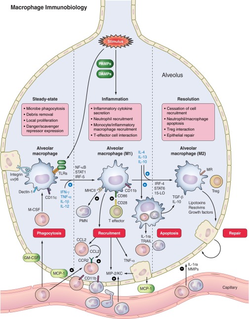
Acute respiratory distress syndrome (ARDS) is a devastating disease with distinct pathological stages. Fundamental to ARDS is the acute onset of lung inflammation as a part of the body's immune response to a variety of local and systemic stimuli. In patients surviving the inflammatory and subsequent fibroproliferative stages, transition from injury to resolution and recovery is an active process dependent on a series of highly coordinated events regulated by the immune system. Experimental animal models of acute lung injury (ALI) reproduce key components of the injury and resolution phases of human ARDS and provide a methodology to explore mechanisms and potential new therapies. Macrophages are essential to innate immunity and host defense, playing a featured role in the lung and alveolar space. Key aspects of their biological response, including differentiation, phenotype, function, and cellular interactions, are determined in large part by the presence, severity, and chronicity of local inflammation. Studies support the importance of macrophages to initiate and maintain the inflammatory response, as well as a determinant of resolution of lung inflammation and repair. We will discuss distinct roles for lung macrophages during early inflammatory and late resolution phases of ARDS using experimental animal models. In addition, each section will highlight human studies that relate to the diverse role of macrophages in initiation and resolution of ALI and ARDS.
Keywords: ARDS; activation; inflammation; macrophage.
Figures
References
-
- Adamson IY, Bowden DH. Role of monocytes and interstitial cells in the generation of alveolar macrophages II. Kinetic studies after carbon loading. Lab Invest 42: 518–524, 1980 - PubMed
-
- Aggarwal A, Baker CS, Evans TW, Haslam PL. G-CSF and IL-8 but not GM-CSF correlate with severity of pulmonary neutrophilia in acute respiratory distress syndrome. Eur Respir J 15: 895–901, 2000 - PubMed
-
- Aggarwal NR, D'Alessio FR, Eto Y, Chau E, Avalos C, Waickman AT, Garibaldi BT, Mock JR, Files DC, Sidhaye V, Polotsky VY, Powell J, Horton M, King LS. Macrophage A2A adenosinergic receptor modulates oxygen-induced augmentation of murine lung injury. Am J Respir Cell Mol Biol 48: 635–646, 2013 - PMC - PubMed
-
- Allen GL, Menendez IY, Ryan MA, Mazor RL, Wispe JR, Fiedler MA, Wong HR. Hyperoxia synergistically increases TNF-α-induced interleukin-8 gene expression in A549 cells. Am J Physiol Lung Cell Mol Physiol 278: L253–L260, 2000 - PubMed
Publication types
MeSH terms
Grants and funding
LinkOut - more resources
Full Text Sources
Other Literature Sources
Medical