

Objective assessment of the corneal endothelium in Fuchs' endothelial dystrophy
- PMID: 24508788
- PMCID: PMC3936508
- DOI: 10.1167/iovs.13-13041
Objective assessment of the corneal endothelium in Fuchs' endothelial dystrophy
Abstract
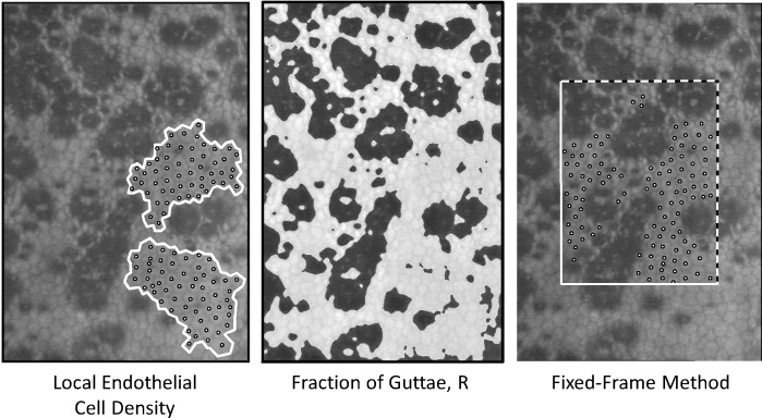
Purpose: To develop a standardized method of endothelial cell density (ECD) assessment in Fuchs' endothelial dystrophy that maximizes the sample area and uses the clearest endothelial cells in confocal images.
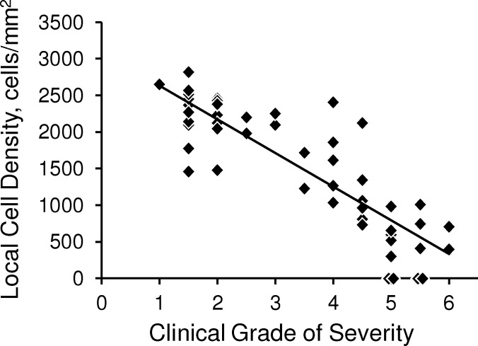
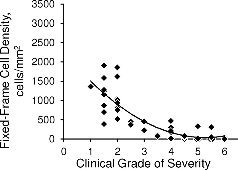
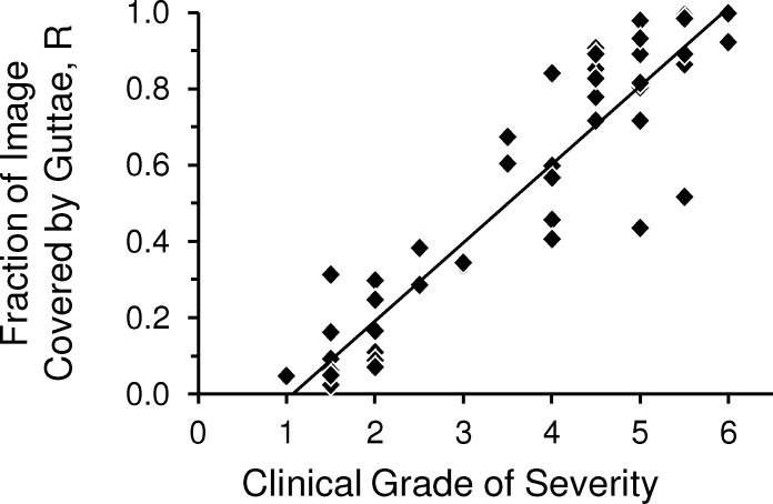
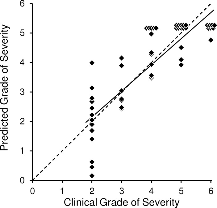
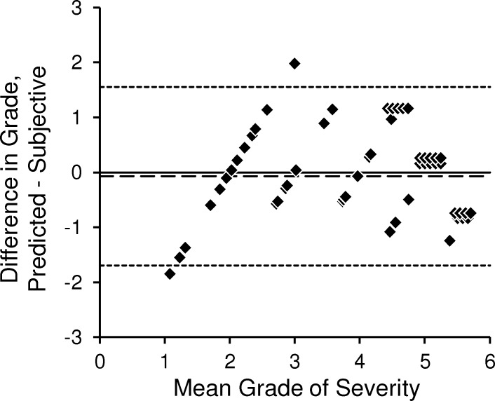
Methods: The corneal endothelium of 51 eyes from 30 patients, with varying degrees of Fuchs' endothelial dystrophy, was examined using confocal microscopy. In two or three distinct images of the central endothelium, local contiguous cell density was determined using a variable frame method. The effective ECD was the product of the local cell density and the fraction of the image that was free of guttae. Two examiners assessed the severity of disease in each eye during slit-lamp examination and assigned a severity grade of 1 to 6. In a second group of 55 eyes with Fuchs' dystrophy from 30 patients, the clinical grade was predicted from the effective ECD and the regression coefficients of the first group and compared to the subjective clinical grade assigned by one examiner.
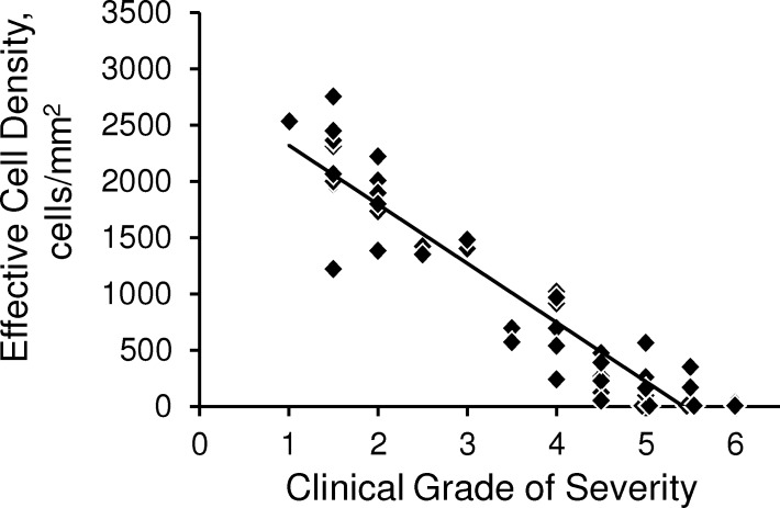
Results: The effective ECD decreased linearly with subjective grade (r = -0.93, P < 0.001). The grade predicted from the effective ECD differed from the subjective clinical grade by -0.1 ± 0.8 (mean difference ± standard deviation).
Conclusions: The effective ECD in confocal images provides an objective means of assessing the corneal endothelium in Fuchs' dystrophy and might be a useful tool in clinical studies.
Keywords: Fuchs' endothelial dystrophy; corneal endothelium; endothelial cell density; guttae.
Figures
Similar articles
-
Regional variability in corneal endothelial cell density between guttae and non-guttae areas in Fuchs endothelial corneal dystrophy.Can J Ophthalmol. 2019 Oct;54(5):570-576. doi: 10.1016/j.jcjo.2018.12.009. Epub 2019 Jun 17. Can J Ophthalmol. 2019. PMID: 31564347 Free PMC article.
-
Corneal abnormalities early in the course of Fuchs' endothelial dystrophy.Ophthalmology. 2014 Dec;121(12):2325-33. doi: 10.1016/j.ophtha.2014.07.001. Epub 2014 Aug 22. Ophthalmology. 2014. PMID: 25156138 Free PMC article.
-
Confocal microscopy in cornea guttata and Fuchs' endothelial dystrophy.Br J Ophthalmol. 1999 Feb;83(2):185-9. doi: 10.1136/bjo.83.2.185. Br J Ophthalmol. 1999. PMID: 10396196 Free PMC article.
-
Imaging the Corneal Endothelium in Fuchs Corneal Endothelial Dystrophy.Semin Ophthalmol. 2019;34(4):340-346. doi: 10.1080/08820538.2019.1632355. Epub 2019 Jun 19. Semin Ophthalmol. 2019. PMID: 31215821 Free PMC article. Review.
-
Cataract surgery in Fuchs' dystrophy.Curr Opin Ophthalmol. 2005 Aug;16(4):241-5. doi: 10.1097/01.icu.0000172828.39608.7c. Curr Opin Ophthalmol. 2005. PMID: 16000897 Review.
Cited by
-
Corneal Hydration Control in Fuchs' Endothelial Corneal Dystrophy.Invest Ophthalmol Vis Sci. 2016 Sep 1;57(11):5060-5065. doi: 10.1167/iovs.16-20205. Invest Ophthalmol Vis Sci. 2016. PMID: 27661858 Free PMC article.
-
Phase 2, Randomized, Open-Label Parallel-Group Study of Two Dosing Regimens of Netarsudil for the Treatment of Corneal Edema Due to Fuchs Corneal Dystrophy.J Ocul Pharmacol Ther. 2022 Dec;38(10):657-663. doi: 10.1089/jop.2022.0069. Epub 2022 Nov 3. J Ocul Pharmacol Ther. 2022. PMID: 36327101 Free PMC article. Clinical Trial.
-
Regional variability in corneal endothelial cell density between guttae and non-guttae areas in Fuchs endothelial corneal dystrophy.Can J Ophthalmol. 2019 Oct;54(5):570-576. doi: 10.1016/j.jcjo.2018.12.009. Epub 2019 Jun 17. Can J Ophthalmol. 2019. PMID: 31564347 Free PMC article.
-
A multi-ancestry GWAS of Fuchs corneal dystrophy highlights the contributions of laminins, collagen, and endothelial cell regulation.Commun Biol. 2024 Apr 6;7(1):418. doi: 10.1038/s42003-024-06046-3. Commun Biol. 2024. PMID: 38582945 Free PMC article.
-
[The German version of the Visual Function and Corneal Health Status (V‑FUCHS): a Fuchs dystrophy-specific visual disability instrument].Ophthalmologe. 2020 Feb;117(2):140-146. doi: 10.1007/s00347-019-0938-7. Ophthalmologe. 2020. PMID: 31342164 German.
References
-
- Adamis AP, Filatov V, Tripathi BJ, Tripathi RC. Fuchs' endothelial dystrophy of the cornea. Surv Ophthalmol. 1993; 38: 149–168 - PubMed
-
- Brooks AM, Grant G, Gillies WE. A comparison of corneal endothelial morphology in cornea guttata, Fuchs' dystrophy and bullous keratopathy. Aust N Z J Ophthalmol. 1988; 16: 93–100 - PubMed
-
- Brooks AM, Grant GB, Gillies WE. The identification of corneal guttae. Cornea. 1991; 10: 249–260 - PubMed
-
- Rodrigues MM, Krachmer JH, Hackett J, Gaskins R, Halkias A. Fuchs' corneal dystrophy. A clinicopathologic study of the variation in corneal edema. Ophthalmology. 1986; 93: 789–796 - PubMed
-
- Bergmanson JP, Sheldon TM, Goosey JD. Fuchs' endothelial dystrophy: a fresh look at an aging disease. Ophthalmic Physiol Opt. 1999; 19: 210–222 - PubMed
