

The immunosuppressant FTY720 prolongs survival in a mouse model of diet-induced coronary atherosclerosis and myocardial infarction
- PMID: 24508946
- PMCID: PMC4574629
- DOI: 10.1097/FJC.0000000000000031
The immunosuppressant FTY720 prolongs survival in a mouse model of diet-induced coronary atherosclerosis and myocardial infarction
Abstract
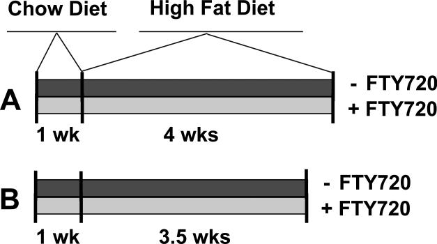
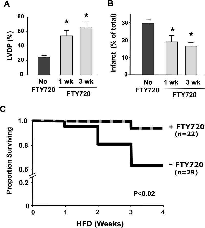
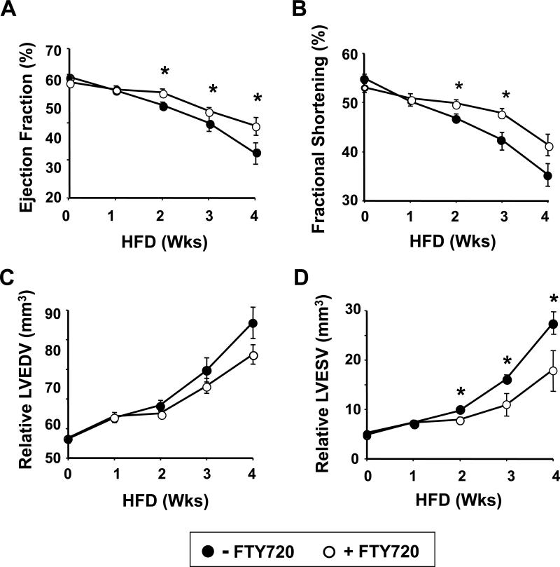
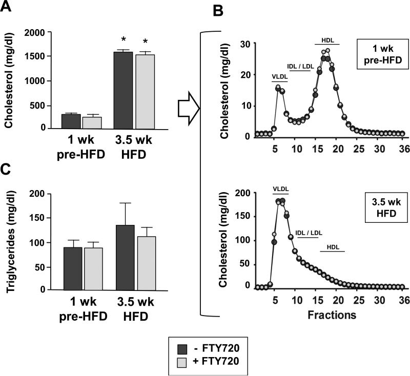
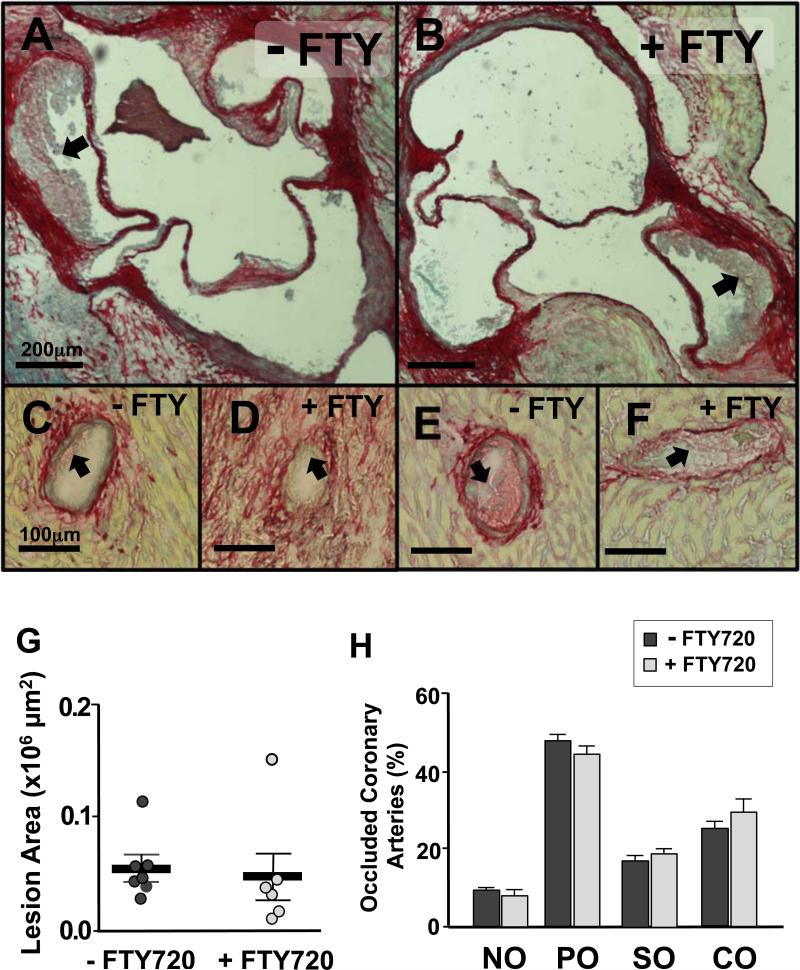
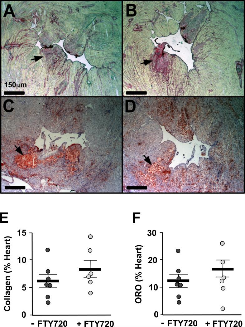
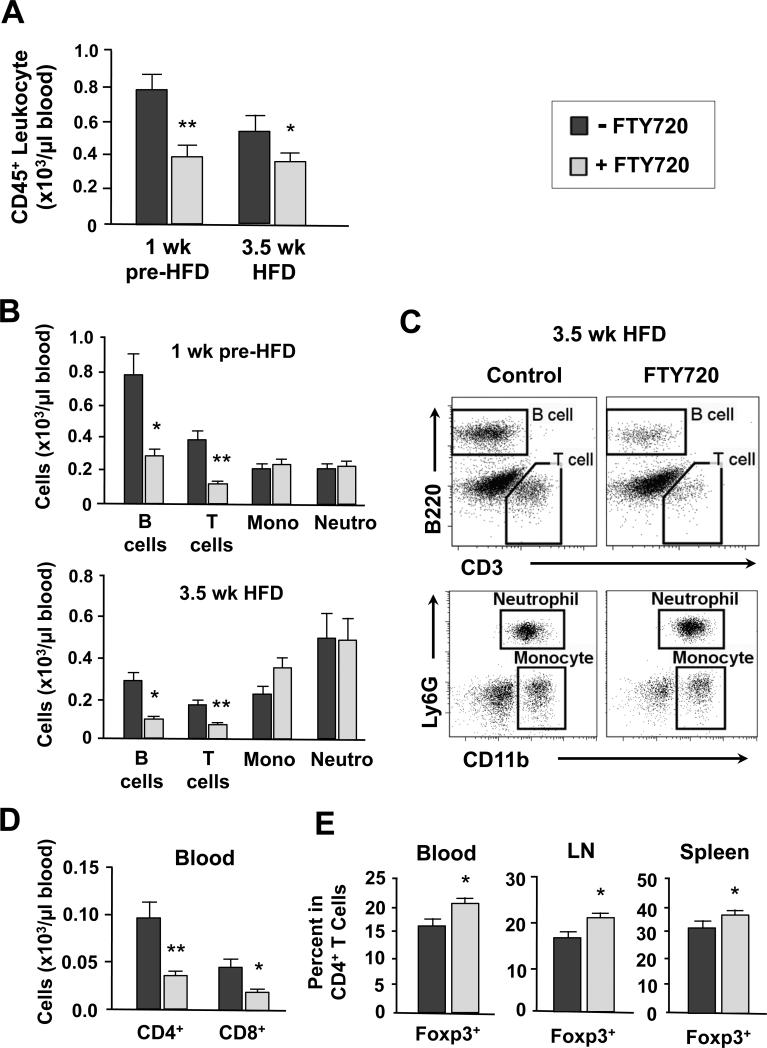
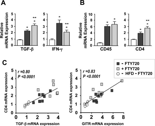
FTY720, an analogue of sphingosine-1-phosphate, is cardioprotective during acute injury. Whether long-term FTY720 affords cardioprotection is unknown. Here, we report the effects of oral FTY720 on ischemia/reperfusion injury and in hypomorphic apoE mice deficient in SR-BI receptor expression (ApoeR61(h/h)/SRB1(-/- mice), a model of diet-induced coronary atherosclerosis and heart failure. We added FTY720 (0.3 mg·kg(-1)·d(-1)) to the drinking water of C57BL/6J mice. After ex vivo cardiac ischemia/reperfusion injury, these mice had significantly improved left ventricular (LV) developed pressure and reduced infarct size compared with controls. Subsequently, ApoeR61(h/h)/SRB1(-/-) mice fed a high-fat diet for 4 weeks were treated or not with oral FTY720 (0.05 mg·kg(-1)·d(-1)). This sharply reduced mortality (P < 0.02) and resulted in better LV function and less LV remodeling compared with controls without reducing hypercholesterolemia and atherosclerosis. Oral FTY720 reduced the number of blood lymphocytes and increased the percentage of CD4+Foxp3+ regulatory T cells (Tregs) in the circulation, spleen, and lymph nodes. FTY720-treated mice exhibited increased TGF-β and reduced IFN-γ expression in the heart. Also, CD4 expression was increased and strongly correlated with molecules involved in natural Treg activity, such as TGF-β and GITR. Our data suggest that long-term FTY720 treatment enhances LV function and increases longevity in mice with heart failure. These benefits resulted not from atheroprotection but from systemic immunosuppression and a moderate reduction of inflammation in the heart.
Figures
Similar articles
-
Immunosuppression With FTY720 Reverses Cardiac Dysfunction in Hypomorphic ApoE Mice Deficient in SR-BI Expression That Survive Myocardial Infarction Caused by Coronary Atherosclerosis.J Cardiovasc Pharmacol. 2016 Jan;67(1):47-56. doi: 10.1097/FJC.0000000000000312. J Cardiovasc Pharmacol. 2016. PMID: 26322923 Free PMC article.
-
Oral FTY720 administration induces immune tolerance and inhibits early development of atherosclerosis in apolipoprotein E-deficient mice.Int J Immunopathol Pharmacol. 2012 Apr-Jun;25(2):397-406. doi: 10.1177/039463201202500209. Int J Immunopathol Pharmacol. 2012. PMID: 22697071
-
Pharmacological pre- and post-conditioning with the sphingosine-1-phosphate receptor modulator FTY720 after myocardial ischaemia-reperfusion.Br J Pharmacol. 2010 Jul;160(5):1243-51. doi: 10.1111/j.1476-5381.2010.00767.x. Br J Pharmacol. 2010. PMID: 20590616 Free PMC article.
-
Ascomycete derivative to MS therapeutic: S1P receptor modulator FTY720.Prog Drug Res. 2008;66:361, 363-81. doi: 10.1007/978-3-7643-8595-8_8. Prog Drug Res. 2008. PMID: 18416311 Review.
-
Overview of FTY720 clinical pharmacokinetics and pharmacology.Ther Drug Monit. 2004 Dec;26(6):585-7. doi: 10.1097/00007691-200412000-00001. Ther Drug Monit. 2004. PMID: 15570180 Review.
Cited by
-
Sphingosine-1-phosphate signaling and the gut-liver axis in liver diseases.Liver Res. 2019 Mar;3(1):19-24. doi: 10.1016/j.livres.2019.02.003. Epub 2019 Mar 4. Liver Res. 2019. PMID: 31360579 Free PMC article.
-
Apolipoprotein E and Atherosclerosis: From Lipoprotein Metabolism to MicroRNA Control of Inflammation.J Cardiovasc Dev Dis. 2018 May 23;5(2):30. doi: 10.3390/jcdd5020030. J Cardiovasc Dev Dis. 2018. PMID: 29789495 Free PMC article. Review.
-
Epigenetic Regulation of the N-Terminal Truncated Isoform of Matrix Metalloproteinase-2 (NTT-MMP-2) and Its Presence in Renal and Cardiac Diseases.Front Genet. 2021 Feb 25;12:637148. doi: 10.3389/fgene.2021.637148. eCollection 2021. Front Genet. 2021. PMID: 33732288 Free PMC article.
-
Pig and Mouse Models of Hyperlipidemia and Atherosclerosis.Methods Mol Biol. 2022;2419:379-411. doi: 10.1007/978-1-0716-1924-7_24. Methods Mol Biol. 2022. PMID: 35237978
-
Sphingosine-1-Phosphate Receptor 1 Regulates Cardiac Function by Modulating Ca2+ Sensitivity and Na+/H+ Exchange and Mediates Protection by Ischemic Preconditioning.J Am Heart Assoc. 2016 May 20;5(5):e003393. doi: 10.1161/JAHA.116.003393. J Am Heart Assoc. 2016. PMID: 27207969 Free PMC article.
References
-
- Kimura T, Sato K, Kuwabara A, Tomura H, Ishiwara M, Kobayashi I, Ui M, Okajima F. Sphingosine 1-phosphate may be a major component of plasma lipoproteins responsible for the cytoprotective actions in human umbilical vein endothelial cells. J Biol Chem. 2001;276:31780–31785. - PubMed
-
- Zhang J, Honbo N, Goetzl EJ, Chatterjee K, Karliner JS, Gray MO. Signals from type 1 sphingosine 1-phosphate receptors enhance adult mouse cardiac myocyte survival during hypoxia. Am J Physiol Heart Circ Physiol. 2007;293:H3150–3158. - PubMed
-
- Rosen H, Goetzl EJ. Sphingosine 1-phosphate and its receptors: an autocrine and paracrine network. Nat Rev Immunol. 2005;5:560–570. - PubMed
-
- Means CK, Xiao CY, Li Z, Zhang T, Omens JH, Ishii I, Chun J, Brown JH. Sphingosine 1-phosphate S1P2 and S1P3 receptor-mediated Akt activation protects against in vivo myocardial ischemia-reperfusion injury. Am J Physiol Heart Circ Physiol. 2007;292:H2944–2951. - PubMed
-
- Jin ZQ, Karliner JS, Vessey DA. Ischaemic postconditioning protects isolated mouse hearts against ischaemia/reperfusion injury via sphingosine kinase isoform-1 activation. Cardiovasc Res. 2008;79:134–140. - PubMed
Publication types
MeSH terms
Substances
Grants and funding
LinkOut - more resources
Full Text Sources
Other Literature Sources
Medical
Research Materials
Miscellaneous
