. 2014 Apr:69:289-99.
doi: 10.1016/j.freeradbiomed.2014.01.039. Epub 2014 Feb 6.

Metal chelator combined with permeability enhancer ameliorates oxidative stress-associated neurodegeneration in rat eyes with elevated intraocular pressure

Affiliations

Metal chelator combined with permeability enhancer ameliorates oxidative stress-associated neurodegeneration in rat eyes with elevated intraocular pressure

P Liu et al. Free Radic Biol Med. 2014 Apr.

Abstract

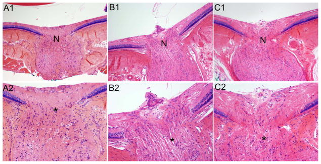
Because as many as half of glaucoma patients on intraocular pressure (IOP)-lowering therapy continue to experience optic nerve toxicity, it is imperative to find other effective therapies. Iron and calcium ions play key roles in oxidative stress, a hallmark of glaucoma. Therefore, we tested metal chelation by means of ethylenediaminetetraacetic acid (EDTA) combined with the permeability enhancer methylsulfonylmethane (MSM) applied topically on the eye to determine if this noninvasive treatment is neuroprotective in rat optic nerve and retinal ganglion cells exposed to oxidative stress induced by elevated IOP. Hyaluronic acid (HA) was injected into the anterior chamber of the rat eye to elevate the IOP. EDTA-MSM was applied topically to the eye for 3 months. Eyeballs and optic nerves were processed for histological assessment of cytoarchitecture. Protein-lipid aldehyde adducts and cyclooxygenase-2 (COX-2) were detected immunohistochemically. HA administration increased IOP and associated oxidative stress and inflammation. Elevated IOP was not affected by EDTA-MSM treatment. However, oxidative damage and inflammation were ameliorated as reflected by a decrease in formation of protein-lipid aldehyde adducts and COX-2 expression, respectively. Furthermore, EDTA-MSM treatment increased retinal ganglion cell survival and decreased demyelination of optic nerve compared with untreated eyes. Chelation treatment with EDTA-MSM ameliorates sequelae of IOP-induced toxicity without affecting IOP. Because most current therapies aim at reducing IOP and damage occurs even in the absence of elevated IOP, EDTA-MSM has the potential to work in conjunction with pressure-reducing therapies to alleviate damage to the optic nerve and retinal ganglion cells.

Keywords: Chelation therapy; EDTA; Free radicals; Glaucoma; Glaucomatous neurodegeneration; Hyaluronic acid; Inflammation; Intraocular pressure; Metal chelation; Methylsulfonylmethane; Ocular hypertension; Oxidative damage; Oxidative stress; Reactive oxygen species; Retinal ganglion cell.


Figures

Figure 1
Figure 1
EDTA-MSM treatment had no effect on IOP. Graph of IOP measured weekly over the 12 weeks of this experiment. There was no difference between the groups before injection (week 0). There were no statistical differences between the PBS-injected eyes treated with PBS or EDTA-MSM at any time point (repeated measures two-way ANOVA), so these values were pooled for further statistical analysis. Both HA-injected groups had significantly higher IOP values than the PBS injected group from the first week onward. In week one, the IOP of the HA+EDTA-MSM treated eyes was significantly higher than that of the HA+PBS eyes (p < 0.001). At later time points, there was no significant difference between the HA-injected eyes treated with PBS or EDTA-MSM.
Figure 2
Figure 2
EDTA-MSM ameliorated cytoarchitectural disruption of the optic nerve induced by HA injection. Longitudinal sections of paraffin-embedded rat eyeballs including the optic nerve head and initial segment were stained with hematoxylin and eosin. Upper panel: 100X magnification, Lower panel: 200X magnification. N: Typical narrow neck as fibers cross the scleral plane, the rat equivalent of the lamina cribrosa in humans. *Center of optic nerve. (A) PBS-injected eye. Fibers in optic nerves from control eyes had a dense and regular texture. (B) HA injected eye. Nerve fibers were loosely packed and disorganized in HA-injected eyes. Highly focal swelling of nerve fibers was observed in these eyes. The narrowed neck of the optic nerve as it passes through the sclera was widened. Disorganization of astrocytic nuclei along with increased cellularity was seen in nerves from HA-injected eyes as compared to control and HA+EDTA-MSM, while swelling was much less in PBS and HA+EDTA-MSM eyes. (C) HA injected eye treated topically with EDTA-MSM. Fibers in optic nerves had a dense and regular texture similar to that seen in control eyes. The optic nerve fibers as they cross the sclera plane resembled the normally narrow neck. Organization and cellularity of glial nuclei also appeared normal. Within the eyeball itself, no significant thinning of the nerve fiber layer of the retina or optic nerve head excavation was observed.
Figure 3
Figure 3
EDTA-MSM ameliorated the edema of optic nerve under HA-induced ocular hypertension. Transverse frozen section of optic nerve, stained with hematoxylin and eosin. Upper panel: 40X magnification; Lower panel: 200X magnification. (A) Optic nerve of control eye without elevated IOP. The texture of the nerve is regular with densely packed fibers. (B) Optic nerve from HA-injected eyes. Edema of the optic nerve in these eyes as compared to control eyes can readily be observed in cross-sections of the optic nerve. Vacuolization is apparent at the higher magnification. (C) Optic nerve of HA-injected eyes treated with EDTA-MSM. Optic nerve fibers are regular and dense with no apparent edema or vacuolization.
Figure 4
Figure 4
EDTA-MSM treatment mitigates demyelination in the optic nerve associated with increased IOP. Longitudinal sections of the optic nerve are stained with Luxol fast blue and counterstained with cresyl violet. 200X magnification. (A) Control optic nerve from eye injected with PBS. Axons are densely stained indicating robust myelination typical of the optic nerve caudal to the lamina cribrosa. (B) Optic nerve from eye injected with HA. The intensity of Luxol blue stain in optic nerve from untreated eyes with is greatly reduced compared with control, suggesting that demyelination has occurred in these nerves. (C) Optic nerve from eye injected with HA and treated with EDTA-MSM. Though not as robust as in the control optic nerve, myelin staining in EDTA-MSM treated nerves is darker than seen in untreated nerves.
Figure 5
Figure 5
Treatment with EDTA-MSM reduced protein-HHE adducts in the optic nerves and retinas of HA-injected eyes. Anti-protein-HHE immunohistochemistry counterstained with hematoxylin. Upper panel: optic nerve head and longitudinal section of initial segment of optic nerve, 100X magnification; lower panel: cross-section of retina, 600X magnification. (A, D) Control optic nerve and retina. Staining for protein-HHE adducts is not visible in control optic nerve or in retina. (B, E) Aggregates of HHE-protein adducts are visible within the optic nerve from HA-injected eyes. In the retina, protein-HHE aggregated in photoreceptor layer (arrow), with some staining in the outer nuclear layer and in retinal ganglion cells. (C, F) EDTA-MSM treated optic nerve and retina. Treatment with EDTA-MSM nearly eliminated these protein adducts in the optic nerves of treated HA-injected eyes. EDTA-MSM also ameliorated accumulation of protein-HHE adducts in the photoreceptor layer, where it was weak in treated HA-injected eyes. Some staining remained in the outer nuclear layer and in ganglion cells.
Figure 6
Figure 6
Treatment with EDTA-MSM reduced protein-HNE adducts in the optic nerves and retinas of HA-injected eyes. Anti-protein-HNE immunohistochemistry counterstained with hematoxylin. Upper panel: longitudinal section of optic nerve, 100X magnification; lower panel: cross-section of retina, 600X magnification. (A, D) Control optic nerve and retina. Protein HNE-adducts were observed in the optic nerves and the photoreceptor layer of the retina in all groups of eyes, including controls. (B, E) Protein-HNE staining was much more intense in the optic nerves and retina of eyes injected with HA than in controls. Staining was particularly intense in the photoreceptor layer (arrows). (C, F) Application of EDTA-MSM ameliorated accumulation of protein-HNE adducts, particularly in the photoreceptor layer. In the optic nerve, protein-HNE staining was reduced to approximately the same level seen in control nerves.
Figure 7
Figure 7
Treatment with EDTA-MSM reduced protein-MDA adducts in the optic nerves and retinas of HA-injected eyes. Upper and center panels, Anti protein-MDA immunohistochemistry counterstained with hematoxylin. Upper panel: longitudinal section of optic nerve, 100X magnification; Center panel: cross-section of retina, 600X magnification. Lower panel: fluorescent staining of anti-protein-MDA (red) counterstained with DAPI (blue). Transverse sections of optic nerve, 600X magnification. (A, D, G) Control optic nerve and retina. Protein MDA-adducts were observed in the optic nerves and in all layers of the retina in all groups of eyes, including controls. (B, E, H) Protein-MDA staining was strongly aggregated in optic nerve of eyes injected with HA. In the retina, staining was observed in all layers, but was strongest in the inner nuclear layer. Glial cell nuclei in optic nerves (H: DAPI stain) appear shrunken and condensed compared to control. (C, F, I) Application of EDTA-MSM ameliorated accumulation of protein- MDA adducts, such that staining level is similar to that in control eyes. In the optic nerve, protein-MDA staining was also reduced to approximately the same level seen in control nerves. Glial cell nuclei resembled those seen in control eyes.
Figure 8
Figure 8
EDTA-MSM treatment decreased the activity of COX-2 in HA-injected eyes. Optic nerve cross-section. Upper panel: COX-2 fluorescent immunohistochemistry (green). Lower panel: COX-2 fluorescence (green) co-stained with DAPI (blue). (A, D) Control optic nerve from eye injected with PBS. Little fluorescence from COX-2 was visible in control optic nerve. (B, E) Optic nerve from eye injected with HA. Increased fluorescence corresponding to the increased levels of COX-2 was observed in the optic nerve from HA-injected eyes. (C) Optic nerve from eye injected with HA and treated with EDTA-MSM. Fluorescence levels in treated nerves resembled that of control nerves.
Figure 9
Figure 9
Quantification of positive immunoreactivity of HHE, HNE, MDA, and COX2 (shown in Figs. 5–8) in retinal photoreceptor layer, optic nerve head, or central optic nerve. Immunoreactivity is expressed as the integrated density per unit area (per 1 mm2 for retinal photoreceptor layer and optic nerve transverse sections; per 6 mm2 for optic nerve longitudinal sections) as measured by NIH Image. (A) Quantification of protein- HHE expression in optic nerve head and retina photoreceptor layer shown in Fig 5. (B) Quantification of protein-HNE expression in optic nerve head and retina photoreceptor layer shown in Fig 6. (C) Quantification of protein-MDA expression in retina photoreceptor layer, optic nerve (longitudinal section), and optic nerve (transverse sections) shown in Fig 7. (D) Quantification of COX2 expression in central optic nerve shown in Fig 8. Statistical significance was determined using one-way ANOVA with Tukey’s post test.* p < 0.05, vs. corresponding control; #, p < 0.05, vs. HA alone.
Figure 10
Figure 10
EDTA treatment ameliorated retinal ganglion cell loss in eyes with elevated IOP. Bars show the average of total number of RGC somata in cross sections of eyes from different treatment groups (n = 2–3 eyes/group). Ganglion cell counts were as follows: control without elevated IOP, 521.33 ± 25.15; untreated (HA only) 445.66 ± 47.06; and treated (HA + EDTA-MSM) 576.5 ± 28.99. There is a significant increase in RGC loss in the HA injected group as compared to the control group (one-way ANOVA with Tukeys post test, p = 0.035). EDTA-MSM treatment significantly decreased RGC loss in eyes with elevated IOP (treated) compared to eyes with elevated IOP and no treatment (untreated; p = 0.026). RGC loss in the HA + EDTA-MSM was not significantly different from the control group (p = 0.053). * p < 0.05, vs. corresponding control; ** p < 0.05, vs. HA alone

References

    1. Quigley HA, Broman AT. The number of people with glaucoma worldwide in 2010 and 2020. Br J Ophthalmol. 2006;90:262–267. - PMC - PubMed
    1. Tezel G. Oxidative stress in glaucomatous neurodegeneration: mechanisms and consequences. Prog Ret Eye Res. 2006;25:490–513. - PMC - PubMed
    1. Halliwell B. Reactive oxygen species and the central nervous system. J Neurochem. 1992;59:1609–1623. - PubMed
    1. Loh A, Hadziahmetovic M, Dunaief JL. Iron homeostasis and eye disease. BBA-Gen Subjects. 2009;1790:637–649. - PMC - PubMed
    1. Wang SY, Singh K, Lin SC. The association between glaucoma prevalence and supplementation with the oxidants calcium and iron. Invest Ophthalmol Vis Sci. 2012 - PMC - PubMed

Publication types

MeSH terms