Review
. 2014 Feb:24:125-31.
doi: 10.1016/j.sbi.2014.01.003. Epub 2014 Feb 11.

The ins and outs of serine integrase site-specific recombination

Affiliations
Review

The ins and outs of serine integrase site-specific recombination

Karen Rutherford et al. Curr Opin Struct Biol. 2014 Feb.

Abstract

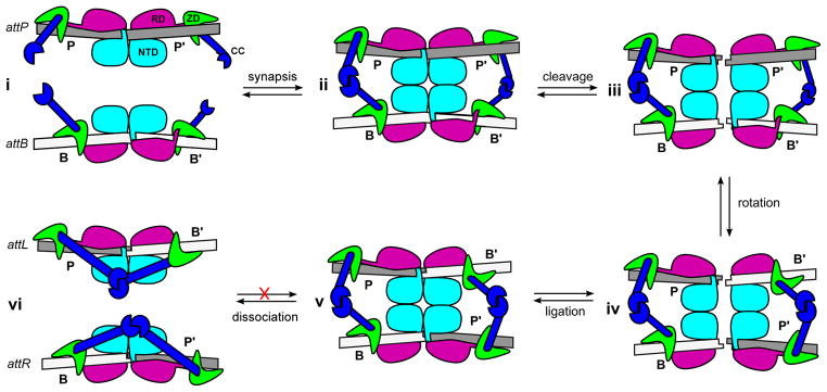
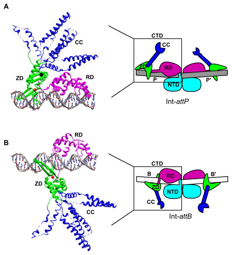
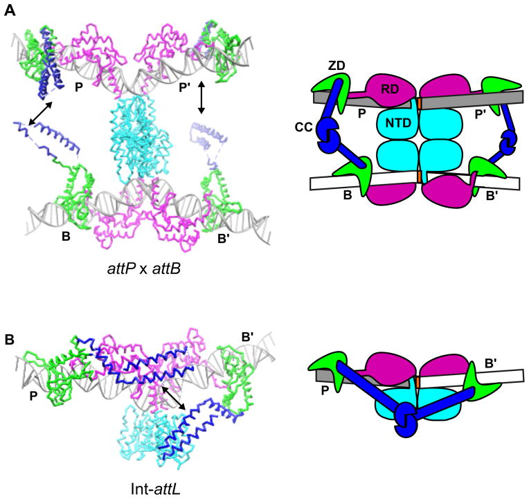
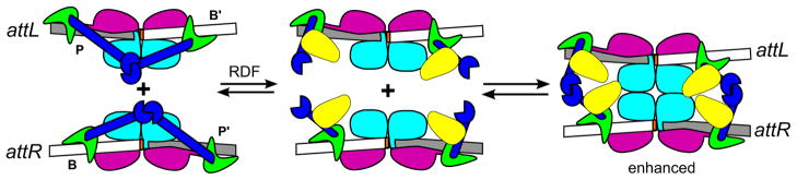
Serine integrases catalyze the integration and excision of phage genomes into and out of bacterial chromosomes in a highly specific and directional manner, making these proteins powerful tools for genome engineering. In 2013, the first structure of a serine integrase-DNA complex was reported. This work revealed how the phage attP sequence is recognized by the integrase and provided important clues about how serine integrases bind to other attachment site sequences. The resulting structural models indicate that distinct spatial arrangements of integrase domains are present for each attachment site complex. Here we describe how serine integrases may exploit this site-dependent domain arrangement to regulate the direction of recombination. We also discuss how phage-encoded recombination directionality factors could change this directionality by altering the nature of inter-subunit interactions.


Figures

Figure 1
Figure 1
Recently proposed model of the integration reaction catalyzed by serine integrases. i) Integrase (Int) dimers bind to specific sequences in the phage (attP) and host (attB) DNA. The Int-attP and Int-attB complexes are conformationally distinct due to different positioning of a zinc ribbon domain (ZD). ii) Int-attP and Int-attB associate to form a synaptic complex that is stabilized by interactions between coiled-coil (CC) motifs. iii) The Int subunits cleave all four DNA strands at the central dinucleotide, forming 5′-phosphoserine linkages between integrase subunits and DNA half-sites (not illustrated) and generating 3′-dinucleotide overhangs. iv) The P′ and B′-linked subunits can exchange places by rotating 180° about a horizontal axis relative to the P and B-linked subunits. v) Base-pairing between the central dinucleotides promotes ligation of the DNA strands, resulting in formation of two new attachment sites, attL and attR. vi) The unique arrangement of ZDs in the attL and attR sites allows the CC motifs to form intra-molecular interactions that prevent the reaction from running efficiently in the reverse direction. NTD: N-terminal catalytic domain (cyan); RD: recombinase domain (magenta); ZD: zinc ribbon domain (green); CC: coiled-coil motif (blue).
Figure 2
Figure 2
Positioning of the zinc ribbon domain distinguishes attP and attB. A) Structure of an Int C-terminal domain (CTD) bound to an attP half-site. The CTD is comprised of a recombinase domain (RD; magenta) and a zinc-ribbon domain (ZD; green) separated by a flexible linker. The RD, linker, and ZD form extensive contacts with bases in the major and minor grooves along one face of the attP half-site. The ZD contains a flexible coiled-coil motif (CC; blue) that does not contact DNA and is likely involved in protein-protein interactions. The CC motifs adopt different trajectories in the four independent complexes in the crystallographic asymmetric unit; these conformers are superimposed here. A coordinated zinc ion in the ZD is shown as a red sphere. A schematic representation of an Int dimer bound to the full attP site is shown on the right, where the region corresponding to the experimental structure is boxed. B) Structural model of an Int-attB half-site complex obtained by shifting the ZD by 5-bp towards the center of the site (see text). A schematic representation of an Int dimer bound to the full attB site is shown on the right (the NTD is not included in the structural model).
Figure 3
Figure 3
The coiled-coil motif regulates synaptic complex formation. A) Structural and schematic models of an attP x attB synaptic complex. The zinc ribbon domains of the juxtaposed half-sites (P/B and P′/B′) are oriented so that their CC-motifs can interact and stabilize the attP x attB complex without inhibiting subunit rotation. B) Structural and schematic models of the Int-attL complex. Whereas the CC-motifs are positioned on opposite faces of the DNA in the Int-attP and Int-attB complexes and would not be expected to interact with one another (see Fig. 2), the CC motifs are positioned on the same face of the complex in Int-attL and could readily interact (black arrow). The same argument can be made for the closely related Int-attR complex. Thus, intra-molecular interactions between coiled-coils in the Int-attL and Int-attR complexes may explain why excision is inhibited in the absence of an RDF protein. Different experimentally observed CC-conformers are shown in A) vs. B). Integrase domains are colored as in Fig. 1.
Figure 4
Figure 4
A plausible model for recombination directionality factor (RDF)-stimulated excision. The RDFs (yellow) could bind to the Int CC motifs and disrupt the intra-molecular interactions responsible for inhibiting attL x attR recombination. The RDFs may also interact with additional integrase domains and in some systems may interact with one another. Integrase domains are colored as in Fig. 1.

References

    1. Groth AC, Calos MP. Phage integrases: biology and applications. J Mol Biol. 2004;335:667–78. - PubMed
    1. Smith MCM, Brown WRA, McEwan AR, Rowley PA. Site-specific recombination by phiC31 integrase and other large serine recombinases. Biochem Soc Trans. 2010;38:388–94. A recent review of serine integrases that covers mechanistic studies through 2010. - PubMed
    1. Grindley N, Whiteson K, Rice P. Mechanisms of site-specific recombination. Annu Rev Biochem. 2006;75:567–605. - PubMed
    1. Li W, Kamtekar S, Xiong Y, Sarkis GJ, Grindley NDF, Steitz TA. Structure of a synaptic gammadelta resolvase tetramer covalently linked to two cleaved DNAs. Sci 80- 2005;309:1210–5. - PubMed
    1. Ritacco CJ, Kamtekar S, Wang J, Steitz TA. Crystal structure of an intermediate of rotating dimers within the synaptic tetramer of the G-segment invertase. Nucleic Acids Res. 2013;41:2673–2682. - PMC - PubMed