. 2015 Mar;261(3):611-8.
doi: 10.1097/SLA.0000000000000572.

Engendering allograft ignorance in a mouse model of allogeneic skin transplantation to the distal hind limb

Affiliations

Engendering allograft ignorance in a mouse model of allogeneic skin transplantation to the distal hind limb

Shailesh Agarwal et al. Ann Surg. 2015 Mar.

Abstract

Objective: The aim of this study was to demonstrate lymphatic isolation in a model of hind limb lymph node (LN) excision, consisting of ipsilateral popliteal and inguinal LN excision and to evaluate the immunologic response to allogeneic skin transplanted onto this region of lymphatic isolation.

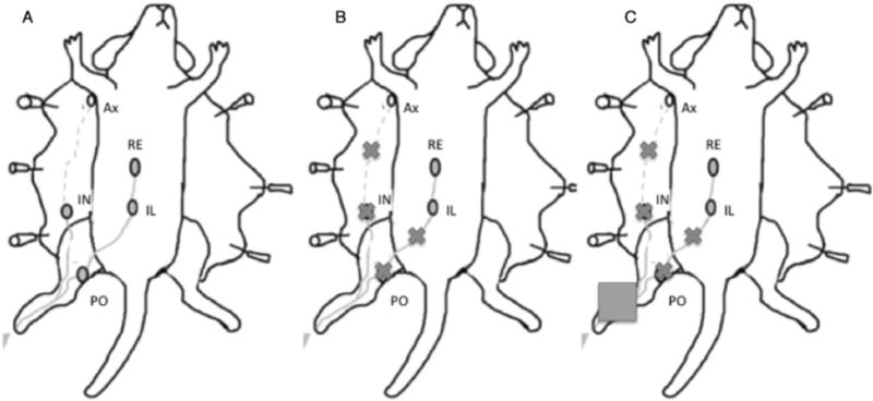
Methods: To study lymphatic flow, C57BL/6 mice underwent lymphadenectomy (n = 5), sham lymphadenectomy (n = 5), or no intervention (n = 5), followed by methylene blue injection. Mice were dissected to determine whether methylene blue traveled to the iliac LN. To study host response to skin transplantation, C57BL/6 mice underwent allogeneic skin transplantation with LN excision (n = 6), allogeneic skin transplantation alone (n = 6), or syngeneic skin transplantation (n = 4). Skin grafts were placed distal to the popliteal fossa and mice were euthanized at day 10. Grafts were stained for endothelial cell and proliferation markers (CD31 and Ki67, respectively). Secondary lymphoid tissues (spleen, ipsilateral axillary LN, and contralateral inguinal LN) were removed and rechallenged with BALB/c alloantigen in vitro with subsequent assay of interferon-γ and interleukin 4 cell expression using ELISPOT technique.

Results: Mice that underwent LN excision had no evidence of methylene blue in the iliac nodes; mice without surgical intervention or with sham LN excision consistently had methylene blue visible in the ipsilateral iliac nodes. Mice treated with allogeneic skin transplantation and LN excision had lower expression of interferon-γ and interleukin 4 in the secondary lymphoid tissues.

Conclusions: Lymph node excision completely interrupts lymphatic flow of the hind limb. This model of lymphatic isolation impairs the ability of the transplant recipient to acutely mount a Th1 or Th2 response to allogeneic skin transplants.


Conflict of interest statement

Disclosure: The authors declare no conflicts of interest.

Figures

FIGURE 1
FIGURE 1
Lymphatic drainage of mouse footpad with experimental models. A, Lymphatic drainage of mouse footpad to the popliteal and inguinal nodes. Lymph then progresses to the iliac node consistently; lymphatic drainage to the axillary node occurs inconsistently. B, Excision of the inguinal and popliteal lymph nodes results in obstructed lymphatic flow to the iliac or axillary lymph nodes. C, Lymphatic restriction of the transplanted skin after inguinal and popliteal lymph node excision requires transplantation to the distal hind limb. (Adapted with modifications from Harrell et al.). Lymph nodes (oval), excised nodes and interrupted lymphatics (“x”), and skin graft (square) are shown. AX indicates axillary; IL, iliac; IN, inguinal; PO, popliteal; RE, renal.
FIGURE 2
FIGURE 2
Lymph node staining with methylene blue after LN excision. A, Naïve mouse with no surgical intervention demonstrates methylene blue drainage to the ipsilateral popliteal, inguinal, and iliac lymph nodes after hind limb injection. B, Mouse with sham surgery demonstrates methylene blue drainage to the ipsilateral popliteal, inguinal, and iliac lymph nodes after hind limb injection. C, Mouse with popliteal and inguinal LN excision demonstrates no methylene blue drainage to the ipsilateral iliac LN. D, Mice receiving no intervention or sham surgery had blue dye in 5/5 popliteal (black), inguinal (light grey), and iliac (grey) nodes. Mice receiving popliteal and inguinal LN excision had blue dye in 0/5 iliac nodes. SLNEx indicates sentinel LN excision.
FIGURE 3
FIGURE 3
A, Day 10 allograft appearance (10×). (Top) Syngeneic graft near 100% survival, CD31 stain shows capillary growth, and Ki67 stain shows graft cell proliferation. (Middle) Allograft without LN excision near 100% survival, CD31 stain shows capillary growth, and Ki67 stain shows graft cell proliferation. (Bottom) Allograft with hind limb LN excision near 100% survival, CD31 stain shows capillary growth, and Ki67 stain shows graft cell proliferation. Therefore, all skin grafts were able to heal onto the recipient site. Bar indicates distance of 100 μm. B, Day 10 allograft appearance (20×). (Top) Syngeneic graft near 100% survival, CD31 stain shows capillary growth, and Ki67 stain shows graft cell proliferation. (Middle) Allograft without LN excision near 100% survival, CD31 stain shows capillary growth, and Ki67 stain shows graft cell proliferation. (Bottom) Allograft with hind limb LN excision near 100% survival, CD31 stain shows capillary growth, and Ki67 stain shows graft cell proliferation. Therefore, all skin grafts were able to heal onto the recipient site. Bar indicates distance of 100 μm. SLNEx indicates sentinel LN excision.
FIGURE 4
FIGURE 4
Secondary lymphoid tissues are primed for response against alloantigen after skin allotransplantation. Elevated IFN-γ expression in lymphocytes from ipsilateral popliteal and inguinal LNs in mice that did not undergo LN excision. Mice that had undergone LN excision did not have popliteal or inguinal LNs that could be assayed. IFN-γ indicates interferon-γ.
FIGURE 5
FIGURE 5
Secondary lymphoid tissue Th1/Th2 priming in mice with LN excision is diminished. A, Decreased IFN-γ expression in splenocytes after skin allograft with LN excision c. B, Decreased IFN-γ expression in lymphocytes from pooled ipsilateral axillary/contralateral inguinal LNs after skin allograft with LN excision. C, Decreased IL-4 expression in splenocytes after allogeneic skin graft with hind limb LN excision. D, Low levels of IL-17 expression in splenocytes from all mice. IFN-γ indicates interferon-γ; SLNEx, sentinel LN excision.

References

    1. Brandacher G, Lee WP, Schneeberger S. Minimizing immunosuppression in hand transplantation. Expert Rev Clin Immunol. 2012;8:673–684. - PubMed
    1. Wu S, Xu H, Ravindra K, et al. Composite tissue allotransplantation: past, present and future-the history and expanding applications of CTA as a new frontier in transplantation. Transplant Proc. 2009;41:463–465. - PMC - PubMed
    1. Ravindra KV, Ildstad ST. Immunosuppressive protocols and immunological challenges related to hand transplantation. Hand Clin. 2011;27:467–479. - PubMed
    1. Ravindra KV, Wu S, McKinney M, et al. Composite tissue allotransplantation: current challenges. Transplant Proc. 2009;41:3519–3528. - PubMed
    1. Swearingen B, Ravindra K, Xu H, et al. Science of composite tissue allotransplantation. Transplantation. 2008;86:627–635. - PMC - PubMed

Publication types