. 2014 Apr;196(8):1514-24.
doi: 10.1128/JB.01441-13. Epub 2014 Feb 7.

The mthA mutation conferring low-level resistance to streptomycin enhances antibiotic production in Bacillus subtilis by increasing the S-adenosylmethionine pool size

Affiliations

The mthA mutation conferring low-level resistance to streptomycin enhances antibiotic production in Bacillus subtilis by increasing the S-adenosylmethionine pool size

Shigeo Tojo et al. J Bacteriol. 2014 Apr.

Abstract

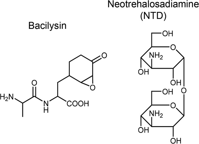
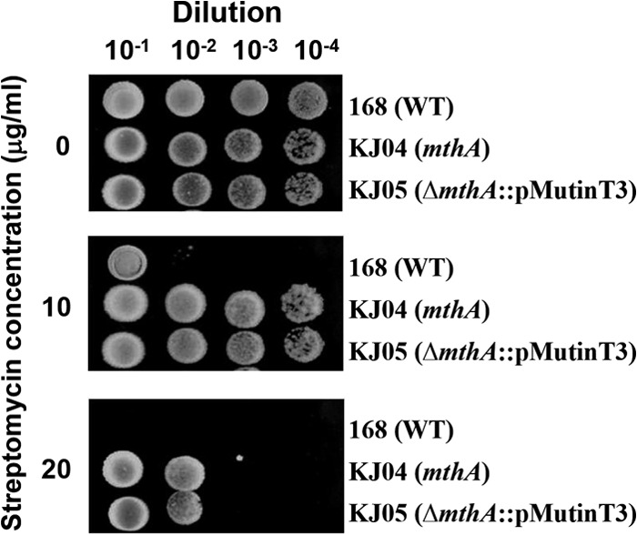
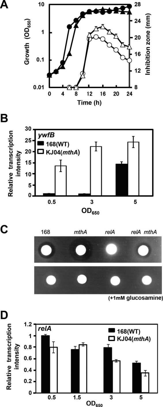
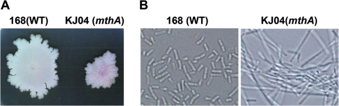
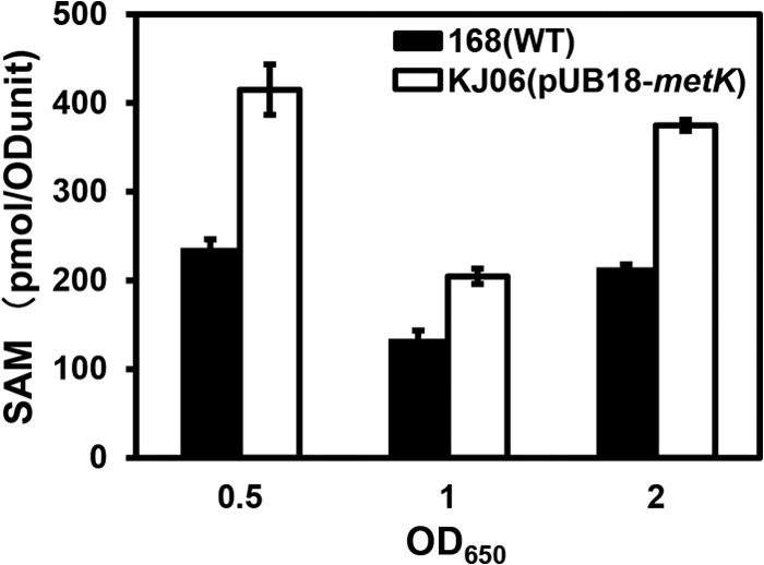
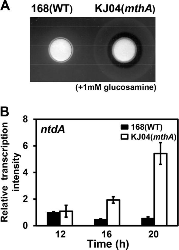
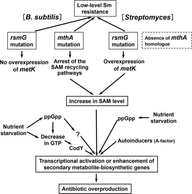
Certain Str(r) mutations that confer low-level streptomycin resistance result in the overproduction of antibiotics by Bacillus subtilis. Using comparative genome-sequencing analysis, we successfully identified this novel mutation in B. subtilis as being located in the mthA gene, which encodes S-adenosylhomocysteine/methylthioadenosine nucleosidase, an enzyme involved in the S-adenosylmethionine (SAM)-recycling pathways. Transformation experiments showed that this mthA mutation was responsible for the acquisition of low-level streptomycin resistance and overproduction of bacilysin. The mthA mutant had an elevated level of intracellular SAM, apparently acquired by arresting SAM-recycling pathways. This increase in the SAM level was directly responsible for bacilysin overproduction, as confirmed by forced expression of the metK gene encoding SAM synthetase. The mthA mutation fully exerted its effect on antibiotic overproduction in the genetic background of rel(+) but not the rel mutant, as demonstrated using an mthA relA double mutant. Strikingly, the mthA mutation activated, at the transcription level, even the dormant ability to produce another antibiotic, neotrehalosadiamine, at concentrations of 150 to 200 μg/ml, an antibiotic not produced (<1 μg/ml) by the wild-type strain. These findings establish the significance of SAM in initiating bacterial secondary metabolism. They also suggest a feasible methodology to enhance or activate antibiotic production, by introducing either the rsmG mutation to Streptomyces or the mthA mutation to eubacteria, since many eubacteria have mthA homologues.


Figures

FIG 1
FIG 1
Structures of bacilysin and neotrehalosadiamine.
FIG 2
FIG 2
Sm susceptibility of the mthA mutant. The B. subtilis strains 168 (wild type [WT]), KJ04 (mthA1 transformant), and KJ05 (mthA disruptant) were grown to stationary phase in L medium at 37°C. Approximately 2 × 10⁸ cells were diluted with distilled water, and 5-μl aliquots of the cell suspension were spotted onto medium containing 0, 10, or 20 μg/ml of Sm, followed by incubation at 37°C for 12 h.
FIG 3
FIG 3
Growth, antibiotic production, and transcriptional analysis of parental (168) and mthA mutant (KJ04) strains. (A) Strains were grown in NG medium at 37°C. Antibiotic production was determined by the paper disk-agar diffusion method and expressed as the size of the inhibition zone around the disk (diameter, 8 mm). Growth (closed symbols) and antibiotic production (open symbols) of strain 168 (circles) and strain KJ04 (triangles) are shown. (B) Transcriptional analysis of the ywfB gene involved in bacilysin biosynthesis. The strains were grown as for panel A, and the levels of expression of the ywfB gene were determined by real-time qPCR. (C) Strains 168 (wild type), KJ04 (mthA), K2-007 (relA), and KO-1234 (relA mthA) were grown for 15 h as for panel A, and bacilysin production was determined by the paper disk-agar diffusion method. (D) Transcriptional analysis of the relA gene involved in ppGpp synthesis. Strains were grown as for panel A, and levels of expression of the relA gene were determined by real-time qPCR. The error bars indicate the standard deviations of the means of three or more samples.
FIG 4
FIG 4
Morphological appearance of parental (168) and mthA mutant (KJ04) strains. (A) Colony morphology observed after 5 days of culture on L agar plates. (B) Microscopic observation (magnification, ×1,000) of cells grown to mid-growth phase (5 h) in L medium.
FIG 5
FIG 5
Outline of SAM-recycling pathways in B. subtilis. The genes and enzymes involved in SAM recycling are as follows: metI, cystathionine γ-synthase; metC and patB, cystathionine β-lyase; metE, methionine synthase; metK, SAM synthetase; speD, SAM decarboxylase; speE, spermidine synthase; mthA, SAH/MTA nucleosidase (mthA is synonymous with mtnA); mtnK, methylthioribose kinase; the mtnA and mtnWXBD gene products, involved in the MTR-to-KMBA recycling pathway; mtnE, aminotransferase; luxS, S-ribosylhomocysteine hydrolase; yrhA (mccA), cystathionine β-synthase; yrhB (mccB), cystathionine γ-lyase and homocysteine γ-lyase; SAM (S-adenosylmethionine); SAH (S-aenosylhomocysteine); KMBA (α-keto-γ-methyl-thiobutyric acid); MTA (methylthioadenosine); MTR (methylthioribose); and SRH (S-ribosylhomocysteine). AI-2 indicates autoinducer-2. The figure was drawn on the basis of work by Hullo et al. (53).
FIG 6
FIG 6
Intracellular SAM level and transcriptional analysis of the metK gene in wild-type (168) and mthA mutant (KJ04) strains. (A) The strains were grown in NG medium at 37°C to an OD650 of 0.5, 1, or 1.5. Intracellular SAM levels were determined by reverse-phase high-performance liquid chromatography. (B) Transcriptional analysis of the metK gene. Strains were grown in NG medium as for panel A. Total RNAs were extracted from the cells and used for real-time qPCR analysis. The transcription level of metK was normalized relative to the amount of 16S rRNA in each RNA sample. The error bars indicate the standard deviations of the means of three or more samples.
FIG 7
FIG 7
Effect of metK overexpression on the intracellular SAM level. Strains were grown in NG medium at 37°C to an OD650 of 0.5, 1, or 2. Intracellular SAM levels were determined by reverse-phase high-performance liquid chromatography. The error bars indicate the standard deviations of the means of three or more samples.
FIG 8
FIG 8
NTD production and transcriptional analysis in parental (168) and mthA mutant (KJ04) strains. (A) Strains were grown in S7N medium at 37°C for 24 h, and NTD production was determined by the paper disk-agar diffusion method. (B) Transcriptional analysis of the ntdA gene. The strains were grown as for panel A for 12, 16, or 20 h, and the level of expression of the ntdA gene was determined by real-time qPCR. The error bars indicate the standard deviations of the means of three or more samples.
FIG 9
FIG 9
Schematic showing the signal transduction pathways in B. subtilis and Streptomyces spp., from the mthA or rsmG mutation to the enhancement of antibiotic production or the activation of silent genes. The scheme was based on the work presented here and previous studies in B. subtilis (12, 18, 23, 29, 31) and Streptomyces spp. (13, 44, 48).

Similar articles

Cited by

References

    1. Schatz A, Waksman SA. 1944. Effect of streptomycin and other antibiotic substances upon Mycobacterium tuberculosis and related organisms. Proc. Soc. Exp. Bio. Med. 57:244–248. 10.3181/00379727-57-14769 - DOI
    1. Klein M, Kimmelman LJ. 1946. The role of spontaneous variants in the acquisition of streptomycin resistance by the Shigellae. J. Bacteriol. 52:471–479 - PMC - PubMed
    1. Honore N, Cole ST. 1994. Streptomycin resistance in mycobacteria. Antimicrob. Agents Chemother. 38:238–242. 10.1128/AAC.38.2.238 - DOI - PMC - PubMed
    1. Honore N, Marchal G, Cole ST. 1995. Novel mutation in 16S rRNA associated with streptomycin dependence in Mycobacterium tuberculosis. Antimicrob. Agents Chemother. 39:769–770. 10.1128/AAC.39.3.769 - DOI - PMC - PubMed
    1. Carter AP, Clemons WM, Brodersen DE, Morgan-Warren RJ, Wimberly BT, Ramakrishnan V. 2000. Functional insights from the structure of the 30S ribosomal subunit and its interactions with antibiotics. Nature 407:340–348. 10.1038/35030019 - DOI - PubMed

Publication types

MeSH terms

LinkOut - more resources