. 2014 Mar;11(2):175-83.
doi: 10.1038/cmi.2013.66. Epub 2014 Feb 10.

HBV life cycle is restricted in mouse hepatocytes expressing human NTCP

Affiliations

HBV life cycle is restricted in mouse hepatocytes expressing human NTCP

Hanjie Li et al. Cell Mol Immunol. 2014 Mar.

Abstract

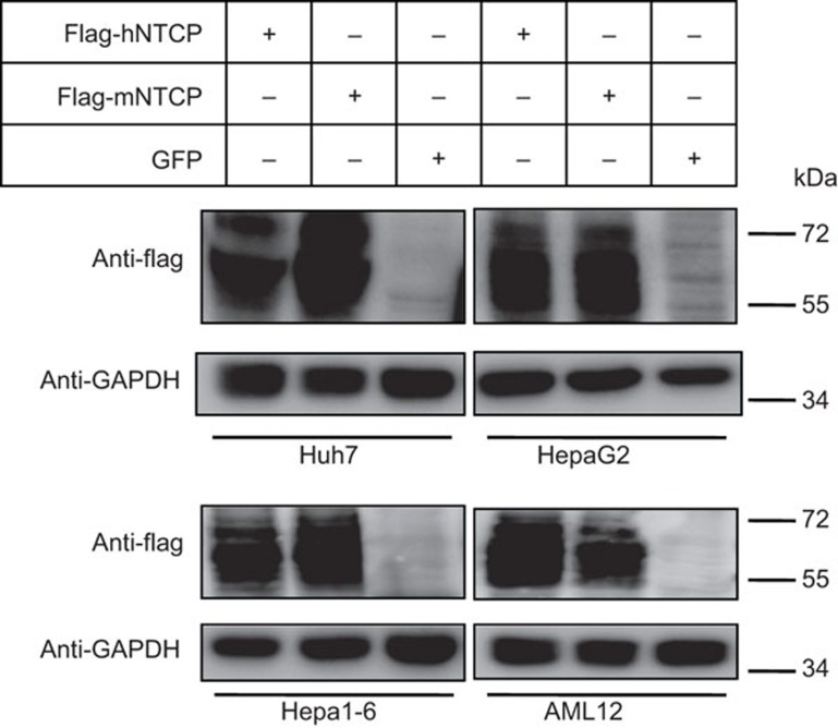
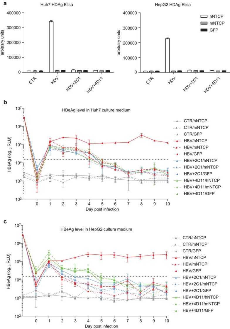
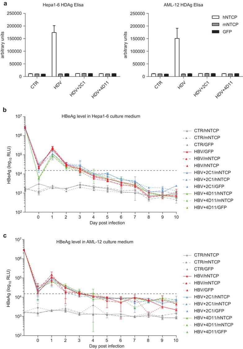
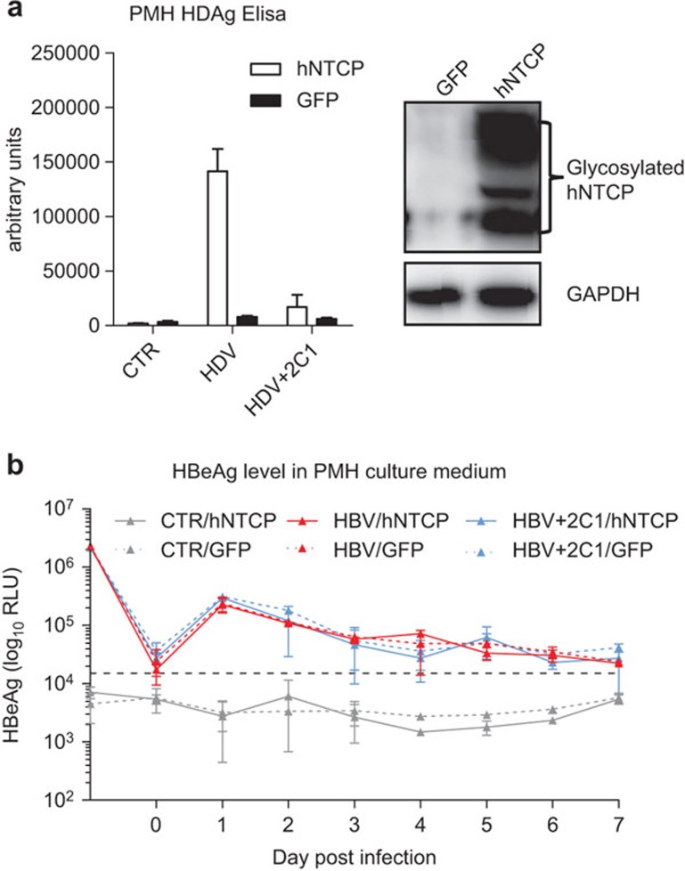
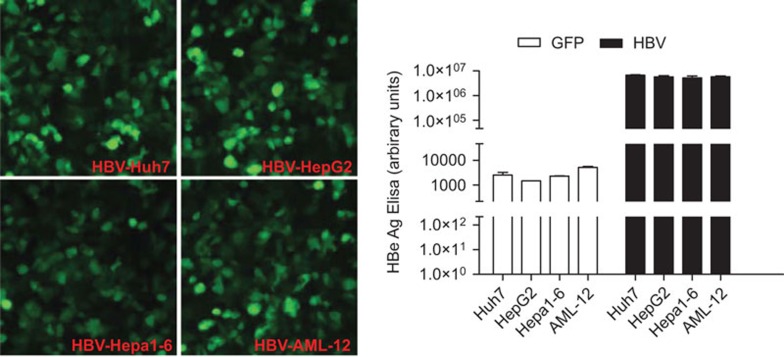
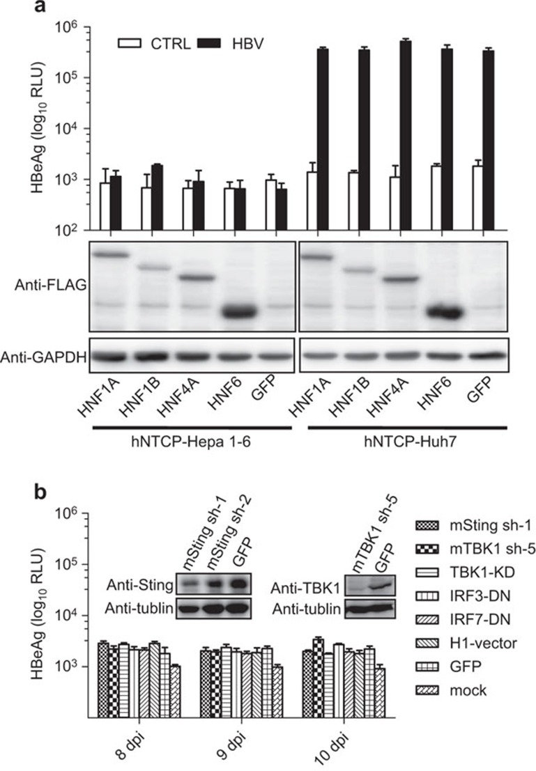
Recent studies have revealed that human sodium taurocholate cotransporting polypeptide (SLC10A1 or NTCP) is a functional cellular receptor for hepatitis B virus (HBV). However, whether human NTCP can support HBV infection in mouse hepatocyte cell lines has not been clarified. Because an HBV-permissible mouse model would be helpful for the study of HBV pathogenesis, it is necessary to investigate whether human NTCP supports the susceptibility of mouse hepatocyte cell lines to HBV. The results show that exogenous human NTCP expression can render non-susceptible HepG2 (human), Huh7 (human), Hepa1-6 (mouse), AML-12 (mouse) cell lines and primary mouse hepatocyte (PMH) cells susceptible to hepatitis D virus (HDV) which employs HBV envelope proteins. However, human NTCP could only introduce HBV susceptibility in human-derived HepG2 and Huh7 cells, but not in mouse-derived Hepa1-6, AML-12 or PMH cells. These data suggest that although human NTCP is a functional receptor that mediates HBV infection in human cells, it cannot support HBV infection in mouse hepatocytes. Our study indicated that the restriction of HBV in mouse hepatocytes likely occurs after viral entry but prior to viral transcription. We have excluded the role of mouse hepatocyte nuclear factors in the restriction of the HBV life cycle and showed that knockdown or inhibition of Sting, TBK1, IRF3 or IRF7, the components of the anti-viral signaling pathways, had no effect on HBV infection in mouse hepatocytes. Therefore, murine restriction factors that limit HBV infection need to be identified before a HBV-permissible mouse line can be created.


Figures

Figure 1
Figure 1
Expression of human NTCP, mouse NTCP or GFP by lentiviral infection in Huh7, HepG2, Hepa1–6 and AML-12 cells. Huh7, HepaG2, Hepa1–6 and AML12 cells were infected with lentivirus expressing human and mouse NTCP with a flag taq at the C terminal, together with GFP as a negative control. Extracts were collected 48 h after infection. Immunoblotting was conducted with anti-flag antibodies to detect NTCP protein levels from the four cell lines. NTCP, sodium taurocholate cotransporting polypeptide.
Figure 2
Figure 2
Human NTCP, but not mouse NTCP, expression in Huh7 and HepG2 cells supports HDV/HBV infection. (a) Huh7 and HepG2 cells were infected with lentivirus expressing human NTCP, mouse NTCP or GFP. Forty-eight hours after infection, the cells were inoculated with or without HDV at 100 mge in the presence or absence of 2C1 and 4D11. Seven days post inoculation, the cells were lysed and HDAg was detected using an ELISA kit. Huh7 (b) and HepG2 (c) cells expressing human NTCP, mouse NTCP or GFP were inoculated with or without HBV at 500 mge in the presence or absence of 2C1 and 4D11. Culture media were changed every day, the supernatants were collected and HBeAg was detected using an ELISA kit. The HBeAg level at day 1 represents the level in the inoculums. The HBeAg level at day 0 represents the level in the fresh media. Data are presented as means±s.d. HBeAg, hepatitis B e antigen; HBV, hepatitis B virus; HDAg, hepatitis D antigen; HDV, hepatitis B virus; mge, multiplicities of genome equivalents; NTCP, sodium taurocholate cotransporting polypeptide.
Figure 3
Figure 3
Human NTCP expression in Hepa1–6 and AML-12 supports HDV infection but not HBV infection. (a) Hepa1–6 and AML-12 cells were infected with lentivirus expressing human NTCP, mouse NTCP or GFP. Forty-eight hours after infection, the cells were inoculated with or without HDV at 100 mge in the presence or absence of 2C1 and 4D11. Seven days post inoculation, the cells were lysed and HDAg was detected using an ELISA kit. Hepa1–6 (b) and AML-12 (c) cells expressing human NTCP, mouse NTCP or GFP were inoculated with or without HBV at 500 mge in the presence or absence of 2C1 and 4D11. Culture media were changed every day, the supernatants were collected and HBeAg was detected using an ELISA kit. The HBeAg level at day 1 represents the level in the inoculums. The HBeAg level at day 0 represents the level in the fresh media. Data are presented as means±s.d. HBeAg, hepatitis B e antigen; HBV, hepatitis B virus; HDAg, hepatitis D antigen; HDV, hepatitis B virus; mge, multiplicities of genome equivalents; NTCP, sodium taurocholate cotransporting polypeptide.
Figure 4
Figure 4
Human NTCP expression in PMHs supports HDV infection, but not HBV infection. (a) PMHs were infected with adenovirus expressing human or GFP. Twenty-four hours after infection, the cells were inoculated with or without HDV at 100 mge in the presence or absence of 2C1. Seven days post inoculation, the cells were lysed and HDAg was detected using an ELISA kit. Rabbit anti-human NTCP polyclonal antibodies were used in the western blotting for detecting human NTCP expression. (b) PMH cells expressing human NTCP or GFP were inoculated with or without HBV at 500 mge in the presence or absence of 2C1. Culture media were changed every day, the supernatants were collected and HBeAg was detected using an ELISA kit. The HBeAg level at day 1 represents the level in the inoculums. The HBeAg level at day 0 represents the level in the fresh media. Data are presented as means±s.d. HBeAg, hepatitis B e antigen; HBV, hepatitis B virus; HDAg, hepatitis D antigen; HDV, hepatitis B virus; mge, multiplicities of genome equivalents; NTCP, sodium taurocholate cotransporting polypeptide; PMH, primary mouse hepatocyte.
Figure 5
Figure 5
Huh7, HepG2, Hepa1–6 and AML-12 cells secrete comparable levels of HBeAg when infected with HBV genome-expressing adenovirus. Huh7, HepG2, Hepa1–6 and AML-12 cells were infected with adenovirus expressing the HBV genome or GFP as a control. The HBV-expressing adenovirus vector also contains a GFP gene allowing us to evaluate the infection efficiency. By titrating the titer of the HBV genome-expressing adenovirus, we ensure that the four cell lines are approximately 80%–100% GFP-positive at a similar intensity 36 h after infection. Sixty hours after infection, the supernatants were collected and measured for HBeAg. Data are presented as means±s.d. HBeAg, hepatitis B e antigen; HBV, hepatitis B virus.
Figure 6
Figure 6
Expressing human HNFs or attenuating the function of key components of anti-viral signaling pathway in human NTCP-expressing Hepa1–6 cannot eliminate the restriction of HBV in mouse hepatocytes. (a) hNTCP-Hepa1–6 and hNTCP-Huh7 cells were infected with lentiviruses expressing HNF1A, HNF1B, HNF4A, HNF6 or GFP. Then, those cells were infected with HBV at 100 mge. Ten days post inoculation, the supernatants were collected and measured for HBeAg levels. (b) hNTCP-Hepa1–6 was infected with lentiviruses expressing mouse Sting shRNA (mSting sh-1), mouse TBK1 shRNA (mTBK1 sh-5), dominant-negative mutants of TBK1 (TBK1-KD), IRF3 (IRF3-DN) and IRF7 (IRF7-DN), GFP and an empty vector. The cells were then infected with HBV at 100 mge. Supernatants at 8, 9 and 10 days post inoculation were collected and measured for HBeAg levels. The knockdown efficiencies of mSting and mTBK1 are shown. Data are presented as means±s.d. HBeAg, hepatitis B e antigen; HBV, hepatitis B virus; HNF, hepatocyte nuclear factor; mge, multiplicities of genome equivalents; NTCP, sodium taurocholate cotransporting polypeptide.

References

    1. Heermann KH, Goldmann U, Schwartz W, Seyffarth T, Baumgarten H, Gerlich WH. Large surface proteins of hepatitis B virus containing the pre-s sequence. J Virol. 1984;52:396–402. - PMC - PubMed
    1. Ott JJ, Stevens GA, Groeger J, Wiersma ST. Global epidemiology of hepatitis B virus infection: new estimates of age-specific HBsAg seroprevalence and endemicity. Vaccine. 2012;30:2212–2219. - PubMed
    1. Hughes SA, Wedemeyer H, Harrison PM. Hepatitis delta virus. Lancet. 2011;378:73–85. - PubMed
    1. Gripon P, Le Seyec J, Rumin S, Guguen-Guillouzo C. Myristylation of the hepatitis B virus large surface protein is essential for viral infectivity. Virology. 1995;213:292–299. - PubMed
    1. Le Seyec J, Chouteau P, Cannie I, Guguen-Guillouzo C, Gripon P. Infection process of the hepatitis B virus depends on the presence of a defined sequence in the pre-S1 domain. J Virol. 1999;73:2052–2057. - PMC - PubMed

MeSH terms