Review
. 2014 Mar;12(3):197-210.
doi: 10.1038/nrmicro3215. Epub 2014 Feb 10.

Molecular mechanisms of varicella zoster virus pathogenesis

Affiliations
Review

Molecular mechanisms of varicella zoster virus pathogenesis

Leigh Zerboni et al. Nat Rev Microbiol. 2014 Mar.

Abstract

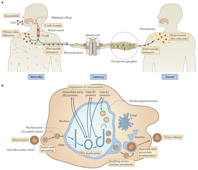
Varicella zoster virus (VZV) is the causative agent of varicella (chickenpox) and zoster (shingles). Investigating VZV pathogenesis is challenging as VZV is a human-specific virus and infection does not occur, or is highly restricted, in other species. However, the use of human tissue xenografts in mice with severe combined immunodeficiency (SCID) enables the analysis of VZV infection in differentiated human cells in their typical tissue microenvironment. Xenografts of human skin, dorsal root ganglia or foetal thymus that contains T cells can be infected with mutant viruses or in the presence of inhibitors of viral or cellular functions to assess the molecular mechanisms of VZV-host interactions. In this Review, we discuss how these models have improved our understanding of VZV pathogenesis.


Conflict of interest statement

Competing interests statement

The authors declare no competing interests.

Figures

Figure 1
Figure 1. VZV life cycle and replication
a | Model of the varicella zoster virus (VZV) life cycle. VZV infects the human host when virus particles reach mucosal epithelial sites of entry. Local replication is followed by spread to tonsils and other regional lymphoid tissues, where VZV gains access to T cells. Infected T cells then deliver the virus to cutaneous sites of replication. VZV establishes latency in sensory ganglia after transport to neuronal nuclei along neuronal axons or by viraemia. Reactivation from latency enables a second phase of replication to occur in skin, which typically causes lesions in the dermatome that is innervated by the affected sensory ganglion. b | Model of VZV replication. Enveloped VZV particles attach to cell membranes, fuse and release tegument proteins. Uncoated capsids dock at nuclear pores, where genomic DNA is injected into the nucleus and circularizes. On the basis of events that have been documented in herpes simplex virus 1 (HSV-1) replication, immediate-early genes are expressed, followed by early and late genes. Nucleocapsids are assembled and package newly synthesized genomic DNA, move to the inner nuclear membrane and bud across the nuclear membrane. Capsids enter the cytoplasm, and virion glycoproteins mature in the trans-Golgi region and tegument proteins assemble in vesicles; capsids undergo secondary envelopment and are transported to cell surfaces, where newly assembled virus particles are released.
Figure 2
Figure 2. VZV T cell tropism
According to the model of varicella zoster virus (VZV) cell-associated viraemia, tonsil T cells are infected following VZV inoculation and replication in respiratory mucosal epithelial cells. T cells traffic into and out of tonsils across the squamous epithelial cells that line the tonsilar crypts (left panel). VZV has increased tropism for activated memory T cells that have skin-homing markers, which are common in tonsils (centre panel). These T cells are programmed for immune surveillance and can transport the virus across capillary endothelial cells into skin. VZV glycoprotein E (gE) (through its unique amino terminus), gI and the viral kinases ORF47 and ORF66 are important for T cell infection. Proteins that regulate cellular gene expression are activated (in the case of signal transducer and activator of transcription 3 (STAT3)) or inhibited (in the case of STAT1) in infected T cells. The microvasculature is extensive at the base of hair follicles, where T cells transit into the surrounding skin and initial VZV replication is observed (right panel).
Figure 3
Figure 3. VZV skin tropism
The schematic illustrates viral factors that ensure spread to the skin surface after varicella zoster virus (VZV) is delivered to cutaneous sites of replication by infected T cells or by retrograde axonal transport from neurons (left-hand side). Two examples of VZV proteins that are important for pathogenesis are shown: ORF61 protein has SUMO-interacting motifs that are important for dispersal of promyelocytic leukaemia nuclear bodies (PML-NBs) and the cytoplasmic domain of glycoprotein B (gB) has an immunoreceptor tyrosine-based inhibition motif that regulates cell–cell fusion and polykaryocyte formation. VZV replication in skin triggers cellular responses, including changes that are induced in infected cells and changes in the uninfected cells adjacent to infected cells. Examples of VZV effects within infected cells are illustrated (right-hand side). VZV induces signal transducer and activator of transcription 3 (STAT3) activation, which triggers the expression of the anti-apoptotic protein survivin and inhibits the expression of interferon-α (IFNα) and STAT1 (REF. 32). In contrast to infected cells, surrounding uninfected cells exhibit upregulation of IFNs, STAT1, which activates IFN-stimulated factors such as PML, and other cell transacivators and innate cytokines.
Figure 4
Figure 4. VZV neurotropism in DRG xenografts
This schematic illustrates active infection of dorsal root ganglia (DRG) which is characterized by the transcription of genes (for example, genes encoding glycoprotein B (gB), immediate early protein 62 (IE62) and IE63) that produce proteins that are required for lytic infection, varicella zoster virus (VZV) genome synthesis, virus assembly in neurons and satellite cells, release of VZV into intracellular spaces and fusion of some neurons and satellite cells (left panel). Virions are captured in cages that are formed by promyelocytic leukaemia nuclear bodies (PML-NBs) in some neurons and satellite cells. By contrast, latency (right panel) is associated with the persistence of VZV genomes and immediate-early (IE) transcripts, whereas late gene transcription, such as transcription of gB, ceases and virion formation ceases. When DRG are infected with VZV mutants in which binding of gE to gI is blocked or in which gI is deleted, the transition to latency is disrupted (right panel; outlined box), infectious virions continue to be produced at low levels and in the case of disrupted binding of gE to gI, tissue destruction is extensive, which is associated with disruption of the cell matrix, elimination of many neurons and the proliferation of satellite cells.

References

    1. Arvin AM, Gilden D. In: Fields Virology. 6. Knipe D, Howley P, editors. Lippincott Williams & Wilkins; 2013. pp. 2015–2057.
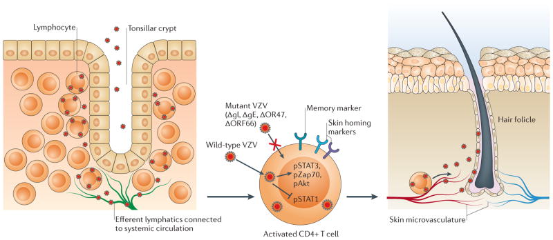
    1. Ku CC, et al. Varicella-zoster virus transfer to skin by T cells and modulation of viral replication by epidermal cell interferon-α. J Exp Med. 2004;200:917–925. This paper shows the role of T cells in the transport of VZV and the control of replication by the potent innate response of skin cells. - PMC - PubMed
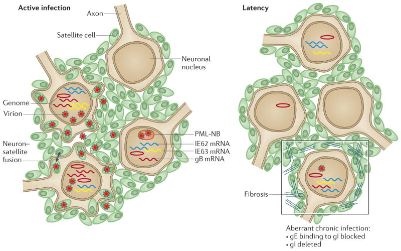
    1. Gilden DH, et al. Varicella-zoster virus DNA in human sensory ganglia. Nature. 1983;306:478–480. This study provides the first evidence of VZV latency in neurons, using in situ hybridization to detect VZV genomes. - PubMed
    1. Takahashi M. Clinical overview of varicella vaccine: development and early studies. Pediatrics. 1986;78:736–741. - PubMed
    1. Gershon AA, Gershon MD. Perspectives on vaccines against varicella-zoster virus infections. Curr Top Microbiol Immunol. 2010;342:359–372. - PMC - PubMed

Publication types