. 2014 Mar;21(3):261-8.
doi: 10.1038/nsmb.2775. Epub 2014 Feb 9.

Structural and mechanistic insight into Holliday-junction dissolution by topoisomerase IIIα and RMI1

Affiliations

Structural and mechanistic insight into Holliday-junction dissolution by topoisomerase IIIα and RMI1

Nicolas Bocquet et al. Nat Struct Mol Biol. 2014 Mar.

Abstract

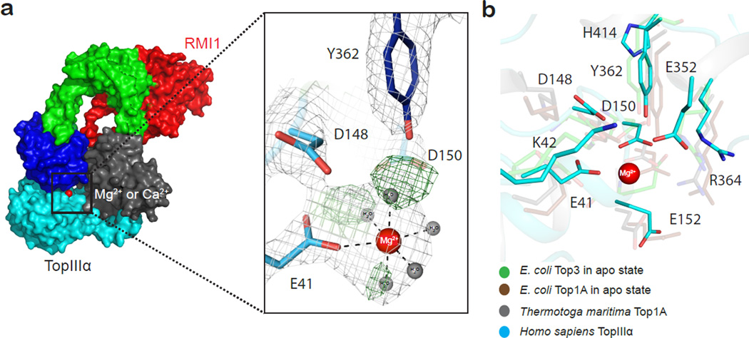
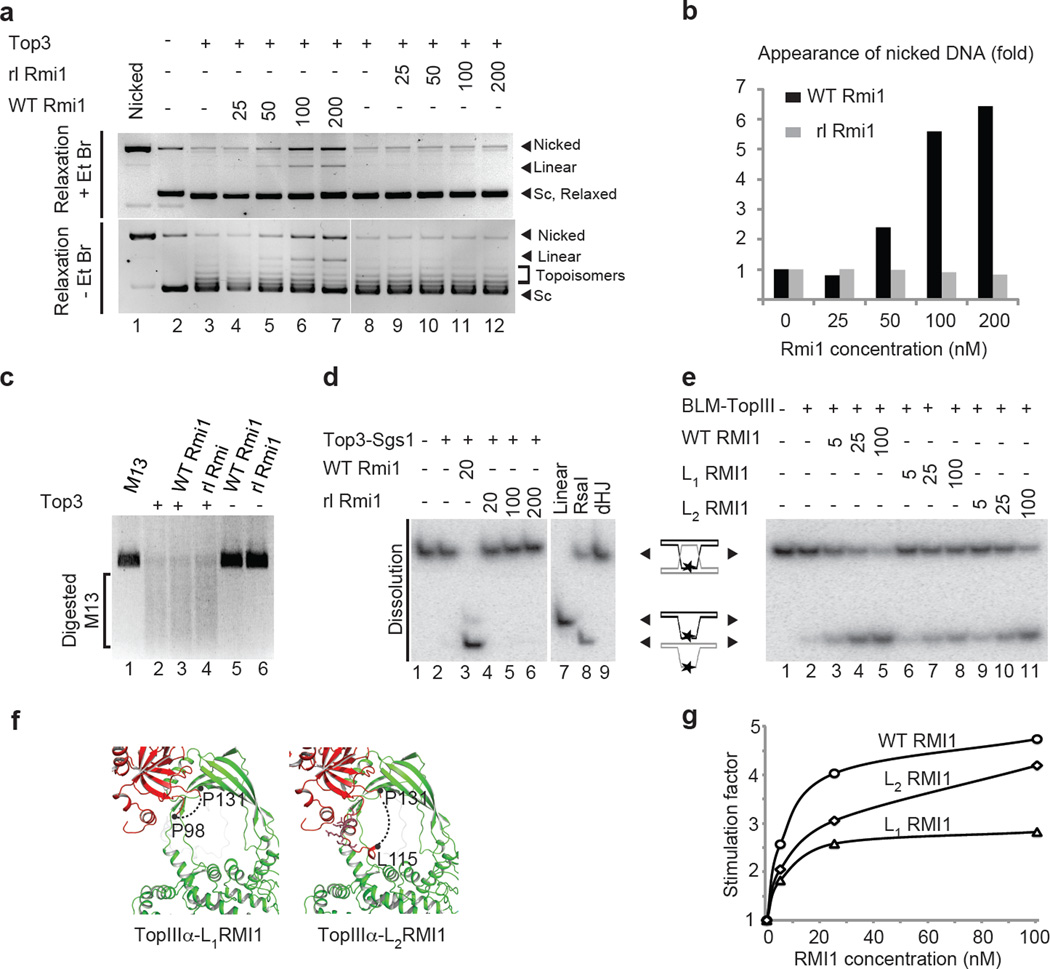
Repair of DNA double-strand breaks via homologous recombination can produce double Holliday junctions (dHJs) that require enzymatic separation. Topoisomerase IIIα (TopIIIα) together with RMI1 disentangles the final hemicatenane intermediate obtained once dHJs have converged. How binding of RMI1 to TopIIIα influences it to behave as a hemicatenane dissolvase, rather than as an enzyme that relaxes DNA topology, is unknown. Here, we present the crystal structure of human TopIIIα complexed to the first oligonucleotide-binding domain (OB fold) of RMI1. TopIII assumes a toroidal type 1A topoisomerase fold. RMI1 attaches to the edge of the gate in TopIIIα through which DNA passes. RMI1 projects a 23-residue loop into the TopIIIα gate, thereby influencing the dynamics of its opening and closing. Our results provide a mechanistic rationale for how RMI1 stabilizes TopIIIα-gate opening to enable dissolution and illustrate how binding partners modulate topoisomerase function.


Figures

Figure 1
Figure 1. Overall architecture and TopIIIα-RMI1 binding interface
a) Domain organization of the human TopIIIα-RMI1 complex. b) Overall architecture of the crystallized complex: (I) Toprim domain (cyan); (II) gate domain with the two topo-fold domains (green); (III) catalytic 5Y-CAP (blue), (IV) non-catalytic CAP domain (grey) with RMI1 shown in red. The catalytic metal is represented by a red sphere. c) Hydrophobic zipper between RMI1 residues originating from OBN and residues originating from the α12 and β10–α12 gate of TopIIIα. d) As in c) but depicting the corresponding hydrogen bonding network. e) Interaction between the RMI1 loop segment and the hydrophobic patch of the TopIIIα gate (helix α19) (near proposed pivot point see Fig 5). RMI1 P98 is engaged in hydrophobic stacking with TopIIIα residues F94, neighboring RMI1 Y100 stacks with TopIIIα F262. f) similar to e) highlighting the corresponding hydrogen-bonding and salt bridge network. RMI1 Q113 is at the center of a hydrogen-bonding network binding TopIIIα E254, H530 and K529, while ionic interaction between RMI1 E119 and TopIIIα R338 lock down the RMI1 insertion loop in position.
Figure 2
Figure 2. Catalytic site of human TopIIIα
a) TopIIIα catalytic site residues and the Mg2+ binding site. Mg2+ is shown as a red sphere, with corresponding water molecules depicted as grey spheres. Simulated annealing composite 2mFo-DFc omit electron density map contoured at 1.5σ (gray). The omit difference map shows peaks (green) for water molecules prior to their inclusion into the model. E41 serves as the sole residue directly contacting Mg2+ while D150 bridges a water molecule from the hydration shell. b) Structural alignment of different catalytic centers taken from the available type IA topoisomerases structures: E. coli Top3 in apo state (PDB code 1D6M35) in cyan, E. coli Top1A in the apo state (PDB code 1ECL) in magenta, Top1A from Thermotoga maritima (PDB code 2GAI) in grey, with human TopIIIα (pdb 4CGY) shown in green.
Figure 3
Figure 3. The RMI1 decatenation loop
a) Comparison between E. coli Top3 (shaded grey) and human TopIIIα (green). RMI1 (shaded red) inserts motifs at sites where E. coli Top3 diverges from human TopIIIα. The two unique E. coli Top3 loop segments shown are the loop between residues 241 and 255, and the decatenation loop (502–519). b) The final TOPO-Ca2+-RMI1 model overlaid with an unbiased 2mFo-DFc electron density envelope calculated prior to modeling the RMI1 decatenation loop, contoured at 1σ. c) Sequence alignments between different yeast strains and the human RMI1. Region in the black box within the S. cerevisiae sequence have been randomized in the rlRmi1 construct (see Methods).
Figure 4
Figure 4. The RMI1 decatenation loop stabilizes the open Top3 and stimulates dHJ dissolution
a) Yeast Top3 relaxation assay using pUC19 DNA to assess the effect of yeast Rmi1 wild-type (WT Rmi1) versus rlRmi1. Increasing concentrations of WT Rmi1 (lanes 4–7) and rlRmi1 (lanes 9–12) (Supplementary fig. 6a) b) Quantitation of the nicked DNA band intensity for WT Rmi1 (grey) and rlRmi1 (black) on EtBr stained gels as shown in a). c) Digest of single stranded M13 substrate with the different complexes following overnight incubation (Supplementary fig. 6b). d) dHJs dissolution assay of a short synthetic dHJ junction, using the yeast Top3, Sgs1 and Rmi1. dHJ substrate was incubated in the absence (lane 1) or presence of Top3 and Sgs1 alone (lane 2). Lanes 3–6 as lane 2, but with addition of GST-WT Rmi1 (lane 3), or increasing concentrations of GST-rlRmi1. A dHJ substrate digested with RsaI (Lane 8) was used as a migration marker indicating the product of dHJ dissolution (Supplementary fig. 6c). e) as in d) with the dHJ substrate incubated in the absence (lane 1) or presence of human TopIIIα and BLM alone (lane 2). Lanes 6–9 were as lane 2, but following addition of WT RMI1 (lanes 3, 4 and 5), L1RMI1(lanes 6, 7 and 8) or L2RMI1 (lanes 9, 10 and 11) at increasing concentrations (Supplementary fig. 6d) f) Scheme representation of the human L1RMI1 and L2RMI1 constructs. Residues conserved residues between yeast and human are represented as sticks. g) Stimulation factor quantitation of e).
Figure 5
Figure 5. Model for the final step of dHJ dissolution catalyzed by TopIIIα-RMI1
A model depicting the TopIIIα-RMI1 catalytic cycle with the decatenation of a hemicatene, the final product of the dHJ dissolution process, taken as an example: TopIIIα (in green) loads onto the DNA substrate (step 1) and introduces a nick in the C-strand (step 2), generating a gap through which the T-strand can be passaged following opening of the gate (step 3). RMI1 (in red) stabilizes step 3 allowing sufficient time to move a DNA strand (T-strand) originating from a second, separate DNA duplex into the gate. Following gate closure (step 4), the substrate is released (step 5) and the decatenation process is completed.

References

References of the main text

    1. Petermann E, Helleday T. Pathways of mammalian replication fork restart. Nature reviews. Molecular cell biology. 2010;11:683–687. - PubMed
    1. Moynahan ME, Jasin M. Mitotic homologous recombination maintains genomic stability and suppresses tumorigenesis. Nature reviews. Molecular cell biology. 2010;11:196–207. - PMC - PubMed
    1. Holliday R. A mechanism for gene conversion in fungi. Genetical research. 2007;89:285–307. - PubMed
    1. Szostak JW, Orr-Weaver TL, Rothstein RJ, Stahl FW. The double-strand-break repair model for recombination. Cell. 1983;33:25–35. - PubMed
    1. Heyer WD, Ehmsen KT, Solinger JA. Holliday junctions in the eukaryotic nucleus: resolution in sight? Trends in biochemical sciences. 2003;28:548–557. - PubMed

References for online methods

    1. Kabsch W. Automatic Processing of Rotation Diffraction Data from Crystals of Initially Unknown Symmetry and Cell Constants. Journal of Applied Crystallography. 1993;26:795–800.
    1. Winn MD, et al. Overview of the CCP4 suite and current developments. Acta crystallographica. Section D, Biological crystallography. 2011;67:235–242. - PMC - PubMed
    1. Karplus PA, Diederichs K. Linking crystallographic model and data quality. Science. 2012;336:1030–1033. - PMC - PubMed
    1. Mccoy AJ, et al. Phaser crystallographic software. Journal of Applied Crystallography. 2007;40:658–674. - PMC - PubMed
    1. Emsley P, Lohkamp B, Scott WG, Cowtan K. Features and development of Coot. Acta crystallographica. Section D, Biological crystallography. 2010;66:486–501. - PMC - PubMed

Publication types

MeSH terms