. 2014 Mar 21;289(12):8532-44.
doi: 10.1074/jbc.M113.526509. Epub 2014 Feb 7.

The integrin β1 subunit regulates paracellular permeability of kidney proximal tubule cells

Affiliations

The integrin β1 subunit regulates paracellular permeability of kidney proximal tubule cells

Bertha C Elias et al. J Biol Chem. .

Abstract

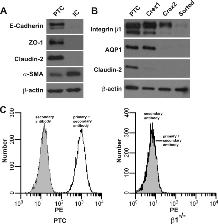
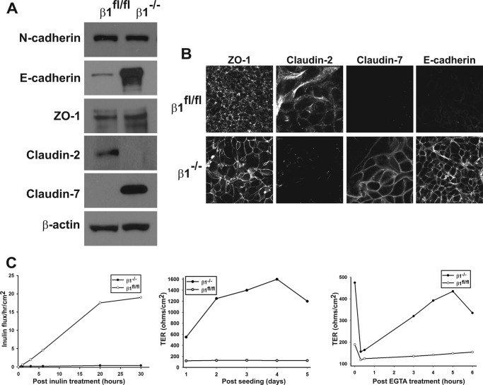
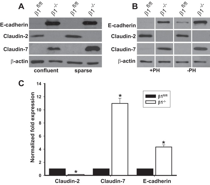
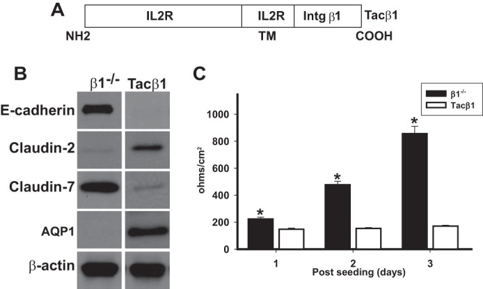
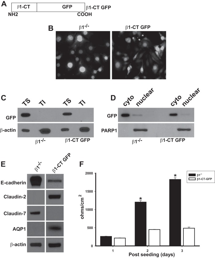
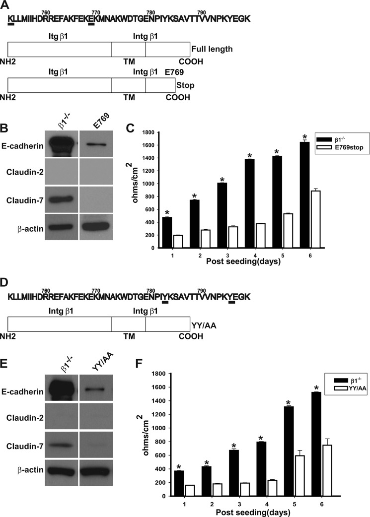
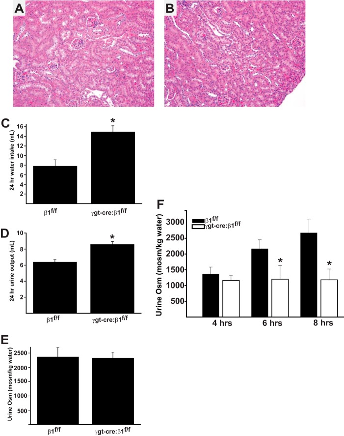
Epithelial cells lining the gastrointestinal tract and kidney have different abilities to facilitate paracellular and transcellular transport of water and solutes. In the kidney, the proximal tubule allows both transcellular and paracellular transport, while the collecting duct primarily facilitates transcellular transport. The claudins and E-cadherin are major structural and functional components regulating paracellular transport. In this study we present the novel finding that the transmembrane matrix receptors, integrins, play a role in regulating paracellular transport of renal proximal tubule cells. Deleting the integrin β1 subunit in these cells converts them from a "loose" epithelium, characterized by low expression of E-cadherin and claudin-7 and high expression of claudin-2, to a "tight" epithelium with increased E-cadherin and claudin-7 expression and decreased claudin-2 expression. This effect is mediated by the integrin β1 cytoplasmic tail and does not entail β1 heterodimerization with an α-subunit or its localization to the cell surface. In addition, we demonstrate that deleting the β1 subunit in the proximal tubule of the kidney results in a major urine-concentrating defect. Thus, the integrin β1 tail plays a key role in regulating the composition and function of tight and adherens junctions that define paracellular transport properties of terminally differentiated renal proximal tubule epithelial cells.

Keywords: Adherens Junction; E-cadherin; Epithelial Cell; Renal Physiology; Tight Junctions.


Figures

FIGURE 1.
FIGURE 1.
Integrin β1−/− PTCs lose expression of aquaporin 1 and claudin-2. A, Western blot analysis of cell lysates obtained from kidney PTCs and interstitial cells (IC) demonstrating that PTCs express the epithelial cell markers E-cadherin, ZO-1, and claudin-2, while ICs express smooth muscle actin (SMA). β-Actin was run as a loading control. B and C, PTCs obtained from β1fl/fl mice were infected twice with adeno cre virus to delete β1 integrin. To obtain a pure β1−/− population, they were sorted via FACS with antibodies directed to the extracellular domain of integrin β1. B, immunoblotting of cell lysates demonstrated that integrin β1 expression decreased with sequential adeno-cre infections, and there was no expression in the β1−/− PTC population. Immunoblotting for aquaporin 1 (AQP1) and claudin-2 demonstrates that their expression decreases as the β1 integrin expression decreases. C, left panel demonstrates high surface expression of integrin β1 in β1fl/fl PTCs, whereas the right panel shows there is no β1 surface expression in β1−/− PTCs.
FIGURE 2.
FIGURE 2.
Deleting integrin β1 alters E-cadherin and claudin expression as well as inulin flux and TER. A, cell lysates from β1fl/fl and β1−/− PTCs were analyzed for expression of AJ (E-cadherin and N-cadherin) and TJ (ZO-1 and claudins 2 and 7) proteins by Western blot analysis. B, β1fl/fl and β1−/− PTCs were grown to confluence on transwell inserts and immunostained for ZO-1, E-cadherin, claudin-2, and claudin-7. C, β1fl/fl and β1−/− PTCs were grown on transwell inserts. Inulin clearance (left panel), transepithelial resistance (TER) (middle panel), and reassembly of tight junctions as measured by the calcium switch assay (right panel) were assessed as described under “Experimental Procedures.” An example from a single assay is shown. At least three assays were performed with similar results.
FIGURE 3.
FIGURE 3.
Claudin and E-cadherin expression is transcriptionally regulated by integrin β1 and is independent of confluency and anchorage. A, cell lysates from β1fl/fl and β1−/− PTCs either sparsely grown or grown to confluency were immunoblotted for the proteins shown in the figure. B, β1fl/fl and β1−/− PTCs were grown in the presence or absence of polyhema (PH), and the amount of E-cadherin and claudin-2 and -7 was analyzed by Western blotting. The lanes are separated by lines to demarcate samples run on the same gel but not in the same order as shown in the figure. C, quantitative RT-PCR analyses for claudin-2, claudin-7, and E-cadherin was performed on cDNA synthesized from RNA isolated from β1fl/fl and β1−/− PTCs. The results were normalized to expression by β1fl/fl PTCs. Mean measurements of three independent experiments are shown; *, p ≤ 0.05 between β1fl/fl and β1−/− PTCs.
FIGURE 4.
FIGURE 4.
Transducing β1−/− PTCs with integrin β1 reestablishes a loose epithelium. A, human integrin β1 cDNA was transfected into the β1−/− PTCs, after which they were sorted to obtain a pure population of high expressing cells (RC). Surface expression of integrin β1 was verified by flow cytometry. B, equal amounts of whole cell lysate from β1−/− and RC PTCs were electrophoresed and immunoblotted for E-cadherin, claudin-2, and claudin-7. C, TER was measured on β1−/− and RC PTCs grown to confluency on transwell inserts for 3 days. Mean measurements of three independent experiments are shown; *, p ≤ 0.05 between β1−/− and RC PTCs.
FIGURE 5.
FIGURE 5.
The cytoplasmic tail of integrin β1 is sufficient for changes in regulation of TER, E-cadherin, claudin-2, and claudin-7 in PTCs. A, schematic of the Tacβ1 construct. The extracellular and transmembrane domain of this chimera is from the Tac receptor of human interleukin 2 (IL2R), and the cytoplasmic domain is from the β1 integrin. B, equal amounts of whole cell lysate from PTCs stably expressing Tacβ1 or the IL2R (β1−/−) cells were electrophoresed and immunoblotted for E-cadherin, claudin-2, or claudin-7. Protein loading was controlled for by immunoblotting for β-actin. C, TER was measured on β1−/− and Tacβ1 PTCs grown to confluency on transwell inserts for 3 days. Mean measurements of three independent experiments are shown; *, p ≤ 0.05 between Tacβ1 and β1−/− PTCs.
FIGURE 6.
FIGURE 6.
Free cytoplasmic integrin β1 cytoplasmic domains are sufficient to regulate AJ and TJ composition of PTCs. A, schematic of the integrin β1cytoplasmic tail GFP construct (β1-CT GFP) where the cytoplasmic domain of integrin β1 was cloned in-frame with GFP in the pEGFP-N2 vector. B, β1−/− PTCs transfected with either an empty GFP vector (β1−/−) or β1-CT GFP were sorted via FACS to collect cell populations expressing equal levels of GFP. GFP levels and localization were analyzed in the cells by placing the cells under an epifluorescence microscope. Cells were incubated with DAPI to visualize nuclei. C, equal amounts of Triton-soluble (TS) and insoluble (TI) fractions from β1−/− and β1-CT GFP PTCs were analyzed by Western blot for levels of and localization of GFP. D, equal amounts of cytoplasmic (cyto) and nuclear fractions of β1−/− and β1-CT GFP PTCs were analyzed by Western blot using anti-GFP antibodies. GFP was only detected only in the cytoplasm and not the nucleus in both cell lines. PARP1 was used to verify the nuclear fraction. E, equal amounts of whole cell lysate from β1−/− and β1-CT GFP PTCs were analyzed by Western blot for levels of E-cadherin, claudin-2, or claudin-7. β-Actin was used to verify equal loading. F, TER was measured on stably expressing β1−/− and β1-CT GFP PTCs grown to confluency on transwell inserts for 3 days. Mean measurements of three independent experiments are shown; *, p ≤ 0.05 between β1-CT GFP and β1−/− PTCs.
FIGURE 7.
FIGURE 7.
E-cadherin, claudin-2, and claudin-7 expression is regulated by specific domains of the integrin β1 tail. A, schematic of the full-length and deletion mutant of β1 integrin. A stop codon was introduced after Glu-769 to obtain the Glu-769 construct. B, equal amounts of whole cell lysate from PTCs stably expressing Glu-769 or control vector (β1−/−) were electrophoresed and immunoblotted for E-cadherin, claudin-2, or claudin-7. Protein loading was controlled for by immunoblotting for β-actin. C, TER was measured on PTCs stably expressing the Glu-769 truncation mutant or control vector (β1−/−) grown to confluency on transwell inserts for 6 days. Mean measurements of three independent experiments are shown; *, p ≤ 0.05 between Glu-769 and β1−/− PTCs. D, schematic of the YY/AA mutant. The 2 tyrosine residues Tyr-783 and Tyr-795 in the highly conserved NPXY motifs were mutated to alanine in integrin β1. E, equal amounts of whole cell lysate from PTCs stably expressing the YY/AA mutant or control vector (β1−/−) were electrophoresed and immunoblotted for E-cadherin, claudin-2, or claudin-7. Protein loading was controlled for by immunoblotting for β-actin. F, TER was measured on PTCs stably expressing the YY/AA or control vector (β1−/−) grown to confluency on transwell inserts for 6 days. Mean measurements of three independent experiments are shown; *, p ≤ 0.05 between YY/AA and β1−/− PTCs.
FIGURE 8.
FIGURE 8.
Kidneys of γgt-cre:β1flox/flox mice have morphologically normal kidneys but have an isosmolar diuresis and an inability to concentrate urine after water loading and subsequent deprivation. A and B, histology of kidney cortex of 6 week β1flox/flox (A) and γgt-cre:β1flox/flox (B) mice is normal (200×). C, 24 h water intake (*, p < 0.01) and (D) 24 h urine output is increased in γgt-cre:β1flox/flox mice (*, p < 0.01). E, baseline urine osmolality is similar in β1flox/flox and γgt-cre:β1flox/flox mice. F, mice (6 in each group) were water loaded with an intraperitoneal injection of 2ml of water 18 h prior to and at the commencement of the experiment. At 4 h, γgt-cre:β1flox/flox and β1flox/flox mice diluted their urines appropriately; however, γgt-cre:β1flox/flox mice were unable to concentrate their urines 6 and 8 h after water deprivation (*, p < 0.01).

Similar articles

Cited by

References

    1. Shin K., Fogg V. C., Margolis B. (2006) Tight junctions and cell polarity. Annu. Rev. Cell Dev. Biol. 22, 207–235 - PubMed
    1. Li J., Ananthapanyasut W., Yu A. S. (2011) Claudins in renal physiology and disease. Pediatric Nephrol. 26, 2133–2142 - PMC - PubMed
    1. Niessen C. M., Gottardi C. J. (2008) Molecular components of the adherens junction. Biochim. Biophys. Acta 1778, 562–571 - PMC - PubMed
    1. Epifano C., Perez-Moreno M. (2012) Crossroads of integrins and cadherins in epithelia and stroma remodeling. Cell Adh. Migr. 6, 261–273 - PMC - PubMed
    1. Saito M., Tucker D. K., Kohlhorst D., Niessen C. M., Kowalczyk A. P. (2012) Classical and desmosomal cadherins at a glance. J. Cell Sci. 125, 2547–2552 - PMC - PubMed

Publication types

LinkOut - more resources