. 2014 Apr 15;74(8):2362-73.
doi: 10.1158/0008-5472.CAN-13-3058. Epub 2014 Feb 7.

Cofilin drives cell-invasive and metastatic responses to TGF-β in prostate cancer

Affiliations

Cofilin drives cell-invasive and metastatic responses to TGF-β in prostate cancer

Joanne Collazo et al. Cancer Res. .

Erratum in

Abstract

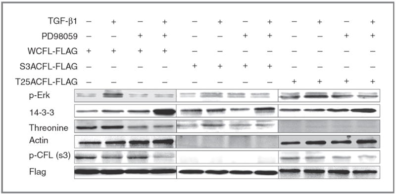
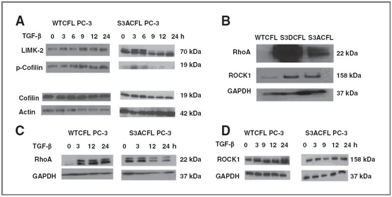
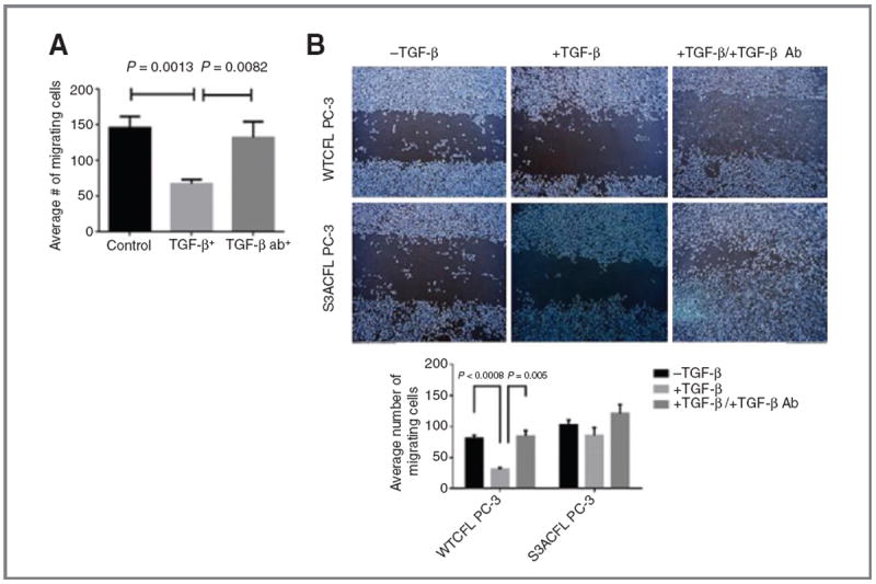
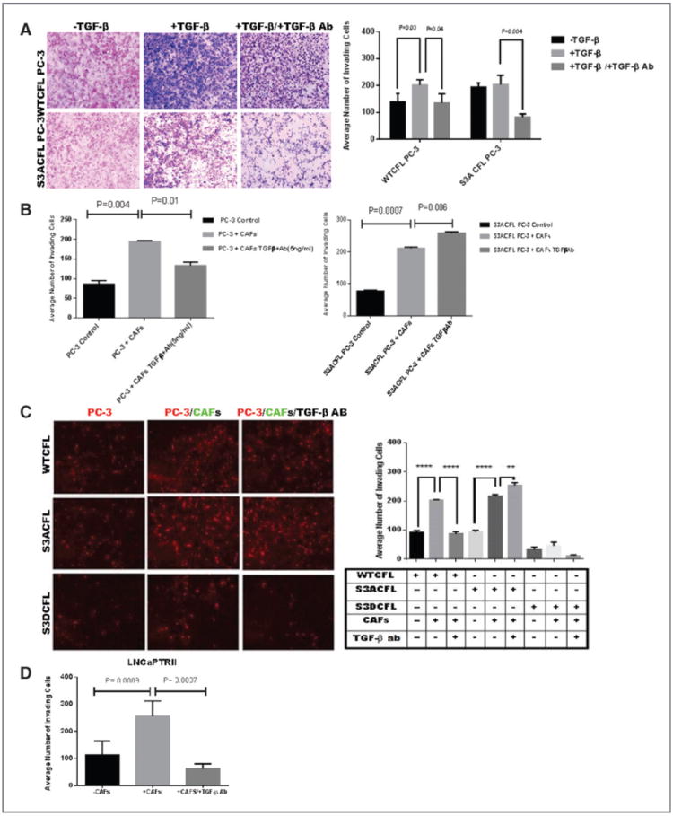
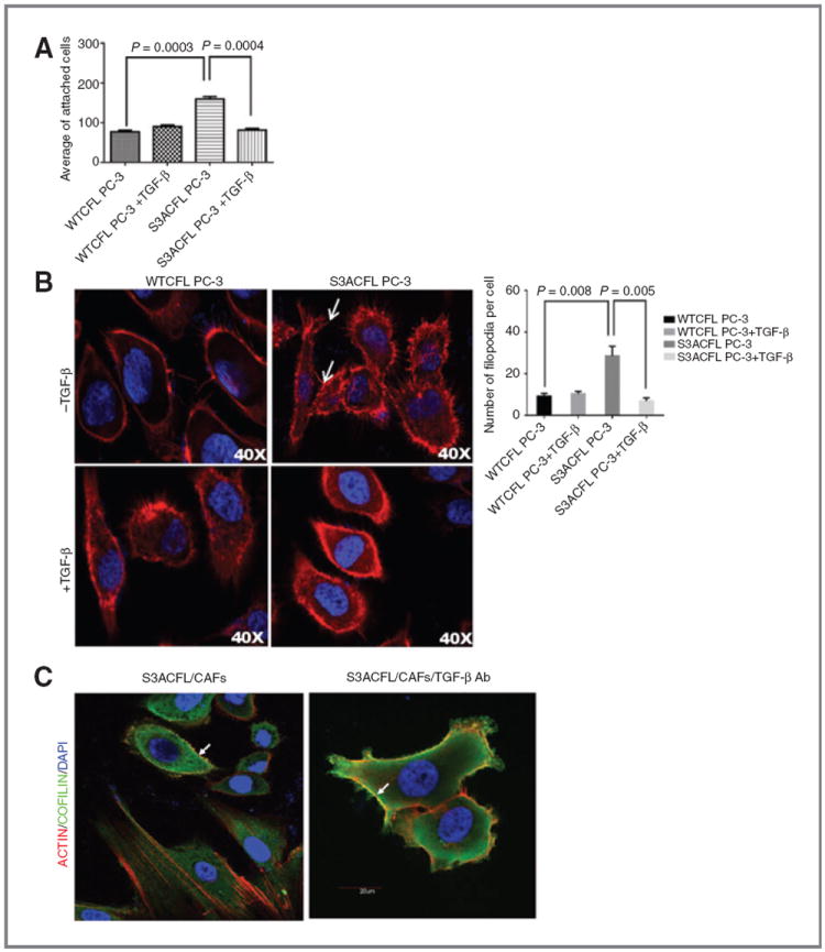
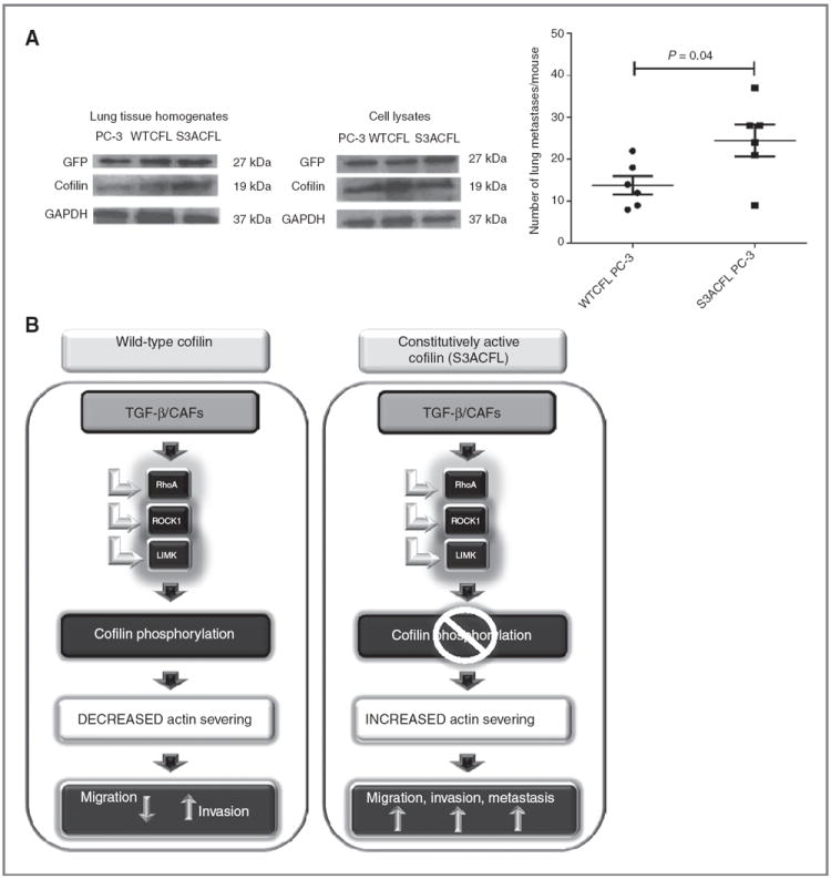
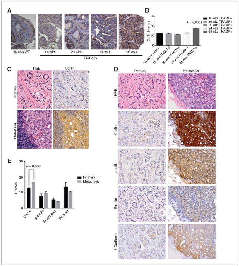
Cofilin (CFL) is an F-actin-severing protein required for the cytoskeleton reorganization and filopodia formation, which drives cell migration. CFL binding and severing of F-actin is controlled by Ser3 phosphorylation, but the contributions of this step to cell migration during invasion and metastasis of cancer cells are unclear. In this study, we addressed the question in prostate cancer cells, including the response to TGF-β, a critical regulator of migration. In cells expressing wild-type CFL, TGF-β treatment increased LIMK-2 activity and cofilin phosphorylation, decreasing filopodia formation. Conversely, constitutively active CFL (SerAla) promoted filipodia formation and cell migration mediated by TGF-β. Notably, in cocultures of prostate cancer epithelial cells and cancer-associated fibroblasts, active CFL promoted invasive migration in response to TGF-β in the microenvironment. Further, constitutively active CFL elevated the metastatic ability of prostate cancer cells in vivo. We found that levels of active CFL correlated with metastasis in a mouse model of prostate tumor and that in human prostate cancer, CFL expression was increased significantly in metastatic tumors. Our findings show that the actin-severing protein CFL coordinates responses to TGF-β that are needed for invasive cancer migration and metastasis.


Conflict of interest statement

Disclosure of Potential Conflicts of Interest

No potential conflicts of interest were disclosed.

Figures

Figure 1
Figure 1
S3A cofilin dephosphorylation specifically directs actin severing response to TGF-β. PC-3 cells were transfected with Flag-tagged WTCFL, S3ACFL, or T25ACFL and were subsequently treated with TGF-β1 (5 ng/mL) for 6 hours in the presence or absence of PD98095 inhibitor. Cell lysates (50 μg of protein) were subjected to immunoprecipitation with anti-Flag antibody and subsequent Western blotting with the indicated antibodies. Phosphorylated proteins p-Erk, protein 14-3-3, actin, and p-cofilin are induced in response to TGF-β in WTFL prostate cancer cells. S3ACFL mutation conferring constitutive cofilin dephosphorylation specifically diminishes p-cofilin and enables active cofilin binding to actin without TGF-β regulation. In contrast, the T25A CFL mutation impairs threonine phosphorylation that does not affect actin binding, and thus both p-cofilin (Ser 3) and actin are detected.
Figure 2
Figure 2
Effect of S3A mutation on cofilin phosphorylation events in PC-3 cells. Effect of TGF-β on cofilin, p-cofilin, and LIMK-2 protein expression in prostate cancer cells. A, upregulation of LIMK-2 protein in mutant S3ACFL PC-3 cells. Treatment with TGF-β (5 ng/mL) increased LIMK-2 and p-cofilin expression in the WT and decreased LIMK-2 expression in the S3ACFL cells. B, Western blotting indicating elevated RhoA and ROCK1 protein in S3ACFL PC-3. C and D, treatment with TGF-β increased RhoA and ROCK1 levels in WTCFL cells and decreased expression of both proteins in S3ACFL cells. Glyceraldehyde-3-phosphate dehydrogenase (GAPDH) was used as loading control.
Figure 3
Figure 3
Cofilin drives TGF-β–mediated prostate cancer migration. A, human prostate cancer cells LNCaP TβRII (normal cofilin) exhibited a significant reduction in cell migration in response to TGF-β. In the presence of a neutralizing antibody against TGF-β, cell migration was restored to untreated control levels. Values shown are the number of migrating cells from two independent experiments performed in triplicate. B, S3ACFL mutation enhances prostate cancer cell migration bypassing TGF-β. Top, representative images of increased cell migration ability for S3ACFL PC-3 cells compared with WTCFL cells (24 hours). Bottom, TGF-β treatment significantly decreased WTCFL cell migration (P < 0.0008), but it had no significant effect in S3ACFL cells. Loss of TGF-β (by neutralizing antibody) restored the WTCFL PC-3 cell migration capacity, whereas it increased S3CFL-mutant cell migration (P = 0.005).
Figure 4
Figure 4
Cofilin navigates invasive response to TGF-β (from stroma microenvironment). A, the invasive response of prostate cancer cells to TGF-β assessed in the Matrigel assay. S3ACFL had no significant effect on PC-3 cell invasion (black barographs). In response to exogenous TGF-β, there was an increase in WTCFL PC-3 cell invasion potential, but not in S3ACFL cells (P = 0.03). In the presence of TGF-β–neutralizing antibody, there was a significant decrease in the invasion potential for both WTCFL and S3ACFL cells (P = 0.04 and 0.004, respectively). B, Matrigel invasion of WTCFL PC-3/CAFs (right) and S3ACFL PC-3/CAFs cocultures (left) after 24 hours. CAFs significantly increased prostate cancer cell invasion for both WTCFL and S3ACFL cells (P = 0.004 and 0.007. Continuous secretion of TGF-β by the reactive microenvironment (in the presence of TGF-β–neutralizing antibody) induced a further increase in the number of invading S3ACFL cells (P = 0.008), whereas it decreased WTCFL cell invasion. Values are the average from two independent experiments in triplicate. C, cocultures of WTCFL, S3ACFL, and S3DCFL with CAFs in the presence or absence of a neutralizing TGF-β antibody. Quantitative assessment of invading cells indicates that only active cofilin (S3A CFL) directs a further increase in TGF-β–mediated cell invasion (derived from CAFs). D, a significantly higher increase in cell invasion is stimulated by CAFs in cocultures with theLNCaP TβRII cells (highly responsive to TGF-β). This was abrogated by the presence of the neutralizing antibody against TGF-β. *, significant difference at P < 0.007.
Figure 5
Figure 5
Active cofilin mediates prostate cancer cell adhesion via cytoskeletal remodeling, an effect impaired by TGF-β. A, effect of S3ACFL on prostate cancer cell adhesion. S3A mutation conferred a significant increase in cell adhesion to fibronectin compared with WTCFL cells (P = 0.0003). TGF-β significantly decreased in S3CFL cell adhesion (P = 0.0004), but not in WTCFL cells. Values shown are the mean (± SEM) of three independent experiments performed in triplicates. Statistical significance set at a P value of <0.005. B, active cofilin enhances filopodia formation; representative images of confocal microscopy (×40) show increased number of filopodia protrusions in S3ACFL PC-3 (arrows) compared with WTCFL cells. Treatment with TGF-β (5 ng/mL; 24 hours) decreased filopodia protrusions in S3ACFL cells. Filopodia quantitated in five random fields were examined for each cell line and values shown are mean ± SEM from three independent experiments (left). Statistical significance is defined at P < 0.01. C, S3ACFL active binding to F-actin at the leading edge of the cells is mediated by TGF-β. Cofilin colocalization with filopodia is dependent on TGF-β derived from surrounding stroma (CAFs). Images of cofilin/rhodamine phalloidin colocalization in S3ACFL prostate epithelial cancer cells cocultured with CAFs. Cofilin (green) colocalizes with filopodia protrusions (arrows). Loss of TGF-β (in the presence of neutralizing antibody) increases actin/cofilin colocalization (yellow) and filopodia protrusions in S3ACFL cells.
Figure 6
Figure 6
Cofilin constitutive activation promotes prostate cancer metastasis. A, male nude mice (n = 12) were inoculated with GFP-labeled PC-3 cells (parental, WTCFL, and S3ACFL) via tail vein injections. Metastatic lesions to the lungs were assessed at 4 weeks after inoculation. S3ACFL cells generated a significantly higher number of metastases compared with control PC-3 cells (P = 0.04). Values show the number of metastatic lesions to the lung/mouse for each cell line. Western blots of mouse lung tissue homogenates and cell lysates indicate the GFP presence in all samples (left). B, schematic diagram illustrating the regulatory impact of cofilin on TGF-β functional switch toward prostate cancer cell migration, invasion, and metastasis. Under constitutive activated cofilin (S3ACFL), TGF-β is produced by the reactive stroma/microenvironment (CAFs), is unable to dephosphorylate cofilin, and increases tumor cell aggressiveness and metastatic potential.
Figure 7
Figure 7
Cofilin overexpression correlates with prostate cancer progression to metastasis A and B, cofilin profiling in TRAMP mouse model. TRAMP transgenic mice develop prostate adenocarcinoma with increasing age, resembling progression of human prostate cancer to metastasis. Prostate sections of increasing grade and metastatic tumors (16–28 weeks) were profiled by immunostaining for cofilin expression; WT mouse prostate tissue (16 weeks) was used as control; magnification, ×40. Quantitative evaluation of CFL immunoreactivity, as determined by the H-scoring, shows a significant increase in metastatic tumors from 28-week-old TRAMP mice (P = 0.001) compared with early-stage tumors. C–E, cofilin expression profile in human prostate cancer. C, hematoxylin and eosin staining and CFL immunostaining in serial sections of prostate tumors; characteristic image of a metastatic lesion to lymph nodes exhibiting intense cofilin immunoreactivity, compared with the primary tumor from the same patient (absence of CFL expression). Magnification, ×100. D, representative images of immunostaining for cofilin, p-cofilin, E-cadherin, and palladin on primary and metastatic prostate cancer. E, quantitative analysis of protein immunoreactivity (from D). There was a significant increase in cofilin levels in metastatic specimens compared with primary tumors (P = 0.005).

Similar articles

Cited by

References

    1. Arnold J, Isaacs JT. Mechanisms involved in the progression of androgen-independent prostate cancers: it is not only the cancer cell’s fault. Endocr Relat Cancer. 2002;9:61–73. - PMC - PubMed
    1. Catalona WJ. Management of cancer of the prostate. N Engl J Med. 1994;331:996–1004. - PubMed
    1. McKenzie S, Kyprianou N. Apoptosis evasion: the role of survival pathways in prostate cancer progression and therapeutic resistance. J Cell Biochem. 2006;97:18–32. - PMC - PubMed
    1. Karantanos T, Thompson TC. GEMMS shine a light on resistance to androgen deprivation therapy for prostate cancer. Cancer Cell. 2013;24:11–3. - PMC - PubMed
    1. Lunardi A, Ala U, Epping MT, Salmera L, Clohessy JG, Webster KA, et al. A co-clinical approach identifies mechanisms and potential therapies for androgen deprivation resistance in prostate cancer. Nat Genetics. 2013;45:747–55. - PMC - PubMed

Publication types

Substances