

Ex vivo Evans blue assessment of the blood brain barrier in three breast cancer brain metastasis models
- PMID: 24510011
- PMCID: PMC4363122
- DOI: 10.1007/s10549-014-2854-5
Ex vivo Evans blue assessment of the blood brain barrier in three breast cancer brain metastasis models
Abstract
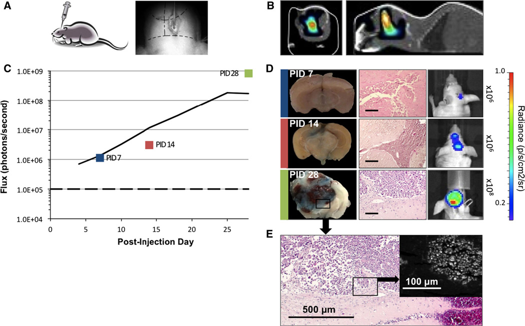
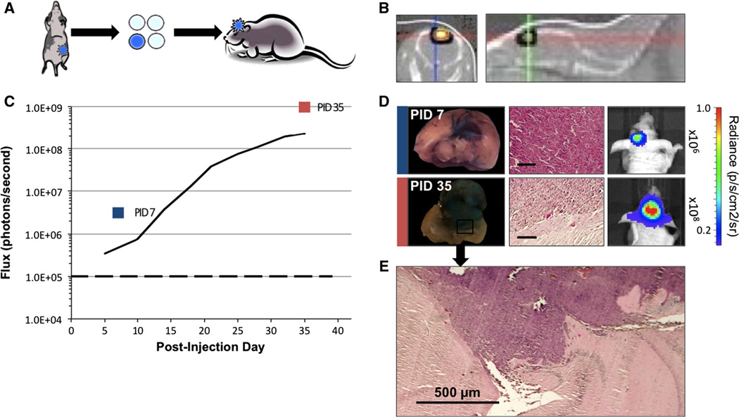
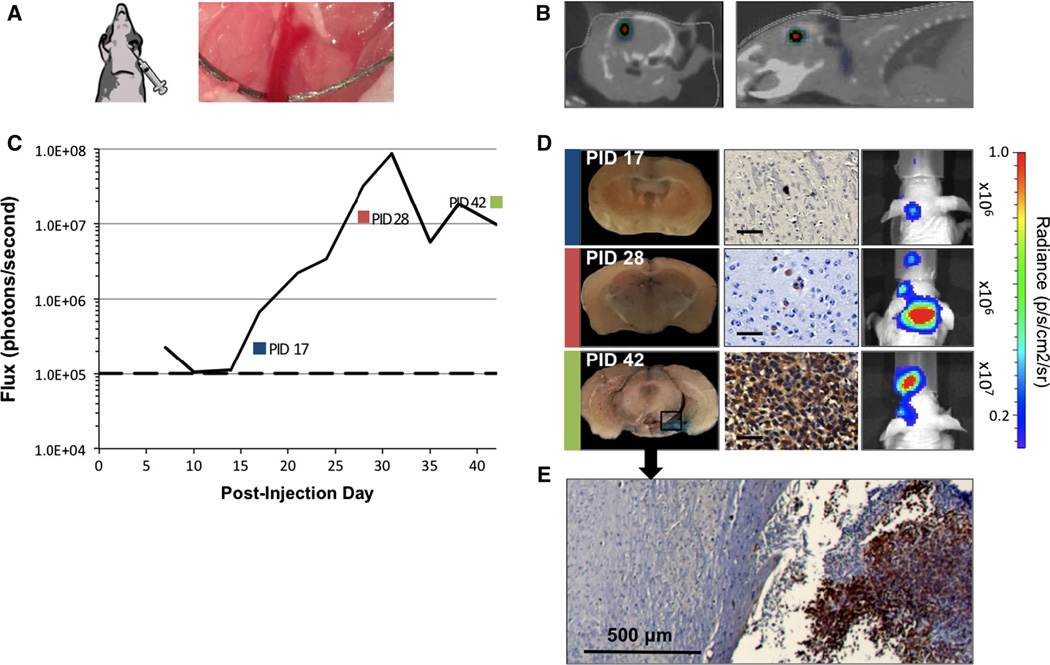
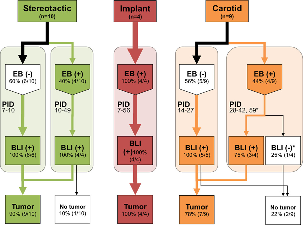
The limited entry of anticancer drugs into the central nervous system represents a special therapeutic challenge for patients with brain metastases and is primarily due to the blood brain barrier (BBB). Albumin-bound Evans blue (EB) dye is too large to cross the BBB but can grossly stain tissue blue when the BBB is disrupted. The course of tumor development and the integrity of the BBB were studied in three preclinical breast cancer brain metastasis (BCBM) models. A luciferase-transduced braintropic clone of MDA-231 cell line was used. Nude mice were subjected to stereotactic intracerebral inoculation, mammary fat pad-derived tumor fragment implantation, or carotid artery injections. EB was injected 30 min prior to euthanasia at various timepoints for each of the BCBM model animals. Serial bioluminescent imaging demonstrated exponential tumor growth in all models. Carotid BCBM appeared as diffuse multifocal cell clusters. EB aided the localization of metastases ex vivo. Tumor implants stained blue at 7 days whereas gross staining was not evident until day 14 in the stereotactic model and day 28 for the carotid model. EB assessment of the integrity of the BBB provides useful information relevant to drug testing in preclinical BCBM models.
Figures
Similar articles
-
Bioluminescent human breast cancer cell lines that permit rapid and sensitive in vivo detection of mammary tumors and multiple metastases in immune deficient mice.Breast Cancer Res. 2005;7(4):R444-54. doi: 10.1186/bcr1026. Epub 2005 Apr 8. Breast Cancer Res. 2005. PMID: 15987449 Free PMC article.
-
In vivo and ex vivo assessment of the blood brain barrier integrity in different glioblastoma animal models.J Neurooncol. 2014 Sep;119(2):297-306. doi: 10.1007/s11060-014-1514-2. Epub 2014 Jul 3. J Neurooncol. 2014. PMID: 24990826
-
A decade of blood-brain barrier permeability assays: Revisiting old traumatic brain injury rat data for new insights and experimental design.Microvasc Res. 2023 Jan;145:104453. doi: 10.1016/j.mvr.2022.104453. Epub 2022 Nov 7. Microvasc Res. 2023. PMID: 36356686 Free PMC article.
-
Biology of breast cancer brain metastases and novel therapies targeting the blood brain barrier: an updated review.Med Oncol. 2023 May 18;40(6):181. doi: 10.1007/s12032-023-02047-0. Med Oncol. 2023. PMID: 37202575 Review.
-
Navigating the Blood-Brain Barrier: Challenges and Therapeutic Strategies in Breast Cancer Brain Metastases.Int J Mol Sci. 2023 Jul 27;24(15):12034. doi: 10.3390/ijms241512034. Int J Mol Sci. 2023. PMID: 37569410 Free PMC article. Review.
Cited by
-
Evaluation the Effect of Sonodynamic Therapy with 5-Aminolevulinic Acid and Sodium Fluorescein by Preclinical Animal Study.Cancers (Basel). 2024 Jan 5;16(2):253. doi: 10.3390/cancers16020253. Cancers (Basel). 2024. PMID: 38254744 Free PMC article.
-
Decreased nonspecific adhesivity, receptor-targeted therapeutic nanoparticles for primary and metastatic breast cancer.Sci Adv. 2020 Jan 15;6(3):eaax3931. doi: 10.1126/sciadv.aax3931. eCollection 2020 Jan. Sci Adv. 2020. PMID: 31998833 Free PMC article.
-
Maintaining unperturbed cerebral blood flow is key in the study of brain metastasis and its interactions with stress and inflammatory responses.Brain Behav Immun. 2017 May;62:265-276. doi: 10.1016/j.bbi.2017.02.012. Epub 2017 Feb 20. Brain Behav Immun. 2017. PMID: 28219803 Free PMC article.
-
Targeting the PI3K/Akt/mTOR pathway with the pan-Akt inhibitor GDC-0068 in PIK3CA-mutant breast cancer brain metastases.Neuro Oncol. 2019 Nov 4;21(11):1401-1411. doi: 10.1093/neuonc/noz105. Neuro Oncol. 2019. PMID: 31173106 Free PMC article.
-
The Role and Mechanism of Borneol to Open the Blood-Brain Barrier.Integr Cancer Ther. 2018 Sep;17(3):806-812. doi: 10.1177/1534735418767553. Epub 2018 Apr 13. Integr Cancer Ther. 2018. PMID: 29652199 Free PMC article.
References
-
- Aragon-Ching JB, Zujewski JA. CNS metastasis: an old problem in a new guise. Clin Cancer Res. 2007;13:1644–1647. - PubMed
-
- Minisini A, Moroso S, Gerratana L, Giangreco M, Iacono D, Poletto E, Guardascione M, Fontanella C, Fasola G, Puglisi F. Risk factors and survival outcomes in patients with brain metastases from breast cancer. Clin Exp Metastasis. 2013 - PubMed
Publication types
MeSH terms
Substances
Grants and funding
LinkOut - more resources
Full Text Sources
Other Literature Sources
Medical
