. 2014 Apr;42(8):5059-71.
doi: 10.1093/nar/gku095. Epub 2014 Feb 7.

Selective nuclear export of specific classes of mRNA from mammalian nuclei is promoted by GANP

Affiliations

Selective nuclear export of specific classes of mRNA from mammalian nuclei is promoted by GANP

Vihandha O Wickramasinghe et al. Nucleic Acids Res. 2014 Apr.

Abstract

The nuclear phase of the gene expression pathway culminates in the export of mature messenger RNAs (mRNAs) to the cytoplasm through nuclear pore complexes. GANP (germinal- centre associated nuclear protein) promotes the transfer of mRNAs bound to the transport factor NXF1 to nuclear pore complexes. Here, we demonstrate that GANP, subunit of the TRanscription-EXport-2 (TREX-2) mRNA export complex, promotes selective nuclear export of a specific subset of mRNAs whose transport depends on NXF1. Genome-wide gene expression profiling showed that half of the transcripts whose nuclear export was impaired following NXF1 depletion also showed reduced export when GANP was depleted. GANP-dependent transcripts were highly expressed, yet short-lived, and were highly enriched in those encoding central components of the gene expression machinery such as RNA synthesis and processing factors. After injection into Xenopus oocyte nuclei, representative GANP-dependent transcripts showed faster nuclear export kinetics than representative transcripts that were not influenced by GANP depletion. We propose that GANP promotes the nuclear export of specific classes of mRNAs that may facilitate rapid changes in gene expression.


Figures
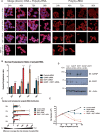
Figure 1.
Figure 1.
GANP depletion displays a less severe mRNA export defect than NXF1 depletion. (A and B) Time course of mRNA export defects in cells depleted for GANP, NXF1 and both GANP and NXF1. RNA FISH was performed and poly(A)+ RNA localization was examined in NXF1, GANP and GANP- and NXF1-depleted HCT116 cells 24–96 h post-transfection. Images are representative of three independent experiments. Nuclei are indicated by DAPI staining and are represented in the merged panel [DNA + poly(A)+RNA]. The scale bar represents 5 µm. A ratio of nuclear to cytoplasmic poly(A)+RNA intensity was taken per cell for ≥200 cells/sample using the ArrayScan VTI automated microscope. Values are the mean of readings from three independent experiments, ± S.E.M. (standard error of the mean) P-values are shown (*P < 0.05; **P < 0.01; ***P < 0.001). (C) Nuclear and cytoplasmic poly(A)+ RNA distribution was also calculated using the ArrayScan VTI automated microscope, where a negative value indicates more intensity in the cytoplasm than in the nucleus, and a positive value indicates more intensity in the nucleus than in the cytoplasm. P-values are shown and represent the mean of readings from three independent experiments, ± S.E.M. (D) Depletion of GANP, NXF1 and both GANP and NXF1. HCT116 cells were depleted of endogenous GANP, NXF1 or both NXF1 and GANP and analysed by immunoblotting for GANP, NXF1 and actin (loading control) 48 h post-transfection. As control, cells were transfected with an siRNA differing from GANP siRNA by two bases. (E) Time course of cell proliferation defects in cells depleted for GANP, NXF1 and both GANP and NXF1. Viable cell number was determined in GANP, NXF1 and GANP-and NXF1-depleted samples 24–96 h post-transfection and represent the mean of independent readings from three independent depletion experiments ± S.E.M.
Figure 2.
Figure 2.
Whole-genome gene expression profiling in cells depleted of GANP or NXF1. (A) GANP functions in the export of a subset of NXF1-containing mRNPs. Whole-genome gene expression profiling was performed using cytoplasmic mRNA-isolated cells depleted of GANP or NXF1 for 72 h as described in ‘Materials and Methods’ section. Each hybridization was performed three times with cytoplasmic RNA isolated from three independent depletion experiments. Each independent output was then compared with its replicates, and an adjusted P-value obtained, allowing determination of genes with significant changes in expression following GANP or NXF1 depletion (adjusted P < 0.05). The total number of downregulated mRNAs in either GANP-(800) or NXF1-(1285) depleted cytoplasmic samples with a log2fold change of >−0.25 is indicated. The total number of upregulated mRNAs in either GANP- or NXF1-depleted cytoplasmic samples is also indicated. (B) Transcripts in which both GANP and NXF1 depletion impair export encode proteins required for mRNA processing, gene expression, nucleic acid metabolism and that are involved in rapid changes of cellular function. A Venn diagram is shown indicating mRNAs downregulated in cytoplasmic samples from cells depleted only of GANP, only of NXF1 or depleted of both NXF1 and GANP (adjusted P < 0.05). For each group, average log2fold change of transcripts in GANP-depleted samples was divided by average log2fold change of same transcripts in NXF1-depleted samples to obtain GANP/NXF1 ratio. Functional annotation was performed on each group of transcripts using the DAVID bioinformatics program. Transcripts enriched for proteins with the following Gene Ontology terms (P < 10−3) are indicated. Functional classes of transcripts in which nuclear export is impaired by both GANP and NXF1 depletion are indicated in bold. Raw data are in Supplementary Figure S2. (C and D) Validation of microarray by qRT-PCR. The mRNA levels of GANP and NXF1 target (C) and NXF1 target genes (D) identified in microarray were quantitated by qRT-PCR from cytoplasmic RNA extracted from control siRNA-treated or NXF1- or GANP-depleted cells. Plots are normalized to control siRNA-treated cells, assigned an arbitrary value of one and represent the mean of triplicate readings from three independent experiments,  ± SD.
Figure 3.
Figure 3.
Expression levels, half-lives, gene length and exon number are correlated with selective impairment of mRNA export following GANP depletion. (A) NXF1-dependent transcripts in which export is impaired by GANP depletion are more highly expressed than those that are insensitive to GANP depletion. The mRNA transcript expression level (RPKM) was calculated for each gene in GANP/NXF1, GANP only and NXF1 only subsets using a publicly available dataset (36) and represented in a box and whiskers plot. Median is indicated by marked line and mean by a plus sign. Statistical significance was determined by a Mann–Whitney test (***P < 0.001). (B) NXF1-dependent transcripts in which nuclear export is impaired by GANP depletion have shorter half-lives. The mRNA half-lives for each transcript in GANP/NXF1, GANP only and NXF1 only subsets were calculated using a publicly available dataset (37) and represented in a box and whiskers plot. Statistical significance was P < 0.001. (C) Gene length is correlated with mRNA export selectivity. The length of each gene (kb) in GANP/NXF1, GANP only and NXF1 only subsets was calculated and represented in a dot plot. The average gene length for genes in each subset is marked by a red line in the plot and also shown numerically. Statistical significance was P < 0.001. (D) Exon number is correlated with mRNA export selectivity. Exon number of each gene in GANP/NXF1, GANP only and NXF1 only subsets was calculated and represented in a dot plot. The average exon number for genes in each subset is marked by a red line in the plot and also shown numerically. Statistical significance was P < 0.001.
Figure 4.
Figure 4.
Transcripts whose nuclear export was impaired following GANP depletion display more rapid nuclear mRNA export kinetics than GANP-independent transcripts. (A and B) A mixture of equal amounts of four fully spliced transcripts, two GANP dependent (EXOSC6 and PDXP) and two GANP independent (GALK1 and TST), was injected into Xenopus oocyte nuclei and their rate of export into the cytoplasm determined. Nuclear (A) and cytoplasmic (B) expression levels were measured for each transcript between 5 and 90 min after nuclear injection and represent the average of triplicate qPCR experiments from nuclear and cytoplasmic RNA harvested from four pooled injections into Xenopus oocyte nuclei per time point.
Figure 5.
Figure 5.
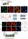
TPR mediates binding of GANP to NPC’s. (A) GANP and TPR co-depletion results in a more severe mRNA export defect than TPR or GANP alone. A ratio of nuclear to cytoplasmic poly(A)+RNA intensity was taken per cell for ≥200 cells/sample using the ArrayScan VTI automated microscope in GANP, TPR and GANP and TPR depleted cells 72 h post-transfection. (B) and (C) GANP co-localizes with TPR in the nuclear interior and at NPCs. Immunofluorescence of HCT116 cells using anti-GANP and anti-TPR, respectively (scale bar, 5 µm), is shown (B) and scanning analysis of GANP and TPR intensity is also shown (C). Nuclei used for scanning and the scanning axis are indicated by white lines. Pairs of nuclei of same scan width as determined by DAPI staining were used for scanning. Nuclear envelope and nuclear interior are indicated, and arrows indicate areas of co-localization in the nuclear interior. (D) and (E) GANP and TPR interact with each other in vivo. (D) Endogenous TPR was immunoprecipitated from nuclear extract of HCT116 cells and blotted for GANP and TPR. (E) Endogenous GANP was immunoprecipitated from nuclear extracts of HCT116 cells with or without RNase treatment and blotted for GANP and TPR. (F) TPR depletion results in loss of GANP from nuclear pores. Immunofluorescence of HCT116 cells with or without TPR depletion using anti-GANP and anti-TPR, respectively (scale bar, 5 µm), is shown. Scanning analysis of GANP and TPR intensity is also shown. (G) ENY2, PCID2 and TPR depletion are often epistatic with GANP depletion. Cytoplasmic expression levels of GANP/NXF1 target transcripts RPS23, SP1 and ARPP-19 and NXF1 target transcripts AXIN2, IDH2 and LLGL were quantitated by qRT-PCR from cytoplasmic RNA extracted from control siRNA-treated, GANP-, ENY2-, PCID2- or TPR-depleted HCT116 cells 72 h post-transfection.
Figure 6.
Figure 6.
Model for GANP’s role in the selective export of a subset of mRNA. We propose that GANP facilitates the nuclear export of specific classes of mRNAs that may enable rapid adaptation to changes in gene expression, thereby optimizing their export to the cytoplasm and ensuring that cells can respond quickly to stimuli. GANP may facilitate export of a subset of mRNA by optimizing its transport from the nuclear interior to nuclear pores, or alternatively could facilitate export through its role as a scaffold protein of the nuclear pore-associated TREX-2 complex, or through its potential role in both compartments.

Similar articles

Cited by

References

    1. Muller-McNicoll M, Neugebauer KM. How cells get the message: dynamic assembly and function of mRNA-protein complexes. Nat. Rev. Genet. 2013;14:275–287. - PubMed
    1. Tutucci E, Stutz F. Keeping mRNPs in check during assembly and nuclear export. Nat. Rev. Mol. Cell Biol. 2011;12:377–384. - PubMed
    1. Rodriguez-Navarro S, Hurt E. Linking gene regulation to mRNA production and export. Curr. Opin. Cell. Biol. 2011;23:302–309. - PubMed
    1. Stewart M. Nuclear export of mRNA. Trends Biochem. Sci. 2010;35:609–617. - PubMed
    1. Kohler A, Hurt E. Exporting RNA from the nucleus to the cytoplasm. Nat. Rev. Mol. Cell Biol. 2007;8:761–773. - PubMed

Publication types

MeSH terms

Associated data