. 2014 Mar;8(2):105-13.
doi: 10.1007/s11832-014-0561-8. Epub 2014 Feb 9.

Use of ultrasonography in developmental dysplasia of the hip

Affiliations

Use of ultrasonography in developmental dysplasia of the hip

Hakan Omeroğlu. J Child Orthop. 2014 Mar.

Abstract

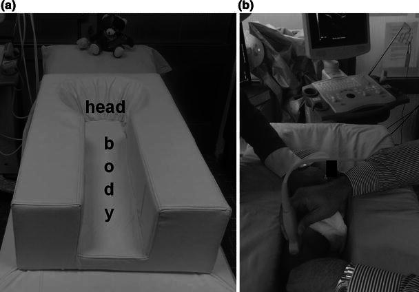
Purpose: Ultrasonography has been used as a diagnostic tool in developmental dysplasia of the hip (DDH) during early infancy since the early 1980s. The aim of this review article is to summarise the technique, benefits and shortcomings of four infantile hip ultrasonography methods, focusing mainly on the Graf method, and to assess the effectiveness of ultrasonographic newborn hip screening programmes.

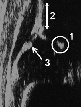
Methods: Several infantile hip ultrasonography methods have been defined to assess the relationship between the femoral head and acetabulum. The Graf, Harcke, Terjesen and Suzuki methods are the universally known ones. The Graf method is composed of a quantitative classification system, while the Harcke and Suzuki methods have qualitative definitions and the Terjesen method contains both quantitative and qualitative descriptions.

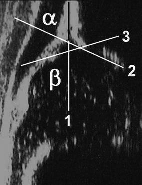
Results: Although the results of several studies assessing the sensitivity and consistency of the ultrasonography methods have still not proven a clear dominance of one of these techniques, the primary advantage of the Graf method is that it has a standardised examination technique, as well as a very well defined numeric hip typing system. The importance of newborn hip screening has been universally accepted, but there is still no strong evidence regarding the superiority of either universal (screening of all newborns) or selective (screening of high-risk newborns) ultrasonographic newborn hip screening programmes.

Conclusions: An effective ultrasonographic method should include simple, precise, quantitative and consistent definitions for a proper examination and diagnosis. Both universal and selective ultrasonographic newborn hip screening programmes have significantly decreased the rate of late detected DDH and lessened the need for surgical treatment.


Figures

Fig. 1
Fig. 1
a The special positioning apparatus that maintains the baby in the lateral decubitus position. b Ultrasonographic examination of the right hip of a baby in the special positioning apparatus
Fig. 2
Fig. 2
Anatomical identification of the structures in an infantile hip sonogram in the frontal plane at rest: 1 chondro-osseous junction, 2 femoral head, 3 synovial fold, 4 joint capsule, 5 acetabular labrum, 6 cartilage roof, 7 lower limb of the ilium and bony roof, 8 bony rim (the point where the concavity of the bony acetabular roof changes to the convexity of the iliac bone or the most lateral point of the acoustic shadow in the bony acetabular roof), 9 perichondrium, 10 iliac bone
Fig. 3
Fig. 3
Standard plane for the Graf method [4]. 1 The lower limb of the ilium is clearly visible. This means that the sectional plane passes through the centre of the acetabulum. 2 A straight iliac wing silhouette exists. This means that the probe is parallel to the iliac bone. 3 The labrum is clearly visible
Fig. 4
Fig. 4
Measurement of the angles in the Graf method [4]. 1 The base line starts from the uppermost point of the proximal perichondrium and is drawn caudally tangential to the iliac bone. 2 The bony roof line starts from the inferior border of the lower limb and is drawn tangentially to the bony roof. 3 The cartilage roof line is drawn between the bony rim and the centre of the labrum. The α angle is measured between lines 1 and 2. The β angle is measured between lines 1 and 3
Fig. 5
Fig. 5
Measurement of the femoral head cover (FHC) [18], where a is the distance between the acetabular fossa and the bony rim, and b is the distance between the acetabular fossa and the lateral joint capsule. FHC = a/b × 100
Fig. 6
Fig. 6
Measurement of the lateral head distance (LHD) [16], which is the distance between the lateral tangent of the femoral head ossification centre and the bony rim

References

    1. Graf R. New possibilities for the diagnosis of congenital hip joint dislocation by ultrasonography. J Pediatr Orthop. 1983;3:354–359. doi: 10.1097/01241398-198307000-00015. - DOI - PubMed
    1. Wientroub S, Grill F. Ultrasonography in developmental dysplasia of the hip. J Bone Joint Surg Am. 2000;82-A:1004–1018. - PubMed
    1. Harcke HT. Imaging methods used for children with hip dysplasia. Clin Orthop Relat Res. 2005;434:71–77. doi: 10.1097/01.blo.0000162411.55355.c8. - DOI - PubMed
    1. Graf R. Hip sonography. Diagnosis and management of infant hip dysplasia. 2. Berlin: Springer; 2006.
    1. Ozçelik A, Akçar N, Inan U, Omeroğlu H. Assessment of the use of hip ultrasonography by Graf’s method between 1 and 6 years of age. J Pediatr Orthop B. 2005;14:97–100. doi: 10.1097/01202412-200503000-00007. - DOI - PubMed