. 2014 Feb 3:75:12.12.1-12.12.29.
doi: 10.1002/0471140864.ps1212s75.

Protein O-mannosylation in metazoan organisms

Affiliations

Protein O-mannosylation in metazoan organisms

Vladislav M Panin et al. Curr Protoc Protein Sci. .

Abstract

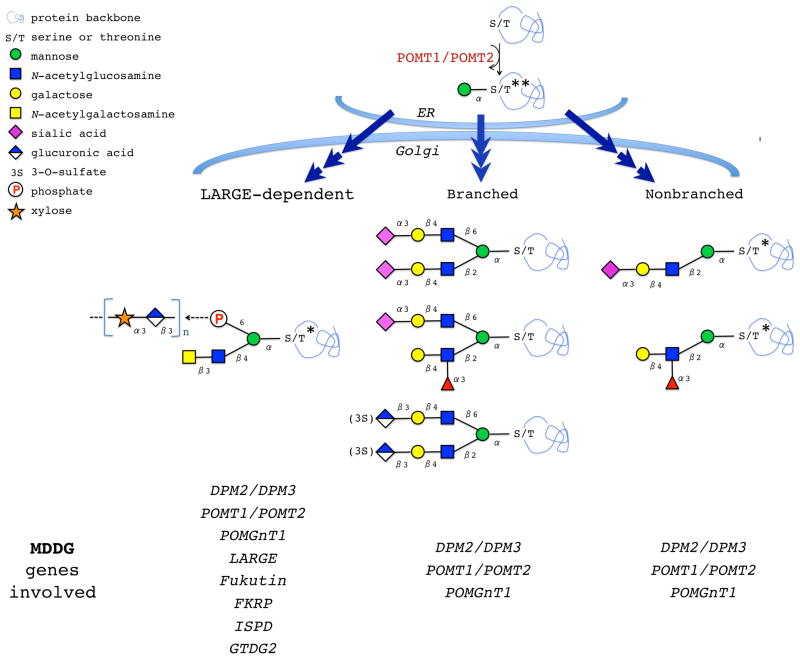
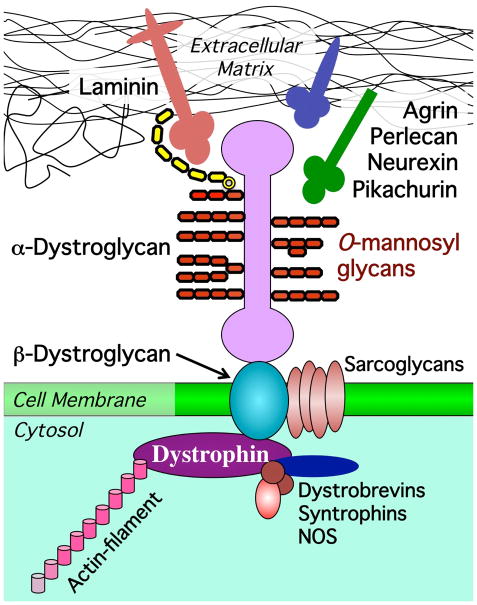
Protein O-mannosylation is a special type of glycosylation that plays prominent roles in metazoans, affecting development and physiology of the nervous system and muscles. A major biological effect of O-mannosylation involves the regulation of α-dystroglycan, a membrane glycoprotein mediating cell-extracellular matrix interactions. Genetic defects of O-mannosylation result in the loss of ligand-binding activity of α-dystroglycan and cause congenital muscular dystrophies termed dystroglycanopathies. Recent progress in mass spectrometry and in vitro analyses has shed new light on the mechanism of α-dystroglycan glycosylation; however, this mechanism is underlain by complex genetic and molecular elements that remain poorly understood. Protein O-mannosylation is evolutionarily conserved in metazoans, yet the pathway is simplified and more amenable to genetic analyses in invertebrate organisms, indicating that genetically tractable in vivo models could facilitate research in this area. This unit describes recent methodological strategies for studying protein O-mannosylation using in vitro and in vivo approaches.

Keywords: O-mannose; Drosophila; O-glycosylation; congenital muscular dystrophy; dystroglycan; mass spectrometry.


Figures

Figure 1
Figure 1. O-mannosylated glycans
Biosynthesis of O-mannosyl glycans is initiated in the ER by a complex of two protein O-mannosyltransferases, POMT1/POMT2. The O-mannose-linked structures undergo further maturation in the Golgi. LARGE-dependent structures are thought to be responsible for Laminin-binding activity of α–Dg. MDDG-associated genes affecting (or predicted to affect) different structures are indicated. *, structures identified on mammalian α-Dystroglycan. **, the structure found on Drosophila Dystroglycan. Only mature structures are shown, however, O-mannosyl glycans with incomplete maturation have been also detected on glycoproteins (Stalnaker et al., 2011b).
Figure 2
Figure 2. O-mannosyl glycans mediate interactions between α–Dg and extracellular matrix
Dystroglycan interacts with basal lamina by binding ECM ligands (Laminin, Neurexin, Agrin, Perlecan, and Pikachurin). β-Dystroglycan is a transmembrane protein that connects extracellular (α-Dystroglycan) and intracellular (Dystrophin) components of the Dystrophin-associated glycoprotein complex (DGC). Dystrophin links the DGC to actin cytoskeleton. Other DGC-associated proteins include sarcoglycans, dystrobrevins, syntrophin, and NOS (nitric oxide synthase). Note that in addition to O-mannosylation, α–Dg has also O-linked mucin-type and N-linked glycans (not shown here). LARGE-dependent carbohydrate chain responsible for Laminin binding is shown in yellow.
Figure 3
Figure 3. Examples of protein O-mannosyltransferase assays (A) and a purified protein substrate for O-mannosylation (B)
A, The activity of microsomal fraction isolated from Drosophila larvae was assayed using a fragment of mucin-type domain of mouse α-DG protein. The activity was assayed for 3 genotypes: Act5C-GAL4/+ (having wild-type level of rt and tw expression), Act5C-GAL4/UAS-dPOMT1-IR (RNAi-mediated knockdown of rt) and Act5C-GAL4/UAS-dPOMT2-IR (RNAi-mediated knockdown of tw). Figure adapted, with permission, from (Ichimiya et al., 2004). B, Coomassie staining of SDS-PAGE gel with purified fragment of Drosophila DG-A-GST protein expressed in E.coli cells (Nakamura et al., 2010b). Lane 1, DG-A-GST released from 10 μl of GST beads; Lane 2, blank; Lane 3, 1 μl of purified DG-A-GST eluted from after dialysis and concentration (estimated amount ~1.5 μg); M, protein molecular mass standards; Lane 4–5, BSA control samples for protein amount quantification, 1 and 2 μg, respectively.
Figure 4
Figure 4. Western and lectin blot analyses of O-mannosylated forms of Drosophila ExDg-FLAG expressed in vivo
A, Western blot detection of ExDG expressed in rt-tw double mutants (rt tw), rt mutants (rt), tw mutants (tw), wildtype background (WT), and backgrounds with ubiquitous ectopic expression of RT (rt+), TW (tw+), or RT-TW co-expression (rt+ tw+). L band represents a highly O-mannosylated glycoform, while S band corresponds to a glycoform without significant O-mannosylation. B, Analysis of ExDg glycosylation by glycosidase treatments. The top panel shows Con A reactivity of purified ExDG after treatments with PNGaseF and α-mannosidase. The S glycoform purified from rt mutant background (left side) loses its Con A reactivity either after the removal of N-linked glycans by PNGaseF or after treatment with α-mannosidase removing α-linked mannose residues, suggesting the absence of O-mannose modifications and efficient removal of oligomannose structures either by trimming N-linked branches with α-mannosidase or by complete elimination of N-linked glycans with PNGaseF. The L glycoform purified from RT-TW co-expression background (right side) retains Con A reactivity after treatment with PNGaseF, α-mannosidase, or both glycosidases, suggesting that L glycoform is O-mannosylated, and that α-mannosidase does not remove O-mannose completely. The bottom panel shows anti-FLAG western blot control corresponding to the lectin blot shown in the top panel. Dashed outline shows the region of the L glycoform on the blots. Figure adapted with permission from (Nakamura et al., 2010b).
Figure 5
Figure 5. Identification of O-linked mannose sites using mass spectrometry
Collision induced MS2 fragmentation of a singly O-mannosylated peptide derived from Drosophila Dystroglycan with m/z of 932.04 results in a predominant neutral loss ion at 850.93. This loss represents a loss of a hexose from the doubly charged peptide. In this example, there are sufficient fragments (b and y) to assign the peptide confidently and there is only one possible site of attachment. For peptides with multiple potential sites, the neutral loss observed upon collision induced fragmentation could be used to trigger an electron transfer dissociation fragmentation for confident site mapping. Figure adapted with permission from (Nakamura et al., 2010b).

References

    1. Abbott KL, Matthews RT, Pierce M. Receptor tyrosine phosphatase beta (RPTPbeta) activity and signaling are attenuated by glycosylation and subsequent cell surface galectin-1 binding. J Biol Chem. 2008;283:33026–33035. - PMC - PubMed
    1. Akasaka-Manya K, Manya H, Nakajima A, Kawakita M, Endo T. Physical and functional association of human protein o-mannosyltransferases 1 and 2. J Biol Chem. 2006;281:19339–19345. - PubMed
    1. Anumula KR, Taylor PB. A comprehensive procedure for preparation of partially methylated alditol acetates from glycoprotein carbohydrates. Analytical biochemistry. 1992;203:101–108. - PubMed
    1. Aoki K, Porterfield M, Lee SS, Dong B, Nguyen K, McGlamry KH, Tiemeyer M. The diversity of O-linked glycans expressed during Drosophila melanogaster development reflects stage- and tissue-specific requirements for cell signaling. J Biol Chem. 2008;283:30385–30400. - PMC - PubMed
    1. Barone R, Aiello C, Race V, Morava E, Foulquier F, Riemersma M, Passarelli C, Concolino D, Carella M, Santorelli F, Vleugels W, Mercuri E, Garozzo D, Sturiale L, Messina S, Jaeken J, Fiumara A, Wevers RA, Bertini E, Matthijs G, Lefeber DJ. DPM2-CDG: A muscular dystrophy-dystroglycanopathy syndrome with severe epilepsy. Ann Neurol. 2012;72:550–558. - PubMed

Publication types

LinkOut - more resources