

Unbiased screen for interactors of leucine-rich repeat kinase 2 supports a common pathway for sporadic and familial Parkinson disease
- PMID: 24510904
- PMCID: PMC3932908
- DOI: 10.1073/pnas.1318306111
Unbiased screen for interactors of leucine-rich repeat kinase 2 supports a common pathway for sporadic and familial Parkinson disease
Abstract
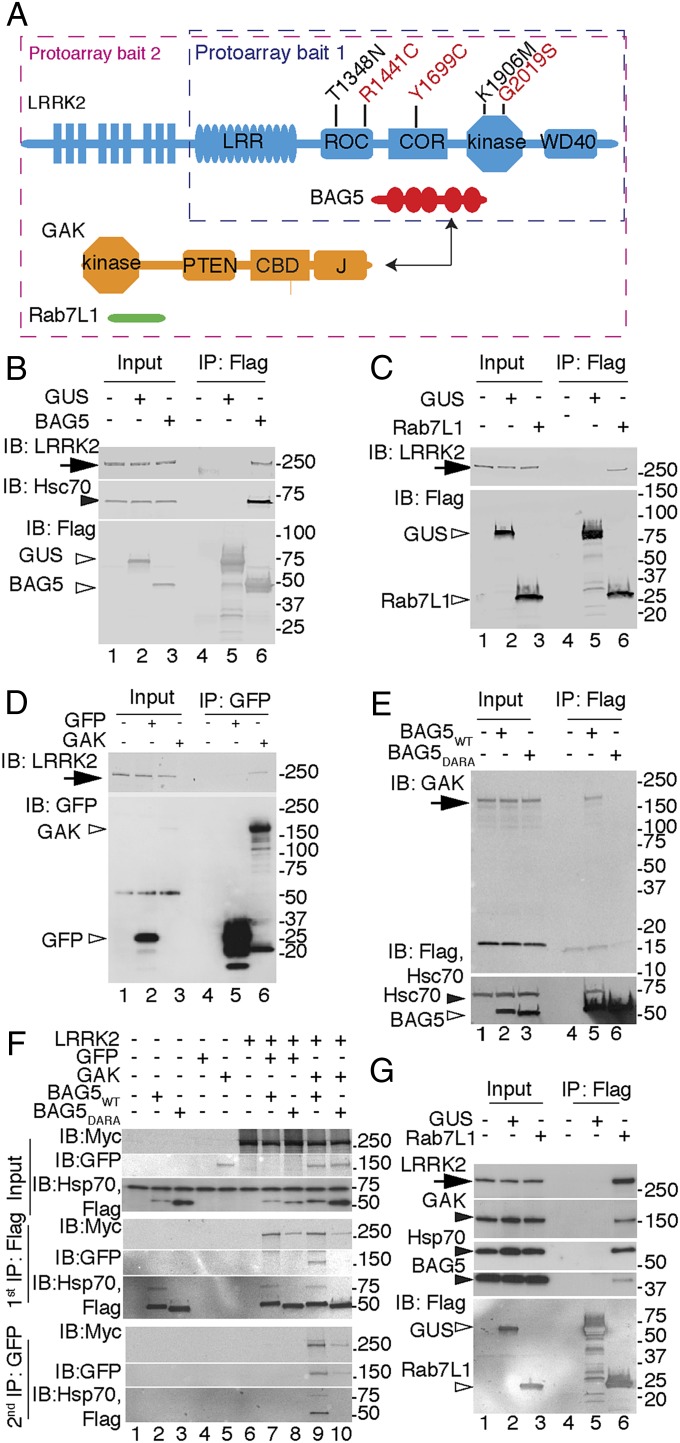
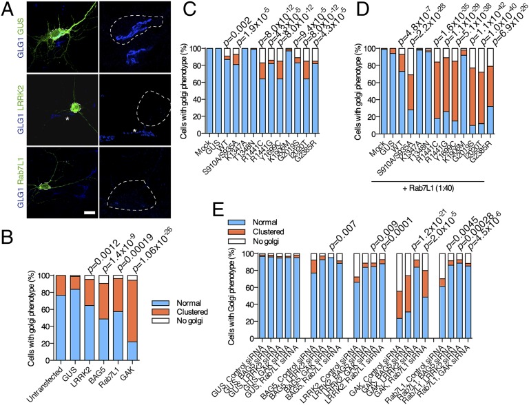
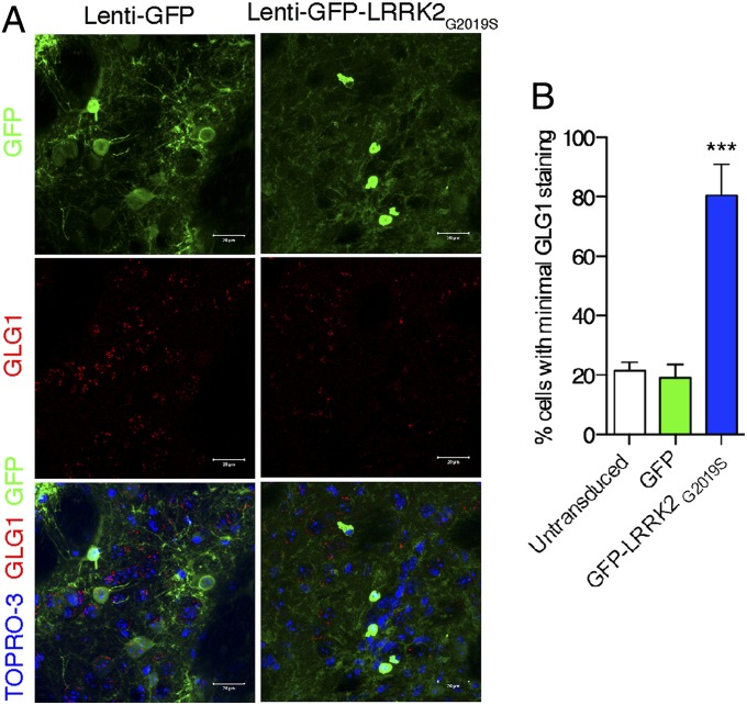
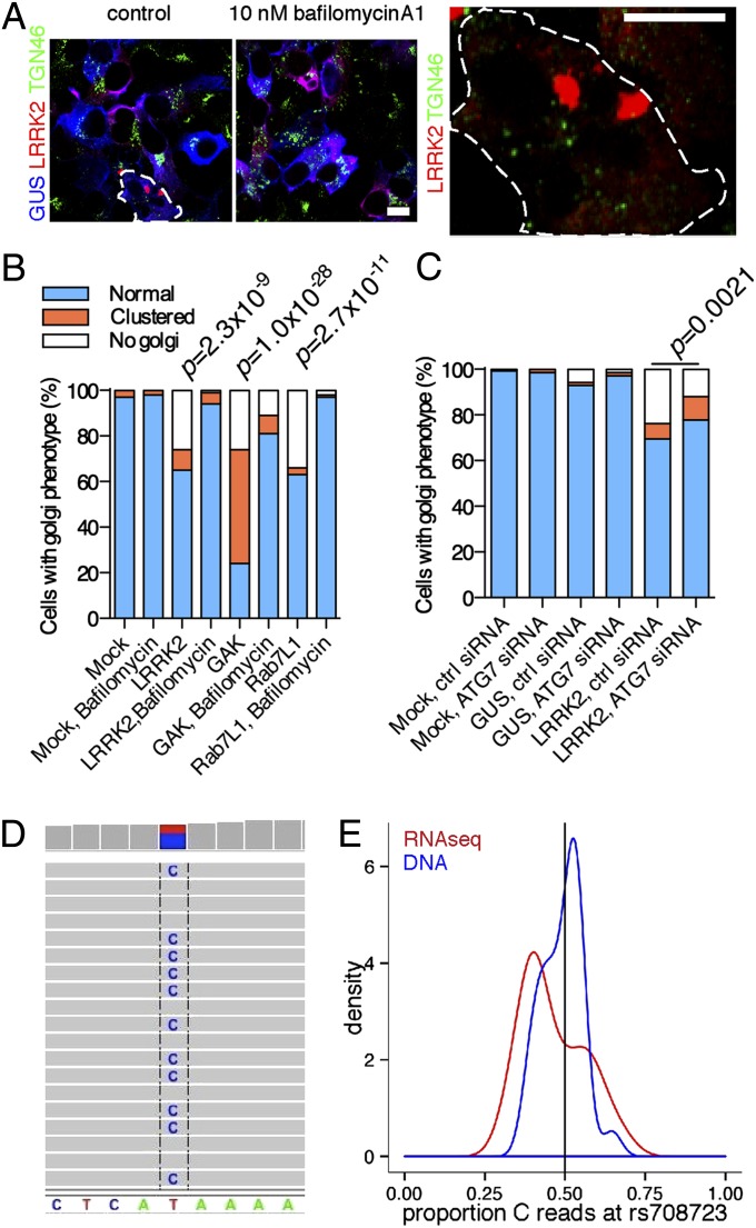
Mutations in leucine-rich repeat kinase 2 (LRRK2) cause inherited Parkinson disease (PD), and common variants around LRRK2 are a risk factor for sporadic PD. Using protein-protein interaction arrays, we identified BCL2-associated athanogene 5, Rab7L1 (RAB7, member RAS oncogene family-like 1), and Cyclin-G-associated kinase as binding partners of LRRK2. The latter two genes are candidate genes for risk for sporadic PD identified by genome-wide association studies. These proteins form a complex that promotes clearance of Golgi-derived vesicles through the autophagy-lysosome system both in vitro and in vivo. We propose that three different genes for PD have a common biological function. More generally, data integration from multiple unbiased screens can provide insight into human disease mechanisms.
Keywords: BAG5; GAK; autophagy; trans-Golgi.
Conflict of interest statement
The authors declare no conflict of interest.
Figures
Comment in
-
Pathway for Parkinson disease.Proc Natl Acad Sci U S A. 2014 Feb 18;111(7):2402-3. doi: 10.1073/pnas.1324284111. Epub 2014 Feb 7. Proc Natl Acad Sci U S A. 2014. PMID: 24510898 Free PMC article. No abstract available.
Similar articles
-
Pathway for Parkinson disease.Proc Natl Acad Sci U S A. 2014 Feb 18;111(7):2402-3. doi: 10.1073/pnas.1324284111. Epub 2014 Feb 7. Proc Natl Acad Sci U S A. 2014. PMID: 24510898 Free PMC article. No abstract available.
-
RAB7L1 interacts with LRRK2 to modify intraneuronal protein sorting and Parkinson's disease risk.Neuron. 2013 Feb 6;77(3):425-39. doi: 10.1016/j.neuron.2012.11.033. Neuron. 2013. PMID: 23395371 Free PMC article.
-
LRRK2 delays degradative receptor trafficking by impeding late endosomal budding through decreasing Rab7 activity.Hum Mol Genet. 2014 Dec 20;23(25):6779-96. doi: 10.1093/hmg/ddu395. Epub 2014 Jul 30. Hum Mol Genet. 2014. PMID: 25080504
-
Cellular functions of LRRK2 implicate vesicular trafficking pathways in Parkinson's disease.Biochem Soc Trans. 2016 Dec 15;44(6):1603-1610. doi: 10.1042/BST20160228. Biochem Soc Trans. 2016. PMID: 27913668 Review.
-
LRRK2 links genetic and sporadic Parkinson's disease.Biochem Soc Trans. 2019 Apr 30;47(2):651-661. doi: 10.1042/BST20180462. Epub 2019 Mar 5. Biochem Soc Trans. 2019. PMID: 30837320 Free PMC article. Review.
Cited by
-
The endoplasmic reticulum contributes to lysosomal tubulation/sorting driven by LRRK2.Mol Biol Cell. 2022 Nov 1;33(13):ar124. doi: 10.1091/mbc.E22-04-0139. Epub 2022 Aug 31. Mol Biol Cell. 2022. PMID: 36044336 Free PMC article.
-
Distinct Roles for RAB10 and RAB29 in Pathogenic LRRK2-Mediated Endolysosomal Trafficking Alterations.Cells. 2020 Jul 17;9(7):1719. doi: 10.3390/cells9071719. Cells. 2020. PMID: 32709066 Free PMC article.
-
Human Induced Pluripotent Stem Cell Phenotyping and Preclinical Modeling of Familial Parkinson's Disease.Genes (Basel). 2022 Oct 25;13(11):1937. doi: 10.3390/genes13111937. Genes (Basel). 2022. PMID: 36360174 Free PMC article. Review.
-
Tied up: Does altering phosphoinositide-mediated membrane trafficking influence neurodegenerative disease phenotypes?J Genet. 2018 Jul;97(3):753-771. J Genet. 2018. PMID: 30027907 Review.
-
A new hypothesis for Parkinson's disease pathogenesis: GTPase-p38 MAPK signaling and autophagy as convergence points of etiology and genomics.Mol Neurodegener. 2018 Aug 2;13(1):40. doi: 10.1186/s13024-018-0273-5. Mol Neurodegener. 2018. PMID: 30071902 Free PMC article. Review.
References
-
- Lill CM, Bertram L. Towards unveiling the genetics of neurodegenerative diseases. Semin Neurol. 2011;31(5):531–541. - PubMed
-
- Satake W, et al. Genome-wide association study identifies common variants at four loci as genetic risk factors for Parkinson’s disease. Nat Genet. 2009;41(12):1303–1307. - PubMed
Publication types
MeSH terms
Substances
Grants and funding
- GGP12237/TI_/Telethon/Italy
- G0701075/MRC_/Medical Research Council/United Kingdom
- G1100643/MRC_/Medical Research Council/United Kingdom
- R01 NS075321/NS/NINDS NIH HHS/United States
- R01 NS041509/NS/NINDS NIH HHS/United States
- 090532/WT_/Wellcome Trust/United Kingdom
- MR/L010305/1/MRC_/Medical Research Council/United Kingdom
- G0700943/MRC_/Medical Research Council/United Kingdom
- U01 NS082151/NS/NINDS NIH HHS/United States
- F-1202/PUK_/Parkinson's UK/United Kingdom
- G-0907/PUK_/Parkinson's UK/United Kingdom
- G1100479/MRC_/Medical Research Council/United Kingdom
- ImNIH/Intramural NIH HHS/United States
LinkOut - more resources
Full Text Sources
Other Literature Sources
Medical
Molecular Biology Databases
Research Materials
