Preparation of multilayered polymeric structures using a novel four-needle coaxial electrohydrodynamic device
- PMID: 24510905
- PMCID: PMC4237175
- DOI: 10.1002/marc.201300777
Preparation of multilayered polymeric structures using a novel four-needle coaxial electrohydrodynamic device
Abstract
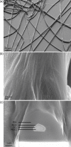
Coaxial four-needle electrohydrodynamic forming is applied for the first time to prepare layered structures in both particle and fiber form. Four different biocompatible polymers, polyethylene glycol, poly (lactic-co-glycolic acid), polycaprolactone, and polymethylsilsesquioxane, are used to generate four distinct layers confirmed using transmission and scanning electron microscopy combined with focused ion beam milling. The incorporation and release of different dyes within the polymeric system of four layers are demonstrated, something that is much desired in modern applications such as the polypill where multiple active pharmaceutical ingredients can be combined to treat numerous diseases.
Keywords: coaxial device; electrohydrodynamic; multilayered structures; polymers.
© 2014 The Authors. Published by WILEY-VCH Verlag GmbH & Co. KGaA, Weinheim.
Figures


Similar articles
-
Preparation of multicompartment sub-micron particles using a triple-needle electrohydrodynamic device.J Colloid Interface Sci. 2013 Nov 1;409:245-54. doi: 10.1016/j.jcis.2013.07.033. Epub 2013 Jul 30. J Colloid Interface Sci. 2013. PMID: 23972499
-
An Adaptive Approach in Polymer-Drug Nanoparticle Engineering using Slanted Electrohydrodynamic Needles and Horizontal Spraying Planes.AAPS PharmSciTech. 2024 Oct 30;25(8):257. doi: 10.1208/s12249-024-02971-y. AAPS PharmSciTech. 2024. PMID: 39477831
-
Electrohydrodynamic preparation of polymeric drug-carrier particles: mapping of the process.Int J Pharm. 2011 Feb 14;404(1-2):110-5. doi: 10.1016/j.ijpharm.2010.11.014. Epub 2010 Nov 17. Int J Pharm. 2011. PMID: 21093562
-
Polymeric Based Therapeutic Delivery Systems Prepared Using Electrohydrodynamic Processes.Curr Pharm Des. 2016;22(19):2873-85. doi: 10.2174/1381612822666160217141612. Curr Pharm Des. 2016. PMID: 26898734 Review.
-
Electrohydrodynamic atomisation driven design and engineering of opportunistic particulate systems for applications in drug delivery, therapeutics and pharmaceutics.Adv Drug Deliv Rev. 2021 Sep;176:113788. doi: 10.1016/j.addr.2021.04.026. Epub 2021 May 4. Adv Drug Deliv Rev. 2021. PMID: 33957180 Review.
Cited by
-
Coaxial Electrohydrodynamic Atomization for the Production of Drug-Loaded Micro/Nanoparticles.Micromachines (Basel). 2019 Feb 14;10(2):125. doi: 10.3390/mi10020125. Micromachines (Basel). 2019. PMID: 30769856 Free PMC article. Review.
-
A combination of experimental and finite element analyses of needle-tissue interaction to compute the stresses and deformations during injection at different angles.J Clin Monit Comput. 2016 Dec;30(6):965-975. doi: 10.1007/s10877-015-9801-9. Epub 2015 Oct 29. J Clin Monit Comput. 2016. PMID: 26515741
-
Advanced Polymers for Three-Dimensional (3D) Organ Bioprinting.Micromachines (Basel). 2019 Nov 25;10(12):814. doi: 10.3390/mi10120814. Micromachines (Basel). 2019. PMID: 31775349 Free PMC article. Review.
-
Novel targeted bladder drug-delivery systems: a review.Res Rep Urol. 2015 Nov 23;7:169-78. doi: 10.2147/RRU.S56168. eCollection 2015. Res Rep Urol. 2015. PMID: 26649286 Free PMC article. Review.
-
Hollow Polycaprolactone Microspheres with/without a Single Surface Hole by Co-Electrospraying.Langmuir. 2017 Nov 21;33(46):13262-13271. doi: 10.1021/acs.langmuir.7b01985. Epub 2017 Nov 8. Langmuir. 2017. PMID: 28901145 Free PMC article.
References
Publication types
MeSH terms
Substances
LinkOut - more resources
Full Text Sources
Other Literature Sources

