Review
. 2014 Jan 29:9:711-26.
doi: 10.2147/IJN.S53717. eCollection 2014.

Nanoparticles for multimodal in vivo imaging in nanomedicine

Affiliations
Review

Nanoparticles for multimodal in vivo imaging in nanomedicine

Jaehong Key et al. Int J Nanomedicine. .

Abstract

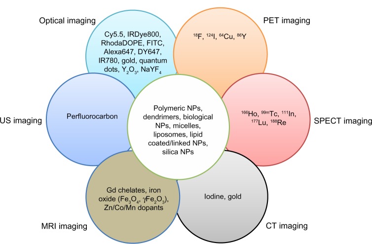
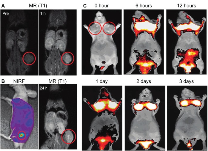
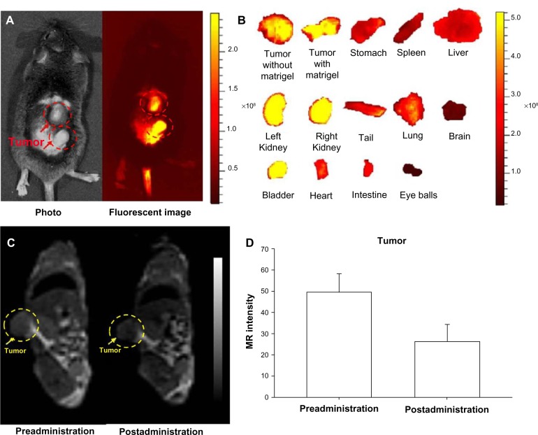
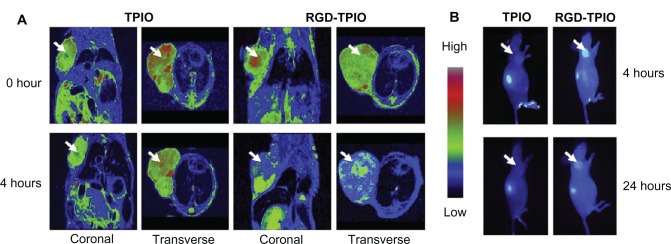
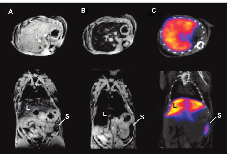
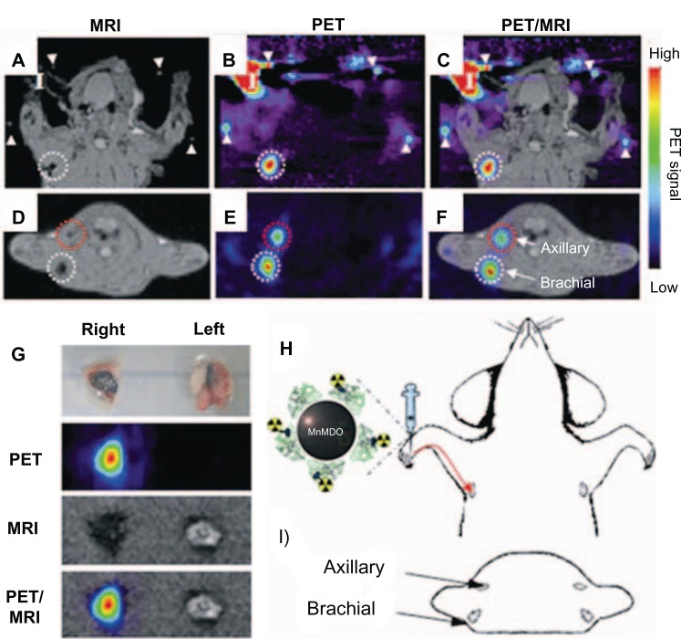
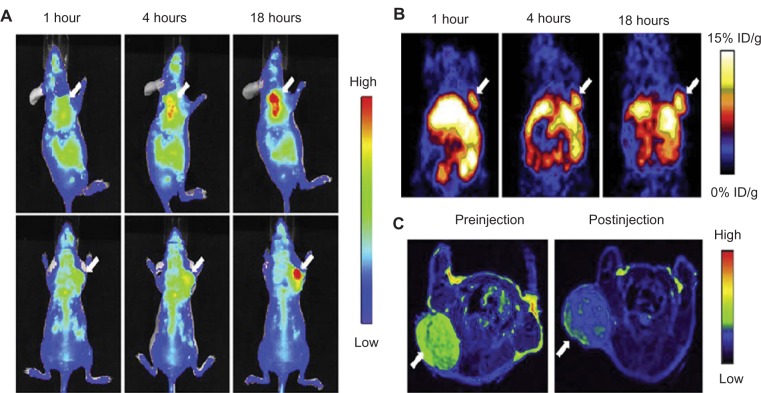
While nanoparticles are usually designed for targeted drug delivery, they can also simultaneously provide diagnostic information by a variety of in vivo imaging methods. These diagnostic capabilities make use of specific properties of nanoparticle core materials. Near-infrared fluorescent probes provide optical detection of cells targeted by real-time nanoparticle-distribution studies within the organ compartments of live, anesthetized animals. By combining different imaging modalities, we can start with deep-body imaging by magnetic resonance imaging or computed tomography, and by using optical imaging, get down to the resolution required for real-time fluorescence-guided surgery.

Keywords: CT; MRI; NIRF; PET; cancer; multimodal imaging; nanomedicine; nanoparticles.


Figures

Figure 1
Figure 1
Incorporation of various contrast agents for multimodality imaging. Notes: Data from Swierczewska et al, Lee et al, and Louie. Abbreviations: DOPE, 1,2-dioleoyl-sn-glycero-3-phosphoethanolamine; FITC, fluorescein isothiocyanate; Gd, gadolinium; PET, positron emission tomography; US, ultrasound; NPs, nanoparticles; SPECT, single-photon emission computed tomography; MRI, magnetic resonance imaging.
Figure 2
Figure 2
(AC) In vivo magnetic resonance (MR)–near-infrared fluorescent (NIRF) dual-modality imaging of SCC7 tumors. Notes: Cy5.5-chitosan nanoparticle-Gd(III) nanoparticles were injected into the SCC7-bearing mice, and the mice were visualized by using MR and NIRF imaging. Red circles indicate tumor sites. (A) In vivo MR imaging showed T1-positive contrast effects 1 hour after injection at the tumor sites. (B) In vivo NIRF imaging showed brighter NIRF intensity at the tumor site. In vivo MR imaging 24 hours after injection showed bright contrast effects at the tumor site. (C) In vivo NIRF imaging showed the accumulated Cy5.5-CNP-Gd(III) nanoparticles over time at the tumor sites. Reproduced with permission Nam T, Park S, Lee SY, et al. Tumor targeting chitosan nanoparticles for dual-modality optical/MR cancer imaging. Bioconjug Chem. 2010;21(4):578–582. Copyright © 2010 American Chemical Society.
Figure 3
Figure 3
(AD) In vivo magnetic resonance (MR)–near-infrared fluorescent (NIRF) dual-modality imaging of murine bladder tumor (MBT-2). SPIO-Cy5.5 conjugated glycol chitosan-based nanoparticles were injected into the tail vein for tumor detection. Notes: (A) In vivo NIRF image; (B) ex vivo NIRF image; (C) in vivo MR image; (D) quantification of intensity at the tumor site. The dashed circles indicate the location of the implanted tumors. Reproduced with permission from Key J, Kim K, Dhawan D, et al. Dual-modality in vivo imaging for MRI detection of tumors and NIRF-guided surgery using multi-component nanoparticles. Vol: Proc SPIE. 2011;79087:90805. Copyright © 2011 Society of Photo Optical Instrumentation engineers.
Figure 4
Figure 4
In vivo magnetic resonance (MR)–near-infrared fluorescent imaging of U87MG tumor. Images were taken before and after injection of nanoparticles (NPs) using MR imaging and optical imaging. Notes: (A) Arginine–glycine–aspartic acid (RGD) triblock copolymer-coated iron oxide (TPIO) NPs showed better T2 contrast effects in MR imaging at the tumor sites; (B) the contrast effect with RGD-TPIO NPs was also validated by optical imaging. Reprinted from Chen K, Xie J, Xu H, et al. Triblock copolymer coated iron oxide nanoparticle conjugate for tumor integrin targeting. Biomaterials. 2009;30(36):6912–6919. With permission from Elsevier. © 2009 Elsevier.
Figure 5
Figure 5
(AC) In vivo magnetic resonance (MR)–single-photon emitted tomography (SPECT) images of axial view (top) and coronal view (bottom). Notes: (A) T2*-weighted MR images before injection of technetium-99m–diethylene triamine pentaacetic acid (99mTc-DTPA)-ale-Endorem; (B) T2*-weighted MR images 15 minutes after injection of 99mTc-DTPA-ale-Endorem; (C) SPECT image 45 minutes after injection of 99mTc-DPA-ale-Endorem; L indicates the liver, S the spleen, showing the accumulation of 99mTc-DPA-ale-Endorem at those organs with MR imaging and SPECT after injection. Reproduced with permission from Torres Martin de Rosales R, TavaréR, Glaria A, Varma G, Protti A, Blower PJ. (99m)Tc-bisphosphonate-iron oxide nanoparticle conjugates for dual-modality biomedical imaging. Bioconjug Chem. 2011;22(3):455–465. Copyright © 2011 American Chemical Society.
Figure 6
Figure 6
(AI) In vivo magnetic resonance (MR) and positron emission tomography (PET) images of sentinel lymph nodes in a rat 1 hour after injection of iodine-124–serum albumin–manganese magnetism-engineered iron oxide into the right forepaw. Notes: Coronal images of (A) in vivo MR, (B) in vivo PET, (C) MR–PET fusion; transverse images of (D) in vivo MR, (E) in vivo PET, (F) MR–PET fusion; (H) explains the coronal images of (AC); (I) describes transverse directions of (DF); (G) ex vivo images of brachial lymph nodes by PET, MR imaging, and PET/MR imaging; (I) in (AC) indicates the injection site. Reproduced with permission by Choi JS, Park JC, Nah H, et al. A hybrid nanoparticle probe for dual-modality positron emission tomography and magnetic resonance imaging. Angew Chem Int Ed Engl. 2008;47(33):6259–6262.
Figure 7
Figure 7
(AC) In vivo optical–positron emission tomography (PET)–magnetic resonance (MR) trimodality imaging by human serum albumin iron oxide nanoparticles. Images of 1, 4, and 18 hours postinjection. (A) In vivo near-infrared fluorescent images of mice; (B) in vivo PET images of mice; (C) in vivo MR images of mice, 18 hours postinjection; white arrows indicate xenograft U87MG tumors. Reprinted from Xie J, Chen K, Huang J, et al. PET/NIRF/MRI triple functional iron oxide nanoparticles. Biomaterials. 2010;31(11):3016–3022. Copyright © 2010, with permission from Elsevier.

References

    1. van Dam GM, Themelis G, Crane LM, et al. Intraoperative tumor-specific fluorescence imaging in ovarian cancer by folate receptor-α targeting: first in-human results. Nat Med. 2011;17(10):1315–1319. - PubMed
    1. Leary JF. Nanotechnology: what is it and why is small so big? Can J Ophthalmol. 2010;45(5):449–456. - PubMed
    1. Weissleder R, Pittet MJ. Imaging in the era of molecular oncology. Nature. 2008;452(7187):580–589. - PMC - PubMed
    1. Hahn M, Singh A, Sharma P, Brown S, Moudgil B. Nanoparticles as contrast agents for in vivo bioimaging: current status and future perspectives. Anal Bioanal Chem. 2011;399(1):3–27. - PubMed
    1. Cheon J, Lee JH. Synergistically integrated nanoparticles as multimodal probes for nanobiotechnology. Acc Chem Res. 2008;41(12):1630–1640. - PubMed

Publication types