. 2014 Feb;2(1):49-61.
doi: 10.1016/j.jchf.2013.08.008.

Human cardiosphere-derived cells from advanced heart failure patients exhibit augmented functional potency in myocardial repair

Affiliations

Human cardiosphere-derived cells from advanced heart failure patients exhibit augmented functional potency in myocardial repair

Ke Cheng et al. JACC Heart Fail. 2014 Feb.

Abstract

Objectives: This study sought to compare the regenerative potency of cells derived from healthy and diseased human hearts.

Background: Results from pre-clinical studies and the CADUCEUS (CArdiosphere-Derived aUtologous stem CElls to reverse ventricUlar dySfunction) trial support the notion that cardiosphere-derived cells (CDCs) from normal and recently infarcted hearts are capable of regenerating healthy heart tissue after myocardial infarction (MI). It is unknown whether CDCs derived from advanced heart failure (HF) patients retain the same regenerative potency.

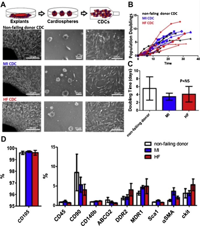
Methods: In a mouse model of acute MI, we compared the regenerative potential and functional benefits of CDCs derived from 3 groups: 1) non-failing (NF) donor: healthy donor hearts post-transplantation; 2) MI: patients who had an MI 9 to 35 days before biopsy; and 3) HF: advanced cardiomyopathy tissue explanted at cardiac transplantation.

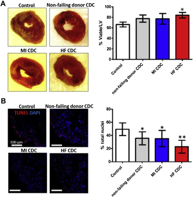
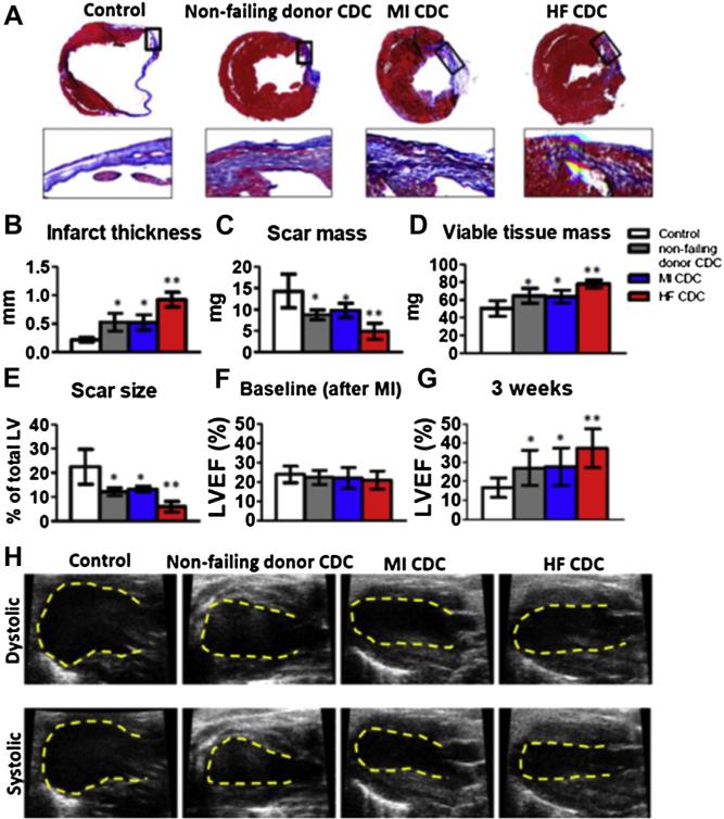
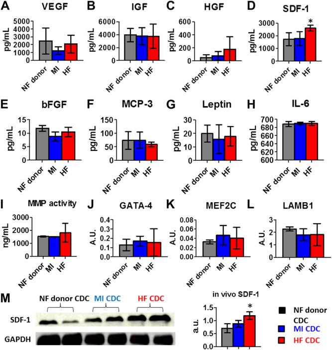
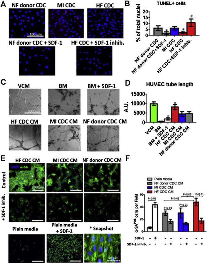
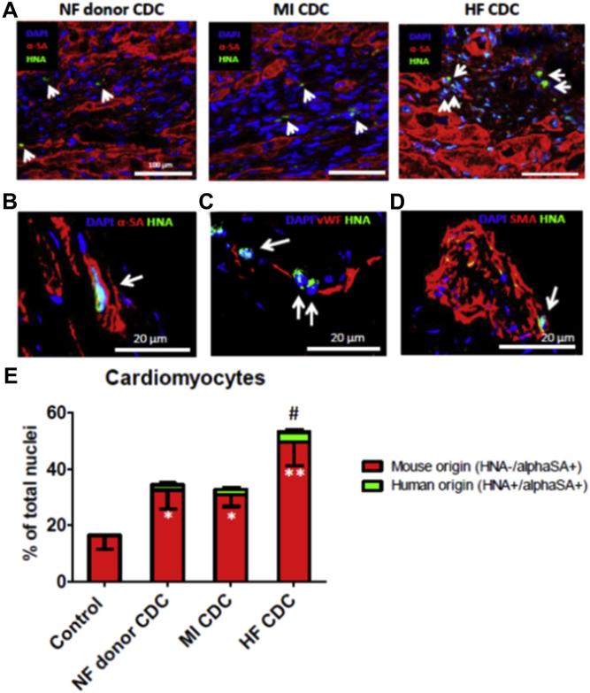
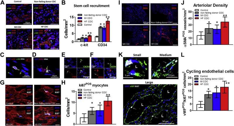
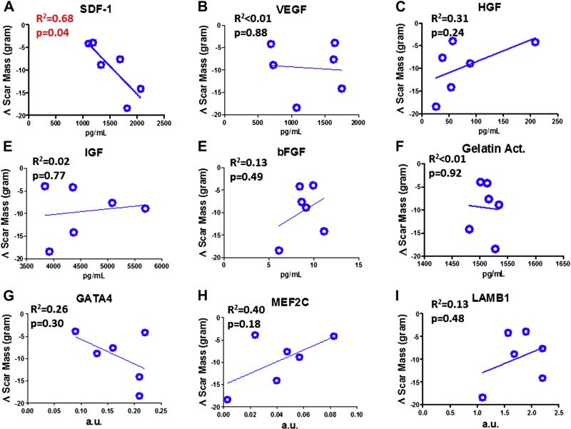
Results: Cell growth and phenotype were identical in all 3 groups. Injection of HF CDCs led to the greatest therapeutic benefit in mice, with the highest left ventricular ejection fraction, thickest infarct wall, most viable tissue, and least scar 3 weeks after treatment. In vitro assays revealed that HF CDCs secreted higher levels of stromal cell-derived factor (SDF)-1, which may contribute to the cells' augmented resistance to oxidative stress, enhanced angiogenesis, and improved myocyte survival. Histological analysis indicated that HF CDCs engrafted better, recruited more endogenous stem cells, and induced greater angiogenesis and cardiomyocyte cell-cycle re-entry. CDC-secreted SDF-1 levels correlated with decreases in scar mass over time in CADUCEUS patients treated with autologous CDCs.

Conclusions: CDCs from advanced HF patients exhibit augmented potency in ameliorating ventricular dysfunction post-MI, possibly through SDF-1–mediated mechanisms.


Figures

Figure 1
Figure 1. Characteristics of CDCs Derived From Patients
(A) Schematic showing the process of deriving cardiosphere-derived cells (CDCs) from myocardial tissue. (B) Population doubling over time in non-failing (NF) donor, myocardial infarction (MI), and heart failure (HF) CDCs. (C) Average doubling time of NF donor, MI, and HF CDCs (n = 6 patient-derived CDC lines). (D) Summary of antigenic phenotype of CDCs. Data are presented as means ± SD.
Figure 2
Figure 2. Acute Cardioprotective Effects of Injected CDCs
(A) Representative macroscopic images of hearts from each experimental group after TTC staining (left). Pale areas are irreversibly injured. Right panel shows pooled data for the percentage viability. (B) Representative confocal images showing terminal deoxynucleotidyl transferase dUTP nick end labeling (TUNEL) staining for apoptotic nuclei in the infarct area 24 h after myocardial infarction (left), and pooled data (right). *p < 0.05 compared with Control; **p < 0.05 compared with all other groups. Data are presented as means ± SD. Abbreviations as in Figure 1.
Figure 3
Figure 3. Therapeutic Benefit of NF Donor, MI, and HF CDCs in Mice With MI
(A) Representative Masson's trichrome–stained myocardial sections 3 weeks after treatment with Control (vehicle only), NF donor, MI, and HF CDCs. Scar tissue and viable myocardium are identified by the blue and red colors, respectively. Snapshots of the infarct border zone (black box area) are presented beneath each group. (B–E) Quantitative analysis of infarct thickness, scar mass, viable tissue mass, and scar size from the Masson's trichrome–stained images (n = 6 animals per group). Left ventricular ejection fraction (LVEF) was measured by echocardiography at baseline (4 h post-MI) (F) and 3 weeks afterwards (G) (Control: n = 11 animals; NF donor, MI, or HF CDC: n = 19 to 24 animals from n = 6 patient-derived CDC lines). Baseline LVEFs were indistinguishable in the 4 groups. (H) Representative echocardiography images. *p < 0.05 compared with Control; **p < 0.05 compared with all other groups. Data are presented as means ± SD. Abbreviations as in Figure 1.
Figure 4
Figure 4. Paracrine Factors and Gene Expression in CDCs
(A–H) Secretion of various cytokines and growth factors from NF donor, MI, and HF CDCs. Concentrations were measured by enzyme-linked immunosorbent assay (ELISA). (I) Proteinolytic activity (MMP2/MMP9) in CDC-conditioned media. (J–L) GATA4, MEF2C, and laminin beta 1 (LAMB1) transcript levels in CDCs measured by reverse transcription–polymerase chain reaction (RT-PCR) (n = 6 for each group). (M) Western blot analysis of myocardial SDF-1 (representative blot, left, and pooled data, right; n = 4 per group). *p < 0.05 compared with the other 2 groups. Data are presented as means ± SD. Abbreviations as in Figure 1.
Figure 5
Figure 5. Resistance to Oxidative Stress, Endothelial Cell Angiogenesis, and Cardiomyocyte Survival and Contractile Assays
(A) Representative confocal fluorescent micrographs showing TUNEL+ nuclei (red) in CDC cultures exposed to 100 μmol/l H2O2. (B) Quantification of the percentages of TUNEL+ cells in NF donor, MI, and HF CDCs (n = 6). *p < 0.05 compared with the NF donor CDC group; #p < 0.05 compared with the HF CDC group. (C) Representative white light images showing tube formation by human umbilical vein endothelial cells (HUVECs) on Matrigel in various types of media. BM = vascular cell basal medium; VCM = vascular cell growth medium. (D) Cumulative tube length measured by Image-Pro Plus (MediaCybernetics, Rockville, Maryland) in each well (n = 3). *p < 0.05 when compared with NF donor CDC CM group; #p < 0.05 when compared with the BM group. A.U. = arbitrary units. (E) Representative confocal images showing neonatal rat cardiomyocytes stained with α-sarcomeric actin in different culture conditions. (F) Numbers of cardiomyocytes per field under each condition (n = 3). Data are presented as means ± SD. CM = conditioned medium; other abbreviations as in Figures 1 and 3.
Figure 6
Figure 6. Engraftment and Differentiation of Transplanted Cells and New Cardiomyocyte Formation
(A) Representative confocal images showing the engraftment of transplanted CDCs (positive for human nuclei antigen [HNA]; green, indicated by white arrows) in the infarct border zone. Cardiomyocytes were stained with alpha-sarcomeric actin (α-SA; red). (B–D), Co-expression of HNA (green) with α-SA, von Willebrand factor (vWF), alpha-smooth muscle actin (SMA) in HF CDCs (white arrows) in mouse hearts. (E) Quantification of endogenous (α-SA+/HNA) and exogenous (α-SA+/HNA+) cardiomyocytes in the peri-infarct area. *p < 0.05 compared with Control; **p < 0.05 compared with all other groups; #p < 0.05 compared with the NF donor CDC or MI CDC group. Data are presented as means ± SD. Abbreviations as in Figure 1.
Figure 7
Figure 7. Recruitment of Stem Cells, Cardiomyocyte Cell-Cycle Re-Entry, and Angiogenesis
(A) Representative confocal images showing c-kit+ (green) and CD34+ (magenta) stem cells in the infarct border zone. Cardiomyocytes were stained with alpha-sarcomeric actin (α-SA; red). (B) Quantitation of c-kit+ and CD34+ cells (n = 3 hearts per group). *p < 0.05 compared with Control; **p < 0.05 compared with all other groups. Large magnification revealed interactions (yellow arrow) between transplanted HF CDCs with c-kit+ (C) or CD34+ cells (D). (E) Co-expression of CXCR4 (green) in c-kit+ cells (red). (F) Co-expression of CXCR4 (green) in CD34+ cells (red). (G) Cardiomyocytes (stained with α-SA; red) containing Ki67+ nuclei (white) in the infarct border zone. (H) Quantitation of Ki67+ cardiomyocytes in each group (n = 3 hearts per group). (I) Arteriolar structures stained with SMA (red) in the infarct border zone. (J) Quantitation of arteriolar density in each group (n = 3 hearts per group). (K) cycling endothelial cells stained with ki67 (white) and vWF (green). (L) Quantitation of cycling endothelial cells in each group (n = 3 hearts per group). *p < 0.05 compared with Control; **p < 0.05 compared with all other groups. Data are presented as means ± SD. Abbreviations as in Figures 1 and 6.
Figure 8
Figure 8. Correlation of CDC Properties With Scar Mass Changes in CADUCEUS Patients
Linear regression analysis was performed to reveal the relationship between various paracrine factors/gene expression levels and the changes of the patients’ cardiac scar tissue mass over the 6-month follow-up. Only SDF-1 levels reveal a significant correlation with decreasing scar mass. Data are presented as means ± SD. a.u. = arbitrary units; CADUCEUS = CArdiosphere-Derived aUtologous stem CElls to reverse ventricUlar dysfunction.

Comment in

References

    1. Makkar RR, Smith RR, Cheng K, et al. Intracoronary cardiosphere-derived cells for heart regeneration after myocardial infarction (caduceus): a prospective, randomised phase 1 trial. Lancet. 2012;379:895–904. - PMC - PubMed
    1. White AJ, Smith RR, Matsushita S, et al. Intrinsic cardiac origin of human cardiosphere-derived cells. Eur Heart J. 2013;34:68–75. - PubMed
    1. Li TS, Cheng K, Malliaras K, et al. Direct comparison of different stem cell types and subpopulations reveals superior paracrine potency and myocardial repair efficacy with cardiosphere-derived cells. J Am Coll Cardiol. 2012;59:942–53. - PMC - PubMed
    1. Smith RR, Barile L, Cho HC, et al. Regenerative potential of cardiosphere-derived cells expanded from percutaneous endomyocardial biopsy specimens. Circulation. 2007;115:896–908. - PubMed
    1. Cheng K, Shen D, Smith J, et al. Transplantation of platelet gel spiked with cardiosphere-derived cells boosts structural and functional benefits relative to gel transplantation alone in rats with myocardial infarction. Biomaterials. 2012;33:2872–9. - PMC - PubMed

Publication types

MeSH terms

Substances