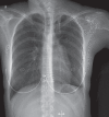
Vanishing lung syndrome
- PMID: 24511569
- PMCID: PMC3938235
- DOI: 10.1155/2014/583697
Vanishing lung syndrome
Figures


References
-
- Roberts L, Putman CE, Chen JTT, Goodman LR, Ravin CE. Vanishing lung syndrome: Upper lobe bullous pneumopathy. Revista interamericana de radiología. 1987;12:249–55.
-
- Stern EJ, Webb WR, Weinacker A, Müller NL. Idiopathic giant bullous emphysema (vanishing lung syndrome): Imaging findings in nine patients. Am J Roentgenol. 1994;162:279–82. - PubMed
-
- Sharma N, Justaniah AM, Kanne JP, Gurney JW, Mohammed TL. Vanishing lung syndrome (giant bullous emphysema): CT findings in 7 patients and a literature review. J Thorac Imaging. 2009;24:227–30. - PubMed
Publication types
MeSH terms
LinkOut - more resources
Full Text Sources
Other Literature Sources

