Review
. 2014 Apr:35:152-61.
doi: 10.1016/j.matbio.2014.01.015. Epub 2014 Feb 7.

Versican and the control of inflammation

Affiliations
Review

Versican and the control of inflammation

Thomas N Wight et al. Matrix Biol. 2014 Apr.

Abstract

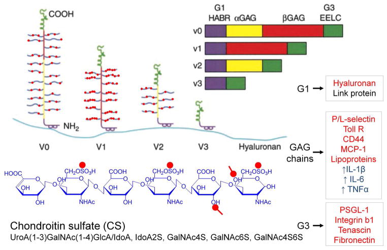
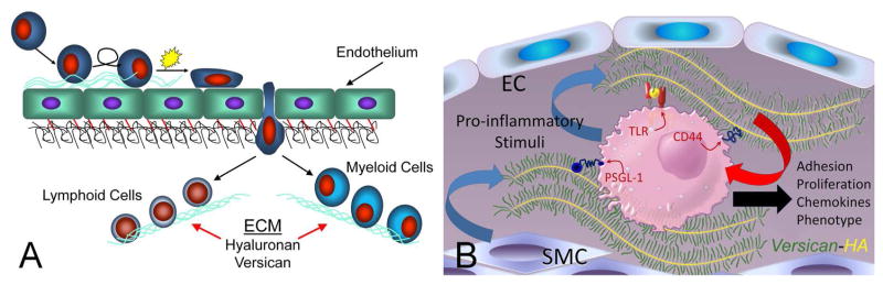
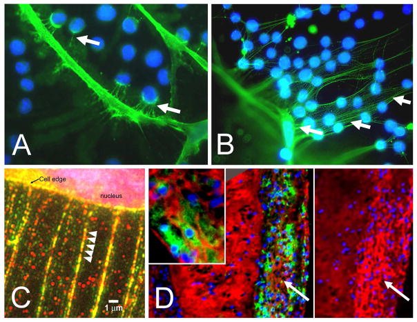
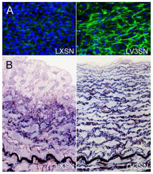
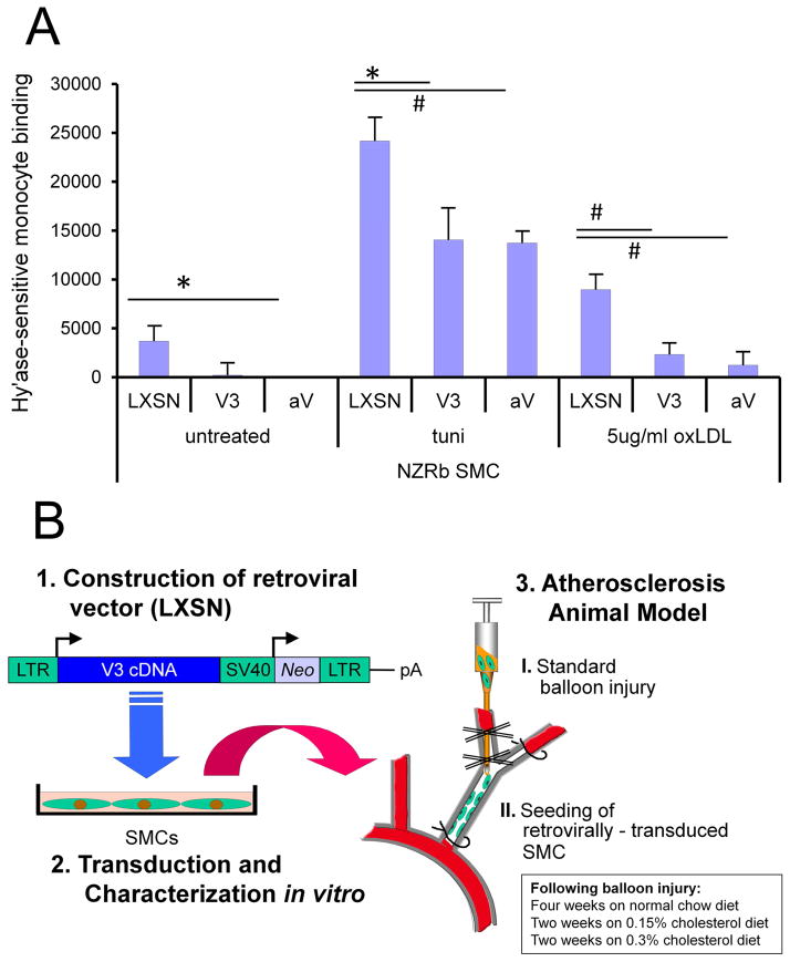
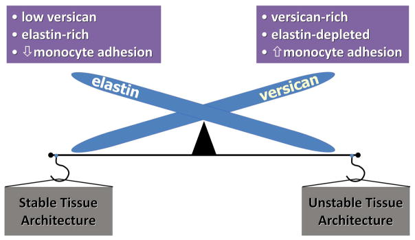
Versican is an extracellular matrix (ECM) proteoglycan that interacts with cells by binding to non-integrin and integrin receptors and to other ECM components that associate with the cell surface. Recent studies have shown also that versican interacts with myeloid and lymphoid cells promoting their adhesion and production of inflammatory cytokines. Versican is produced by stromal cells, as well as leukocytes, and is markedly increased in inflammation. Inflammatory agonists, such as double-stranded RNA mimetics (e.g., poly I:C), stimulate stromal cells, smooth muscle cells and fibroblasts, to produce fibrillar ECMs enriched in versican and hyaluronan (HA) that interact with leukocytes promoting their adhesion. Interference with the incorporation of versican into this ECM blocks monocyte adhesion and dampens the inflammatory response. Tumor cells also express elevated levels of versican which interact with myeloid cells to promote an inflammatory response, through stimulating cytokine release, and metastasis. In addition, myeloid cells, such as macrophages in tumors, synthesize versican which affects tumor cell phenotypes, inflammation, and subsequent metastasis. Versican, by binding to hyaluronan, influences T lymphocyte phenotypes and in part controls the ability of these cells to synthesize and secrete cytokines that influence the immune response. Collectively, these studies indicate that versican as an ECM molecule plays a central role in inflammation and as a result it is emerging as a potential target promising wide therapeutic benefits.

Keywords: Hyaluronan; Immunity; Inflammation; Macrophages; T lymphocytes; Versican.


Figures

Figure 1
Figure 1
A model of the different isoforms generated by alternative splicing of the mRNA transcript for versican. All isoforms interact with hyaluronan and thus are capable of forming different sized versican-HA aggregates. Different colors denote specific domains in the gene and in the protein product. Purple = hyaluronan binding region (HABR); yellow = α GAG exon and protein product; red = β GAG exon and protein product; green = two epidermal growth factor repeats (EE), a lectin binding domain (L) and a complement regulatory region. Inflammatory molecules that bind to different domains of versican are shown in the boxes to the right, marked in red. Those marked in blue indicate macrophage responses to treatment with versican. Structure of the CS GAG is shown at the bottom in blue with red dots denoting negatively charged residues. Reprinted (with modifications) from Current Opin Cell Biol, 14(5), Thomas N. Wight, Versican: a versatile extracellular matrix proteoglycan in cell biology, pp. 617–23, Copyright (2002), with permission from Elsevier.
Figure 2
Figure 2
A. Extravasation of myeloid and lymphoid cells across the endothelium into the underlying tissue where they interact with the ECM enriched in versican and HA. (B. ECM and immune cell regulation. Leukocytes come into contact with the ECM as they invade tissue as part of the inflammatory phase of tissue repair. Certain types of ECMs, including those that contain versican and hyaluronan, interact with myeloid and lymphoid cells through specific cell surface receptors such as, PSGL-1, TLR2, and CD44 to promote their adhesion, accumulation, and activation. Such matrices may exhibit either pro- or anti-inflammatory properties. These versican-enriched ECMs may be produced by either stromal cells or the myeloid and lymphoid cells themselves. Image shown in B kindly provided by Dr. Charles W. Frevert, University of Washington, Seattle, WA. Panel A is from Wight TN, “The biomatrix of the vascular system and the control of cell phenotype,” Hyaluronan: From Basic Science to Clinical Applications, vol 5, Structure and Function of Biomatrix: Control of Cell Behavior and Gene Expression, Balazs, EA, Editor. Matrix Biology Institute, Edgewater, NJ, 2012, pp. 315–340. Reprinted with permission of the Matrix Biology Institute.
Figure 3
Figure 3
A. Cell processes of a cultured human lung fibroblast stained for HA (green) with U937 monocytes added (blue nuclei) showing interaction of the monocytes with the fibroblast surface by HA-positive fibrils. B. Increased adhesion of monocytes to HA-positive fibrils of lung fibroblasts treated with poly I:C. C. Poly I:C-treated fibrobasts stained for HA (red) and versican (green) showing that the strands emanating from the cell surface contain both HA and versican (A–C, from Evanko et al., 2009). D. A section from an atherosclerotic lesion in a Watanabe rabbit immunostained for macrophages (green) and affinity-stained for HA (red). Inset of the area indicated by the arrow shows that the HA is organized into cables. Panel to the right is the adjacent section immunostained for versican. These images show that macrophages accumulate in regions of the lesion that contain both hyaluronan and versican, but not in regions devoid of versican (Sakr and Wight, unpublished observation).
Figure 4
Figure 4
A. Cultures of ASMCs transduced by an empty retroviral vector (LXSN) or one that contains the V3 gene (LV3SN) and immunostained for tropoelastin (green). Figure kindly supplied by Michel Gooden and Robert Vernon. B. A section from a balloon-injured carotid artery seeded with ASMCs transduced with an empty retroviral vector (LXSN; left panel) or seeded with ASMCs transduced with a retroviral vector containing the V3 gene (LV3SN; right panel). Multiple laminae of elastic fibers are seen in the vessel seeded with the V3-transduced cells. (From Merrilees M.J. et al, 2011).
Figure 5
Figure 5
A. Monocyte adherence in vitro. Hyaluronidase sensitive (U937) monocyte binding to 21-day cultures of empty vector control (LXSN) and V3 ASMC in untreated cultures, and cultures stimulated with 5 μg/mL tunicamycin (tuni) or 5 μg/mL oxLDL for 20 hours, showing reduced binding to matrix generated by V3-expressing ASMC. Degree of binding of monocytes in the V3-expressiong cultures was similar that when versican expression was blocked by versican antisense (AV). B. Experimental protocol to test impact of V3 expression on lipid and macrophages in an animal model of atherosclerosis. Rabbit ASMCs were transduced with a retroviral vector containing the V3 gene and then seeded into balloon-injured rabbit carotid artery. The animals were then placed on a normal chow diet for 4 weeks and then a cholesterol-enriched diet for an additional 4 weeks. The carotid arteries were then processed for histological analysis using histomorphometric measures for evidence of lipid and macrophage accumulation. (From Merrilees M.J. et al, 2011).
Figure 6
Figure 6
Cross section of vector control (A and C) and V3-seeded (B and D) carotid arteries of rabbits fed a high fat diet after balloon-injury and cell seeding. All sections have been immunostained for macrophages (brown). Control vessels (A & C) shows extensive macrophage accumulation, while V3 cell-seeded vessel is devoid of macrophages. (From Merrilees M.J. et al, 2011).
Figure 7
Figure 7
Reciprocal relationship of versican, elastin, and inflammation. Organs such as skin, lung, and blood vessels that are enriched in elastic fibers are low in versican and resistant to monocyte accumulation, leading to a stable tissue architecture with anti-inflammatory properties. When tissues from these organs are enriched in versican, as occurs in disease, they are low in elastic fibers, and show increased macrophage accumulation and inflammation, leading to unstable tissue architecture.

References

    1. Adair-Kirk TL, Senior RM. Fragments of extracellular matrix as mediators of inflammation. Int J Biochem Cell Biol. 2008;40:1101–1110. - PMC - PubMed
    1. Apte SS. A disintegrin-like and metalloprotease (reprolysin-type) with thrombospondin type 1 motif (ADAMTS) superfamily: functions and mechanisms. J Biol Chem. 2009;284:31493–31497. - PMC - PubMed
    1. Arroyo AG, Iruela-Arispe ML. Extracellular matrix, inflammation, and the angiogenic response. Cardiovasc Res. 2010;86:226–235. - PMC - PubMed
    1. Asplund A, Friden V, Stillemark-Billton P, Camejo G, Bondjers G. Macrophages exposed to hypoxia secrete proteoglycans for which LDL has higher affinity. Atherosclerosis. 2011;215:77–81. - PubMed
    1. Asplund A, Stillemark-Billton P, Larsson E, Rydberg EK, Moses J, Hulten LM, Fagerberg B, Camejo G, Bondjers G. Hypoxic regulation of secreted proteoglycans in macrophages. Glycobiology. 2009;20:33–40. - PubMed

Publication types

MeSH terms