

Short-term effects of an endotoxin on substantia nigra dopamine neurons
- PMID: 24513404
- PMCID: PMC3970422
- DOI: 10.1016/j.brainres.2014.02.005
Short-term effects of an endotoxin on substantia nigra dopamine neurons
Abstract
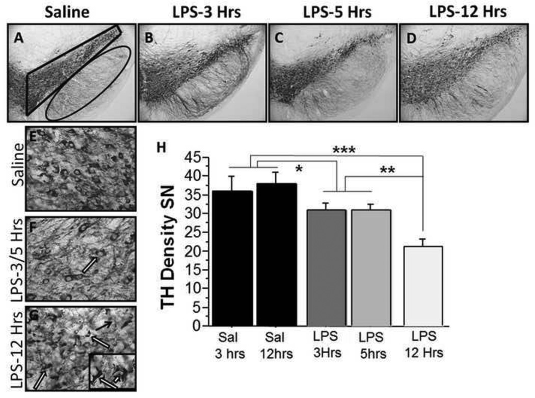
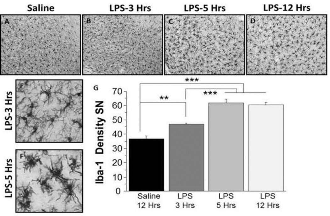
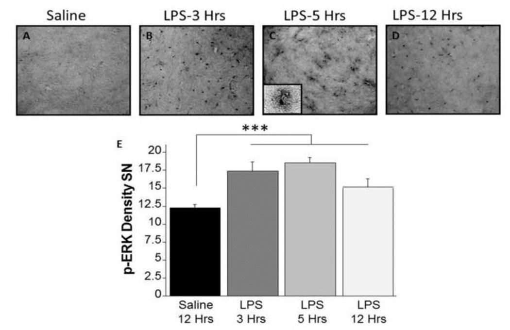
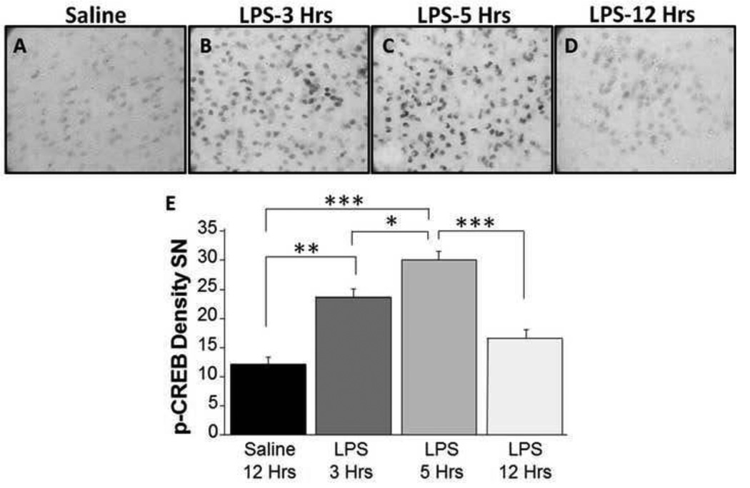
Inflammation has been implicated in the pathology of several neurodegenerative diseases, including Parkinson׳s disease (PD). Studies using the endotoxin lipopolysaccharide (LPS), a potent inflammogen, show that systemic insults can trigger prolonged microglial activation and pro-inflammatory cytokine production leading to degeneration of substantia nigra (SN) dopamine (DA) neurons, mimicking idiopathic PD. Because rapid effects of LPS on SN neurons had not been investigated previously, the focus of this study is to assess time-dependent alterations in SN neuroinflammation, DAergic neurons, and neuronal signaling cascades following LPS administration. LPS (5mg/kg, i.p.) or saline (0.9% NaCl) was administered to 8-month-old male mice. At 3h, 5h, and 12h post-injection, the morphology of the SN was assessed using antibodies directed against tyrosine hydroxylase (TH, DAergic marker), Iba-1 (pan-microglial marker), phospho-ERK, and phospho-CREB (signaling). LPS administration significantly reduced TH-immunoreactivity (ir) at all time-points with the greatest reduction observed at 12h post-injection. Reduced TH-ir was accompanied by a significant increase in activated microglia at all time-points following LPS. By 12h post-injection, LPS-treated mice exhibited activated as well as reactive microglia, which can result in neuronal damage. These data demonstrate that the initial reduction in TH-ir observed after an LPS injection was not concomitant with morphological alterations in microglial cells, even though a significant increase in phospho-ERK was observed in glial cells as soon as 3h post-injection. It is possible that the initial alteration in DA phenotype (TH reduction) may perpetuate an inflammatory response that persists and leads to further DAergic damage.
Keywords: Dopamine; Inflammation; Lipopolysaccharide; Substantia nigra.
Published by Elsevier B.V.
Figures
Similar articles
-
Endotoxin induces a delayed loss of TH-IR neurons in substantia nigra and motor behavioral deficits.Neurotoxicology. 2008 Sep;29(5):864-70. doi: 10.1016/j.neuro.2008.02.014. Epub 2008 Mar 13. Neurotoxicology. 2008. PMID: 18471886 Free PMC article.
-
The acute and the long-term effects of nigral lipopolysaccharide administration on dopaminergic dysfunction and glial cell activation.Eur J Neurosci. 2005 Jul;22(2):317-30. doi: 10.1111/j.1460-9568.2005.04220.x. Eur J Neurosci. 2005. PMID: 16045485
-
Human neuromelanin induces neuroinflammation and neurodegeneration in the rat substantia nigra: implications for Parkinson's disease.Acta Neuropathol. 2008 Jul;116(1):47-55. doi: 10.1007/s00401-008-0361-7. Epub 2008 Mar 15. Acta Neuropathol. 2008. PMID: 18343932
-
Anti-inflammatory effects of BHBA in both in vivo and in vitro Parkinson's disease models are mediated by GPR109A-dependent mechanisms.J Neuroinflammation. 2015 Jan 17;12:9. doi: 10.1186/s12974-014-0230-3. J Neuroinflammation. 2015. PMID: 25595674 Free PMC article.
-
The endotoxin hypothesis of neurodegeneration.J Neuroinflammation. 2019 Sep 13;16(1):180. doi: 10.1186/s12974-019-1564-7. J Neuroinflammation. 2019. PMID: 31519175 Free PMC article. Review.
Cited by
-
The role of inflammation in core features of depression: Insights from paradigms using exogenously-induced inflammation.Neurosci Biobehav Rev. 2018 Nov;94:219-237. doi: 10.1016/j.neubiorev.2018.09.006. Epub 2018 Sep 7. Neurosci Biobehav Rev. 2018. PMID: 30201219 Free PMC article. Review.
-
Vagus nerve stimulation improves locomotion and neuronal populations in a model of Parkinson's disease.Brain Stimul. 2017 Nov-Dec;10(6):1045-1054. doi: 10.1016/j.brs.2017.08.008. Epub 2017 Aug 24. Brain Stimul. 2017. PMID: 28918943 Free PMC article.
-
Associations between inflammation and striatal dopamine D2-receptor availability in aging.J Neuroinflammation. 2025 Jan 30;22(1):24. doi: 10.1186/s12974-025-03355-0. J Neuroinflammation. 2025. PMID: 39885603 Free PMC article.
-
Profiling Inflammatory Extracellular Vesicles in Plasma and Cerebrospinal Fluid: An Optimized Diagnostic Model for Parkinson's Disease.Biomedicines. 2021 Feb 25;9(3):230. doi: 10.3390/biomedicines9030230. Biomedicines. 2021. PMID: 33669043 Free PMC article.
-
Inhibition of semicarbazide-sensitive amine oxidase/vascular adhesion protein-1 reduces lipopolysaccharide-induced neuroinflammation.Br J Pharmacol. 2017 Jul;174(14):2302-2317. doi: 10.1111/bph.13832. Epub 2017 Jun 10. Br J Pharmacol. 2017. PMID: 28437839 Free PMC article.
References
-
- Banati RB, Daniel SE, Blunt SB. Glial pathology but absence of apoptotic nigral neurons in long-standing Parkinson's disease. Mov Disord. 1998;13(2):221–227. - PubMed
-
- Boger HA, Middaugh LD, Huang P, Zaman V, Smith AC, Hoffer BJ, Tomac AC, Granholm AC. A partial GDNF depletion leads to earlier age-related deterioration of motor function and tyrosine hydroxylase expression in the substantia nigra. Exp Neurol. 2006;202(2):336–347. - PubMed
-
- Boger HA, Middaugh LD, Patrick KS, Ramamoorthy S, Denehy ED, Zhu H, Pacchioni AM, Granholm AC, McGinty JF. Long-term consequences of methamphetamine exposure in young adults are exacerbated in glial cell line-derived neurotrophic factor heterozygous mice. J Neurosci. 2007;27(33):8816–8825. - PMC - PubMed
-
- Castano A, Herrera AJ, Cano J, Machado A. Lipopolysaccharide intranigral injection induces inflammatory reaction and damage in nigrostriatal dopaminergic system. Journal of Neurochemistry. 1998;70(4):1584–1592. - PubMed
-
- Fiedorowicz A, Figiel I, Kamińska B, Zaremba M, Wilk S, Oderfeld-Nowak B. Dentate granule neuron apoptosis and glia activation in murine hippocampus induced by trimethyltin exposure. Brain Res. 2001;912(2):116–127. - PubMed
Publication types
MeSH terms
Substances
Grants and funding
LinkOut - more resources
Full Text Sources
Other Literature Sources
Miscellaneous
