Review
. 2014 May;1840(5):1554-65.
doi: 10.1016/j.bbagen.2013.10.032. Epub 2013 Oct 26.

Aquaporin and brain diseases

Affiliations
Review

Aquaporin and brain diseases

Jérôme Badaut et al. Biochim Biophys Acta. 2014 May.

Abstract

Background: The presence of water channel proteins, aquaporins (AQPs), in the brain led to intense research in understanding the underlying roles of each of them under normal conditions and pathological conditions.

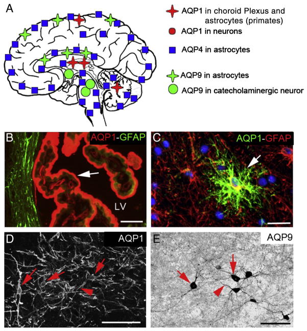
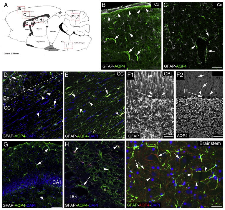
Scope of review: In this review, we summarize some of the recent knowledge on the 3 main AQPs (AQP1, AQP4 and AQP9), with a special focus on AQP4, the most abundant AQP in the central nervous system.

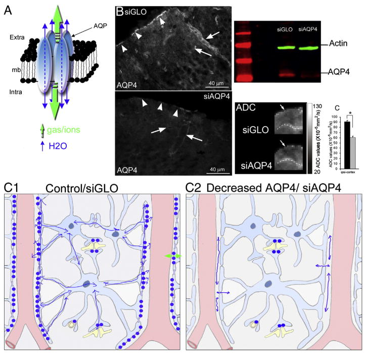
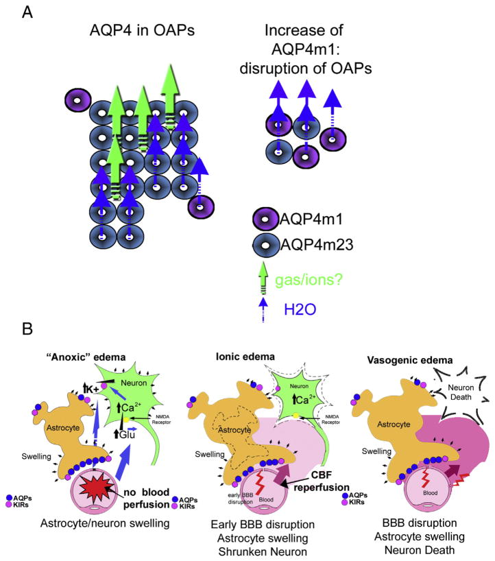
Major conclusions: AQP4 was most studied in several brain pathological conditions ranging from acute brain injuries (stroke, traumatic brain injury) to the chronic brain disease with autoimmune neurodegenerative diseases. To date, no specific therapeutic agents have been developed to either inhibit or enhance water flux through these channels. However, experimental results strongly underline the importance of this topic for future investigation. Early inhibition of water channels may have positive effects in prevention of edema formation in brain injuries but at later time points during the course of a disease, AQP is critical for clearance of water from the brain into blood vessels.

General significance: Thus, AQPs, and in particular AQP4, have important roles both in the formation and resolution of edema after brain injury. The dual, complex function of these water channel proteins makes them an excellent therapeutic target. This article is part of a Special Issue entitled Aquaporins.

Keywords: Blood–brain barrier; Edema; Neuroimaging; Neuroinflammation; Neurovascular unit; Water channel.


Figures

Fig. 1
Fig. 1
Aquaporin 1, 4 and 9 distributions in the brain. (A) Schematic drawing of aquaporin 1, 4 and 9 distributions in the brain. AQP1 is mainly observed in the choroid plexus and in some neurons. AQP1 is present in some astrocyte in primates. AQP4 is present in astrocytes in all brain structures with different levels and patterns of expression (see Fig. 2). AQP9 is mostly present in astrocytes and catecholaminergic neurons. (B) AQP1 immunostaining (red) in choroid plexus epithelium (arrow) located in the lateral ventricle (LV). The border of the LV is outlined by the GFAP staining (green), specific marker of the astrocytes. (C) AQP1 staining (green) in monkey cerebellum is co-localized (arrow) with GFAP staining (in red), suggesting the presence of AQP1 in a sub-population of astrocytes in primates. (D) AQP1 labeling is present in some neurons of the septum in the rat brain. (E) AQP9 immunoreactivity in catecholaminergic neurons of the ventral tegmental area. Bars: B, D = 50 μm; C, D = 40 μm.
Fig. 2
Fig. 2
Variety in astrocyte AQP4 distribution. (A) Sagittal drawing of rat brain with the location of the pictures of AQP4 distribution in different brain areas. (B) AQP4 labeling (green) in the parietal cortex (Cx) is abundant on the glia limitans (arrow heads), revealed in gray by the GFAP staining. The AQP4 labeling is underlining the blood vessels (arrows) by its concentration in the astrocyte endfeet stained by the anti-GFAP (gray). (C) AQP4 distribution in the deeper cortex layer showed the “polarization” of the AQP4 labeling around the blood vessels (arrows). The double staining of AQP4 and GFAP exhibits its presence on the astrocyte endfeet (arrows). (D) AQP4 staining (green) in the corpus callosum (CC) is abundant around the blood vessel (arrows) and at distance from the perivascular space (arrowheads). In the white matter structure, AQP4 exhibits a different pattern of staining, with distribution of the protein on the astrocyte processes (gray, GFAP staining), possibly in association with node of Ranvier. (E) AQP4 staining (green) at higher magnification in the corpus callosum (CC) shows the co-localization with GFAP staining (gray, arrows and arrowheads). In contrast with the Cx, the AQP4 staining is spread in all the structure with “patchy” distribution, following the direction of the neuronal processes. (F1, F2) GFAP (F1) and AQP4 (F2) staining in the cerebellum differs from the cortex, with an abundant staining around all the neurons of the granular layer as well as around the Purkinje cell bodies (P, arrowheads). AQP4 is observed around the blood vessels in the molecular layer, where the radial glia is located. (G) AQP4 labeling (green) in the location of CA1 of the hippocampus outlines the blood vessels but it is also present in astrocyte processes in the stratum radiatum layer. (H) AQP4 labeling (green) in proximity of the dentate gyrus (DG) in the hippocampus. AQP4 is around the blood vessels (arrow) and in astrocyte processes remote of the perivascular space (arrowheads). (I) AQP4 labeling (red) shows staining around the neuronal cell bodies (arrows) as well as in contact with blood vessels (arrowheads). AQP4 (red) is co-localized with the GFAP staining (green). Bars: B, C, D, E, H = 50 μm; F1, F2 = 40 μm; G = 100 μm; I = 25 μm.
Fig. 3
Fig. 3
AQP4 water diffusion in the brain. (A) Schematic drawing of the aquaporin homo-tetramer assembly within the lipid membrane resulting in a central pore permeable to cations and gases (green arrows). Each individual aquaporin facilitates bi-directional water movement depending on the osmotic gradient (blue arrows, adapted from [33]). (B) AQP4 labeling in siGLO and siAQP4 treated rats showed a significant decrease in the intensity of AQP4 staining in the glia limitans and perivascular astrocyte in the cortex of siAQP4 treated rats. AQP4 Western blot (red) showed a significant decrease of the intensity in siAQP4 compared to siGLO treated rats. Decrease of the AQP4 is associated with a decrease of the water diffusion. Enlarged ADC images focusing on the contralateral cortex showed the decrease in ADC in siAQP4 compared to siGLO treated rats. The quantification showed a 50% decrease of the ADC signals where the AQP4 expression is decreased by 30% (modified from [35]). (C) Schema depicting the location of the APQ4 in brain cortex in control/siGLO condition (C1) and after siAQP4 injection (C2). The presence of AQP4 facilitates the water movement in the astrocyte network (C1), and after silencing APQ4 (C2), lower ADC values are caused by decreased water permeability due to a decreased number of AQP4 channels in perivascular space (C2).
Fig. 4
Fig. 4
AQP4 expression in spinal cord in normal and EAE rats. The co-staining AQP4 (A1, B1, in red A3, B3) and GFAP (A2, B2, in green A3, B3) showed in the spinal cord the abundant presence of AQP4 in astrocytes around the blood vessel and in processes (arrowheads, A3, B3). Additional positive AQP4 staining is observed in the astrocyte cell bodies (arrows, A3, B3). As described in literature, AQP4 expression is dramatically increased in the spinal cord of the EAE rats (B) compared to the control (CTL, A). Bars: A, B = 50 μm.
Fig. 5
Fig. 5
Disorganization of AQP4 in orthogonal particles after brain injuries and edema process. (A) AQP4-m1 (purple circles) and AQP4-m23 (blue circles) isoforms contribute together to form orthogonal array particles (OAPs) in astrocyte endfeet in contact with the blood vessels. It was previously shown that higher expression of AQP4-m23 contributes to the formation of large OAPs. However, increase of AQP4-m1 induced disruption of OAPs with a reduction of the size. This modification is observed in pathological conditions such as stroke. Recent knowledge on AQP leads us to hypothesize that the large OAPs contribute to gas and cation diffusion in the astrocyte membranes through central pores (green arrows) (modified from [33]). (B) Schematic drawing of the events happening during edema formation with 3 different edema phases: anoxic, ionic and vasogenic edema. During the injury with decrease of brain perfusion, the first minutes are characterized by anoxic edema. Anoxic edema is characterized as a swelling of the astrocytes and the neuronal dendrites caused by a disruption of the cellular ionic gradients and the entry of ions followed by water entry and leading to cellular swelling. During the ionic edema, astrocytes become swollen and neuronal death starts to occur resulting in shrinkage of the neurons, shear stress and endothelial dysfunctions on the non-perfused vascular tree, which results in early transient leakage of the BBB. Vasogenic edema is a result of disruption of the tight junctions between the endothelial cells, leading to increased permeability of the cerebral blood-vessels to albumin and other plasma proteins, further contributing to swelling of astrocytes and subsequent neuronal cell death (adapted from [33]).

References

    1. Gonen T, Walz T. The structure of aquaporins. Q Rev Biophys. 2006;39:361–396. - PubMed
    1. Badaut J, Lasbennes F, Magistretti PJ, Regli L. Aquaporins in brain: distribution, physiology, and pathophysiology. J Cereb Blood Flow Metab. 2002;22:367–378. - PubMed
    1. Badaut J, Nehlig A, Verbavatz J, Stoeckel M, Freund-Mercier MJ, Lasbennes F. Hypervascularization in the magnocellular nuclei of the rat hypothalamus: relationship with the distribution of aquaporin-4 and markers of energy metabolism. J Neuroendocrinol. 2000;12:960–969. - PubMed
    1. Yang M, Gao F, Liu H, Yu WH, Sun SQ. Temporal changes in expression of aquaporin-3, -4, -5 and -8 in rat brains after permanent focal cerebral ischemia. Brain Res. 2009;1290:121–132. - PubMed
    1. Jung JS, Bhat RV, Preston GM, Guggino WB, Baraban JM, Agre P. Molecular characterization of an aquaporin cDNA from brain: candidate osmoreceptor and regulator of water balance. Proc Natl Acad Sci U S A. 1994;91:13052–13056. - PMC - PubMed

Publication types