Review
. 2014 Aug;1843(8):1433-41.
doi: 10.1016/j.bbamcr.2013.10.013. Epub 2013 Oct 24.

Co-translational protein targeting to the bacterial membrane

Affiliations
Review

Co-translational protein targeting to the bacterial membrane

Ishu Saraogi et al. Biochim Biophys Acta. 2014 Aug.

Abstract

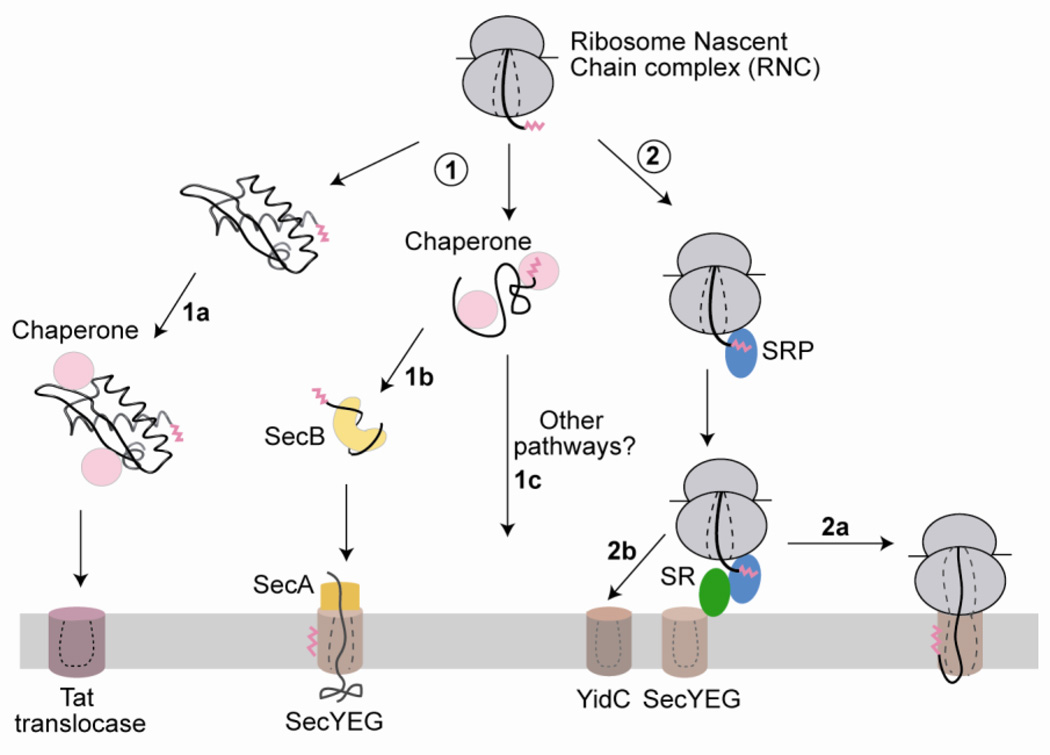
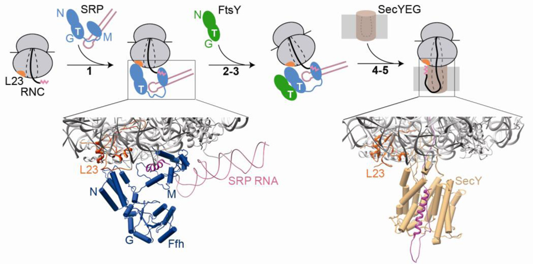
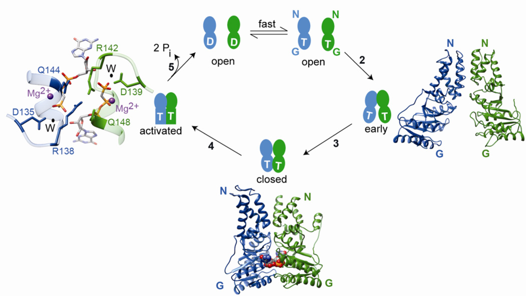
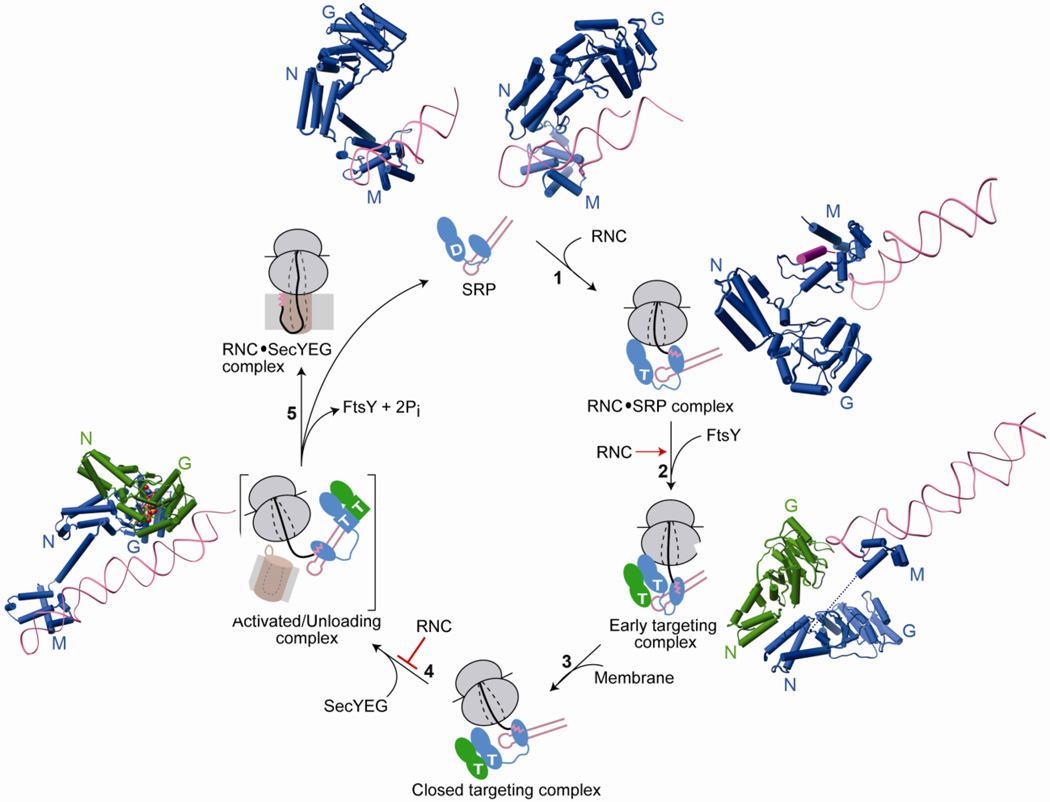
Co-translational protein targeting by the Signal Recognition Particle (SRP) is an essential cellular pathway that couples the synthesis of nascent proteins to their proper cellular localization. The bacterial SRP, which contains the minimal ribonucleoprotein core of this universally conserved targeting machine, has served as a paradigm for understanding the molecular basis of protein localization in all cells. In this review, we highlight recent biochemical and structural insights into the molecular mechanisms by which fundamental challenges faced by protein targeting machineries are met in the SRP pathway. Collectively, these studies elucidate how an essential SRP RNA and two regulatory GTPases in the SRP and SRP receptor (SR) enable this targeting machinery to recognize, sense and respond to its biological effectors, i.e. the cargo protein, the target membrane and the translocation machinery, thus driving efficient and faithful co-translational protein targeting. This article is part of a Special Issue entitled: Protein trafficking and secretion in bacteria. Guest Editors: Anastassios Economou and Ross Dalbey.

Keywords: GTPases; Molecular recognition and regulation; Protein targeting; Ribosome; SecYEG.


Figures

Fig. 1
Fig. 1
A schematic depiction of various targeting pathways for delivering proteins to the bacterial inner membrane. Newly synthesized proteins with N-terminal targeting sequences (magenta) can be targeted either post-translationally (route 1) or co-translationally (route 2). Post-translational targeting (route 1) involves targeting of the nascent protein either in a fully folded state via the Tat pathway (1a) or in an unfolded state via the chaperone SecB and the ATPase SecA (1b). Both pathways may also involve general chaperones (pink) that maintain the proteins in a translocation-competent state. The co-translational targeting pathway (route 2), which primarily handles inner membrane proteins in bacteria, is mediated by the signal recognition particle (SRP, blue) and its receptor (SR, green) (2a). Both SecA (yellow) and SRP deliver proteins to the SecYEG protein-conducting channel and may co-operate in the translocation of membrane proteins with large periplasmic domains. Translating ribosomes may also be directly delivered to the YidC translocase (2b), which may either act independently or in conjunction with SecYEG. Whether additional pathways exist for the targeting of substrates, such as tail-anchored proteins, remains to be determined (1c). The same color scheme is maintained throughout the paper.
Fig. 2
Fig. 2
An overview of co-translational protein targeting by the bacterial SRP. Step 1: a ribosome-nascent chain complex (RNC) displaying an SRP signal sequence (magenta) is recognized by SRP, primarily via interactions of the SRP N-domain with the ribosomal protein L23 (orange), and the SRP M-domain with the signal sequence. The lower panel shows a molecular model of the RNC-SRP complex, derived from docking the individual crystal structures of the ribosome (grey) and SRP into a cryo-electron microscopy reconstruction of the complex (PDB ID: 2j28). For clarity, only the region near the ribosome exit site (boxed in the cartoon) is shown. Steps 2–3: binding of cargo-loaded SRP to the SRP receptor (FtsY), via their homologous NG domains, localizes this complex to the membrane. Steps 4–5: the translating ribosome is transferred to the SecYEG protein-conducting channel (brown) at the membrane, which binds to the same sites on the RNC as the SRP. The lower panel shows a molecular model of RNC bound to SecYEG derived from docking the individual crystal structures of the ribosome and a homology model of SecYEG into a cryo-electron microscopy reconstruction of the complex (PDB ID: 3j00/3j01). The steps are numbered to be consistent with Figs 3 and 4.
Fig. 3
Fig. 3
SRP and SR are multi-state regulatory GTPases that undergo a series of conformational changes during their GTPase cycle. For clarity, only the NG-domains of SRP and SR are shown. T and D represent GTP and GDP, respectively. Under cellular conditions, nucleotide exchange on free SRP and SR is rapid and the proteins exist predominantly in the GTP-bound state. Step 2: SRP and SR GTPases first associate to form an early intermediate, which primarily involves interactions between the two N-domains. The right panel shows a molecular model for the early complex (PDB ID: 2xkv). Step 3: the G-domains of both proteins gain closer approach to one another, forming a closed complex with an extensive binding interface. The bottom panel shows a co-crystal structure of the SRP-FtsY NG domain complex (PDB ID: 1rj9) in the closed/activated conformation. The non-hydrolyzable GTP analog GMPPCP is shown in space filling model. Step 4: rearrangement of the IBD loops optimizes the position of catalytic residues relative to GTP, generating the activated conformation for efficient GTP hydrolysis. The left panel shows a magnification of the composite active site at the dimer interface for GTPase activation. The active site Mg2+ is in magenta, nucleophilic water (W) is in black and the catalytic residues of SRP (blue) and SR (green) are indicated. Step 5: GTP hydrolysis drives the disassembly and recycling of SRP and SR. The steps are numbered to be consistent with Figures 2 and 4.
Fig. 4
Fig. 4
Conformational changes in SRP and SR GTPases are coupled to global reorganization of the SRP particle and are regulated by biological effectors for the pathway. Free SRP exists in a number of conformations in which the NG-domain of Ffh is oriented differently with respect to the M-domain and the SRP RNA. The top panel shows structures of SRP from S. solfataricus (PDB ID: 1qzw, left) and M. jannaschii (PDB ID: 2v3c, right) highlighting its conformational flexibility. The binding of RNC to SRP favors an SRP conformation in which the tetraloop of the SRP RNA is poised to interact with the G-domain of SR (step 1). This interaction strongly stabilizes the early targeting complex resulting in very efficient assembly of this complex (step 2). Top right (PDB ID: 2j28) and bottom right (PDB ID: 2xkv) panels show molecular models of the interaction of RNC with SRP without or with FtsY. Anionic phospholipids in the membrane strongly accelerate the rearrangement of the early targeting complex to the closed state (step 3). Interaction with SecYEG induces the SRP/SR complex into the activated state (step 4) in which the NG-domain complex relocalizes to the distal end of SRP RNA (left panel, PDB ID: 2xxa). This movement is negatively regulated by the RNC allowing a productive search for the translocon. The activated complex is shown in brackets to indicate that it is a proposed intermediate with transient lifetime, and its precise structure is not known. Hydrolysis of GTP triggers disassembly of the GTPase complex while the cargo is transferred to the translocon (step 5).

References

    1. Blobel G. Intracellular Protein Topogenesis. Proc. Nat. Acad. Sci. USA. 1980;77:1496–1500. - PMC - PubMed
    1. Protein targeting, transport and translocation. London: Academic Press; 2002.
    1. Fekkes P, Driessen AJM. Protein targeting to the bacterial cytoplasmic membrane. Microbiology and Molecular Biology Reviews. 1999;63:161–173. - PMC - PubMed
    1. Cross BCS, Sinning I, Luirink J, High S. Delivering proteins for export from the cytosol. Nat. Rev. Mol. Cell. Biol. 2009;10:255–264. - PubMed
    1. Hegde RS, Bernstein HD. The surprising complexity of signal sequences. Trends Biochem. Sci. 2006;31:563–571. - PubMed

Publication types

MeSH terms

LinkOut - more resources