. 2014 Jan 15;385(2):405-16.
doi: 10.1016/j.ydbio.2013.10.021.

A fate-map for cranial sensory ganglia in the sea lamprey

A fate-map for cranial sensory ganglia in the sea lamprey

Melinda S Modrell et al. Dev Biol. .

Abstract

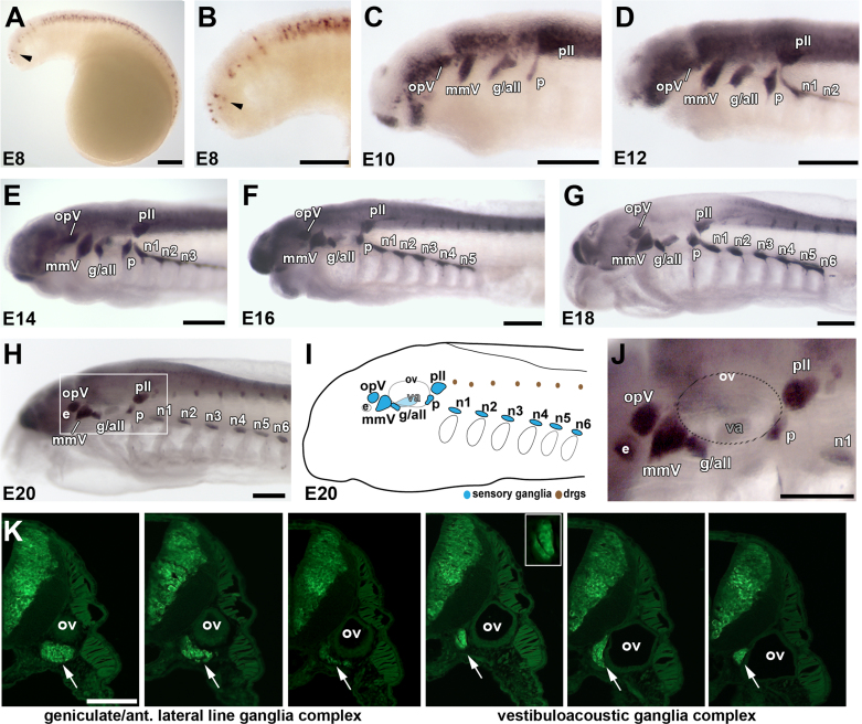
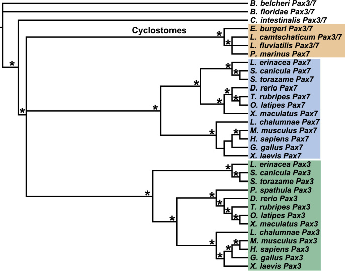
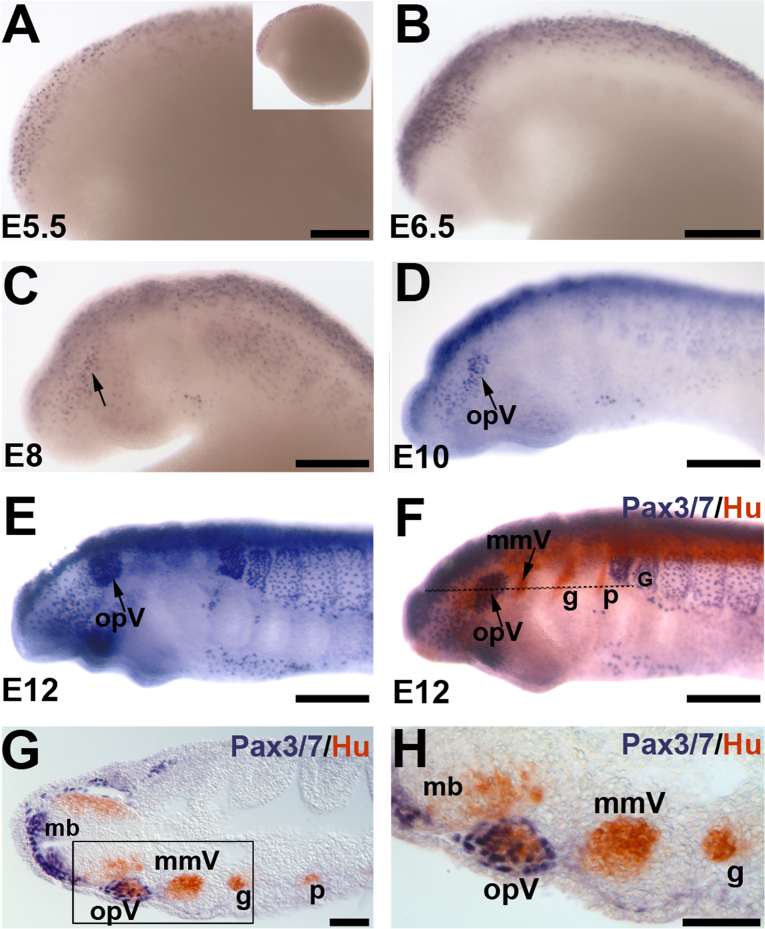
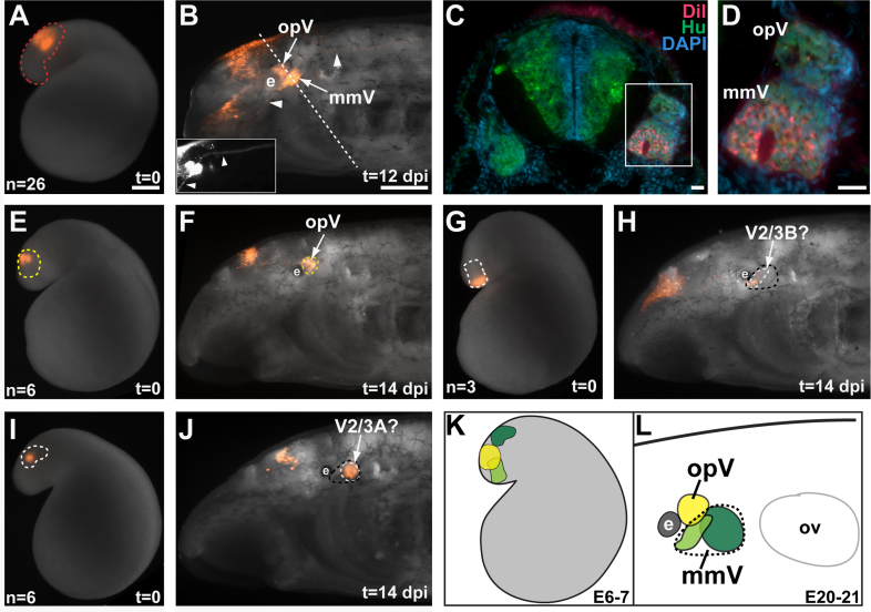
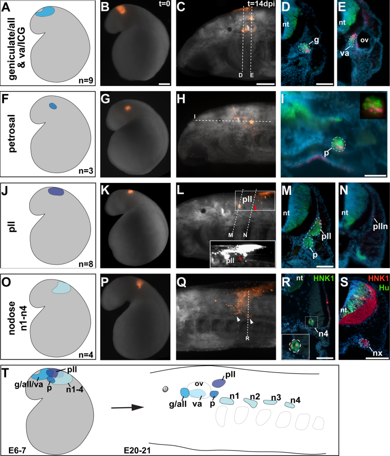
Cranial neurogenic placodes and the neural crest make essential contributions to key adult characteristics of all vertebrates, including the paired peripheral sense organs and craniofacial skeleton. Neurogenic placode development has been extensively characterized in representative jawed vertebrates (gnathostomes) but not in jawless fishes (agnathans). Here, we use in vivo lineage tracing with DiI, together with neuronal differentiation markers, to establish the first detailed fate-map for placode-derived sensory neurons in a jawless fish, the sea lamprey Petromyzon marinus, and to confirm that neural crest cells in the lamprey contribute to the cranial sensory ganglia. We also show that a pan-Pax3/7 antibody labels ophthalmic trigeminal (opV, profundal) placode-derived but not maxillomandibular trigeminal (mmV) placode-derived neurons, mirroring the expression of gnathostome Pax3 and suggesting that Pax3 (and its single Pax3/7 lamprey ortholog) is a pan-vertebrate marker for opV placode-derived neurons. Unexpectedly, however, our data reveal that mmV neuron precursors are located in two separate domains at neurula stages, with opV neuron precursors sandwiched between them. The different branches of the mmV nerve are not comparable between lampreys and gnatho-stomes, and spatial segregation of mmV neuron precursor territories may be a derived feature of lampreys. Nevertheless, maxillary and mandibular neurons are spatially segregated within gnathostome mmV ganglia, suggesting that a more detailed investigation of gnathostome mmV placode development would be worthwhile. Overall, however, our results highlight the conservation of cranial peripheral sensory nervous system development across vertebrates, yielding insight into ancestral vertebrate traits.


Figures

Fig. 1
Fig. 1
Spatiotemporal development of lamprey cranial sensory ganglia. Anterior is to the left for all whole-mount images. (A) and (B) Low-power (A) and higher-power view (B) of an embryo at embryonic day 8 (E8) immunostained for the pan-neuronal Elav-family members HuC/D (Hu). HuC/D expression is strong in neurons within the neural tube, with fainter expression in neurons lateral to the rostral neural tube (arrowhead). (C) By E10, discrete lateral patches of HuC/D expression reveal the primordia of all cranial sensory ganglia except the nodose: the ophthalmic trigeminal ganglion (opV), the maxillomandibular trigeminal ganglion (mmV), the geniculate/anterior lateral line ganglionic complex (g/all), the petrosal ganglion (p) and the posterior lateral line ganglion (pll). (D)–(G) HuC/D immunostaining of embryos at E12 (D), E14 (E), E16 (F) and E18 (G) shows the development of the six nodose ganglia in a rostrocaudal sequence dorsal to the branchial arches and the progressive condensation of all the cranial ganglia. Dorsal root ganglia are also visible from E16, adjacent to the dorsal neural tube. (H)–(J) By E20, all the cranial sensory ganglia have formed. (H) Low-power and (I) schematic view of an E20 embryo, showing the location of cranial sensory ganglia [blue in (I)] and dorsal root ganglia [brown in (I)]. (J) A higher-power view of the boxed area in (H), showing distinct opV and mmV ganglia, the geniculate/anterior lateral line ganglionic complex, the vestibuloacoustic ganglion (medial to the otic vesicle, hence hardly stained in whole-mount), the petrosal ganglion, the posterior lateral line ganglion and the most rostral nodose ganglion (n1). (K) Transverse serial sections immunostained for HuC/D (green), starting near the rostral edge of the otic vesicle [see panel (J) for orientation of otic vesicle, which is indicated by a dotted oval] and progressing caudally through the geniculate/aLL ganglionic complex ventral to the otic vesicle (arrow, left-hand three images) and then the vestibuloacoustic ganglion medial to the otic vesicle (arrow, right-hand three images). In the fourth and fifth images, the developing intracapsular ganglion (second ganglion of the anterior lateral line nerve) may also be visible, medial to the vestibuloacoustic ganglion and slightly separated from it by a thin HuC/D-negative space (inset). Abbreviations: all, anterior lateral line ganglion; drgs, dorsal root ganglia; e, eye; g, geniculate ganglion; mmV, maxillomandibular trigeminal ganglion; n, nodose ganglion; opV, ophthalmic trigeminal (profundal) ganglion; ov, otic vesicle; p, petrosal ganglion; pll, posterior lateral line ganglion; va, vestibuloacoustic ganglion. Scale bars: (A)–(J) 0.2 mm; (K) 50 μm.
Fig. 2
Fig. 2
Cyclostome Pax3/7 subfamily proteins form a well-supported clade, separate from gnathostome Pax3 and Pax7 clades. Maximum a posteriori (MAP) topology, obtained with Bali-Phy software, summarizing all of the phylogenetic analyses performed. Nodes consistently recovered with high support in all Bayesian and coalescence-based analyses are indicated by an asterisk (where the posterior probability is >90). These analyses consistently recovered three main clades of orthologous sequences: (i) a cyclostome clade containing all published hagfish and lamprey Pax3/7 proteins (orange box), (ii) a gnathostome Pax7 clade (blue box) and (iii) a gnathostome Pax3 clade (green box). Relationships between these three clades, however, remain unresolved: the gnathostome Pax3 and Pax7 clades formed a sister group in most analyses but with low support. Within the gnathostome clades, relationships are well resolved for the Pax3 but not the Pax7 subfamily: in some cases, short sequence lengths for Pax7 likely account for a topology that is incongruent with the currently proposed relationships among vertebrates. Generally, however, phylogenetic relationships recovered here are consistent with current understanding of the chordate phylogeny.
Fig. 3
Fig. 3
Pax3/7 is a specific marker for opV placode-derived neurons. Whole-mount immunostaining with the pan-Pax3/7 antibody DP312 (Davis et al., 2005) shows (A) Pax3/7 expression at E5.5 in the anterior-most dorsal neural tube; inset shows entire embryo. (B) By E6.5, more Pax3/7-positive cells appear along the length of the neural tube and in presumptive migrating neural crest cells adjacent to it. (C) At E8, Pax3/7 expression is seen in the neural tube, somites and scattered cells lateral to the neural tube (arrow). (D) By E10, a patch of Pax3/7-positive cells (arrow) is seen lateral to the neural tube in a similar position to the developing opV ganglion (compare with Fig. 1C). (E) By E12, Pax3/7 is strongly expressed in the presumptive opV placode/ganglion (opV, arrow); (F) this is confirmed by double immunostaining for Pax3/7 (blue) and the pan-neuronal marker HuC/D (red). Dotted line shows plane of section in (G) and (H). (G) Coronal section shows Pax3/7 expression in the dorsal neural tube and opV ganglion, but not in the mmV, geniculate or petrosal ganglia. (H) A higher-power view of the boxed region in (E) confirms that Pax3/7 expression is restricted to the developing opV ganglion (and dorsal neural tube cells). Abbreviations: g, geniculate ganglion; mb, midbrain; mmV, maxillomandibular trigeminal ganglion; opV, ophthalmic trigeminal placode/ganglion; p, petrosal ganglion. Scale bars: (A)–(F) 0.2 mm; (G) and (H) 50 μm.
Fig. 4
Fig. 4
Fate-mapping of opV and mmV placode-derived neurons reveals two separate domains of mmV neuron precursors at late neurula stages. Anterior is to the left for all whole-mount images. (A) An E6.5 embryo immediately after DiI injection into a broad patch of anterodorsal head ectoderm within the region outlined in red, which contains both opV and mmV neuron precursors. (B) The same embryo as in (A) at E18 (t=12 dpi), showing DiI labeling in the opV and mmV ganglia. DiI is also visible in the upper lip-innervating mmV nerve (V2/3A, inset, ventral arrowhead) and central projections from the mmV ganglion (inset, dorsal arrowhead). Dotted line shows plane of section in (C) and (D). (C) Merged images of low-power and (D) higher-power views of an oblique section through both the opV and mmV ganglia, immunostained for the neuronal marker HuC/D (green) and counterstained for the nuclear marker DAPI (blue), showing DiI (red) in surface ectoderm and also co-localized with neurons (HuC/D, green) in both the opV and mmV ganglia. (E) An E6.5 embryo immediately after focal DiI injection into the region of head ectoderm outlined in yellow, which contains opV neuron precursors. (F) The same embryo as in (E) at E20 (t=14 dpi), showing restriction of DiI to the opV ganglion. (G) An E6.5 embryo immediately after a focal DiI injection into the region of head ectoderm outlined in white (shaded light green in schematic). (H) The same embryo as in (G) at E20 (t=14 dpi), showing DiI localization to a relatively small, rostral domain (outlined in white) of the mmV ganglion (outlined in black), which may correspond to lower lip/velum-innervating V2/3B neurons (see text). (I) An E6.5 embryo immediately after a focal DiI injection into the region of head ectoderm outlined in white (shaded dark green in schematic). (J) The same embryo as in (I) at E20 (t=14 dpi), showing DiI localization to a larger, caudal domain (outlined in white) of the mmV ganglion (outlined in black), which may correspond to upper lip-innervating V2/3A neurons (see text). (K) Schematic fate-map at E6–7 summarizing the location of opV neuron precursors (yellow) between two separate patches of mmV neuron precursors (light and dark green). (L) Schematic summarizing the fate at E20–21 of DiI-injected cells within the locations shown in panel (K). The opV ganglion (yellow) lies dorsal to the mmV ganglion (dotted black outline). Rostral (light green) and caudal (dark green) subregions of the mmV ganglion are distinguishable in the E6–7 fate-map, which may correspond to V2/3B and V2/3A neurons, respectively (see text). Abbreviations: dpi, days post-injection; e, eye; mmV, maxillomandibular trigeminal ganglion; opV, ophthalmic trigeminal ganglion; ov, otic vesicle; t, time, V2/3A, upper lip-innervating trigeminal neurons; V2/3B, lower lip/velum-innervating trigeminal neurons. Scale bars: (A), (E), (G) and (I) 0.2 mm; (B), (F), (H) and (J) 0.2 mm; (C) and (D) 10 μm.
Fig. 5
Fig. 5
Fate-maps at E6-7 for lamprey epibranchial and lateral line placode-derived ganglia. (A)–(E) Ectoderm in the colored region in (A) was fated to contribute to neurons in the geniculate/anterior lateral line (aLL) ganglionic complex, and in 7/9 cases, also to the vestibuloacoustic/intracapsular ganglionic complex. (B) An embryo shortly after DiI injection at E6.5 (t=0) in the region shown in (A). (C) The same embryo as in (B), at E20 (t=14 dpi), with DiI visible in the geniculate/aLL complex. Dotted lines indicate planes of section in (D) and (E). (D) and (E) Transverse sections through (D) the geniculate/aLL ganglionic complex and (E) the vestibuloacoustic ganglion, immunostained for the neuronal marker HuC/D (green) and counterstained with DAPI (blue), showing co-localization of DiI with HuC/D. (F)–(I) Ectoderm in the colored region in (F) was fated to contribute to neurons in the petrosal ganglion. (G) An embryo shortly after DiI injection at E6.5 (t=0) in the region shown in (F). (H) The same embryo as in (G), at E20 (t=14 dpi), showing DiI in the petrosal ganglion. Dotted line indicates plane of section in (I). (I) Coronal section through the petrosal ganglion, showing co-localization of DiI and HuC/D. (J)–(N) Ectoderm in the colored region in (J) was fated to contribute to neurons in the posterior lateral line (pLL) ganglion. (K) An embryo shortly after DiI injection at E6.5 (t=0) in the region shown in (J). (L) The same embryo as in (K), at E20 (t=14 dpi), showing DiI in the pLL ganglion and the pLL nerve (inset; red arrowheads). (M) Transverse section through the petrosal and pLL ganglia showing DiI specifically in the pLL ganglion. (N) Transverse section further caudally showing DiI in the pLL nerve. (O)–(R) Ectoderm in the colored region in (O) was fated to contribute to neurons in the nodose ganglia. (P) An embryo shortly after DiI injection at E7 (t=0) in the region shown in (O). (Q) The same embryo as in (P), at E21 (t=14 dpi), showing DiI in the third and fourth nodose ganglia (arrowheads). Dotted line indicates plane of section in (R). (R) Transverse section through the fourth nodose ganglion (n4), in this case immunostained for HNK1 (green). Inset shows higher-power view of the ganglion and co-localization of DiI (red) with HNK1 immunoreactivity (green). (S) Transverse section through one of the nodose ganglia in a control embryo immunostained for HNK1 (red) and HuC/D (green), confirming expression of HNK1 in lamprey sensory ganglia. (T) Schematic summary of the fate-map for epibranchial and lateral line placode-derived ganglia at E6–7: the different regions that gave rise to neurons in the corresponding ganglia at E20–21 are indicated in varying shades of blue. Abbreviations: all, anterior lateral line; dpi, days post-injection; e, eye; g, geniculate; ICG, intracapsular ganglion; n, nodose; nt, neural tube; ov, otic vesicle; p, petrosal; pll, posterior lateral line; plln, posterior lateral line nerve; t, time; va, vestibuloacoustic. Scale bars: (B), (G), (K) and (P) 0.2 mm; (C), (H), (L) and (Q) 0.2 mm; (D), (E), (I), (M), (N), (R) and (S) 50 μm.
Fig. 6
Fig. 6
Neural crest-derived cells are found in cranial sensory ganglia and along cranial nerves (presumptive Schwann cells). (A) An E5 embryo immediately after DiI injection (t=0 dpi) into the presumptive rostral hindbrain. (B) At E10 (t=5 dpi), labeled neural crest cells are observed in optic, trigeminal and mandibular arch regions (arrowhead). (C) At E16 (t=11 dpi), DiI labeling is seen in the mmV ganglion (arrow) and on the lower lip/velum-innervating mmV nerve branch (V2/3B, arrowhead). Dotted lines indicate planes of section in (D)–(F). (D)–(F) In transverse sections immunostained for neurofilament (green), DiI (red) is observed in neural crest-derived cells (D) around the eye (white arrowhead) and on the upper lip-innervating mmV nerve branch (V2/3A, blue arrowhead); (E) in the mmV ganglion (white arrowhead) and on the lower lip/velum-innervating mmV nerve branch (V2/3B, blue arrowhead). (F) As expected, DiI labeling is also observed within the neural crest-derived branchial arch basket (yellow arrowhead). (G) An E6.5 embryo one day after DiI injection (t=1 dpi) at late E5 into the dorsal neural tube in the vagal region. (H) The same embryo as in G at E9 (t=3 dpi). DiI-labeled neural crest cells are observed migrating ventrally (arrowhead). (I) The same embryo at E15 (t=9 dpi), showing DiI-labeled neural crest cells (arrowhead) dorsal to the branchial arches. (J)–(L) At E19 (t=13 dpi), immunostaining on transverse sections through the nodose ganglia for the neuronal marker HuC/D (green), counterstained with DAPI (blue), revealed DiI-positive cells (red) in the nodose ganglia [(J), lower-power view; (K) and (L), higher-power views]. Abbreviations: ba, branchial arch basket; dpi, days post-injection; mmV, maxillomandibular trigeminal ganglion; nt, neural tube; t, time; V2/3A, upper lip-innervating mmV nerve branch; V2/3B, lower lip/velum-innervating mmV nerve branch. Scale bars: (A)–(C), (G)–(I) 0.2 mm; (D)–(F) and (J) 50 μm; (K) and (L) 10 μm.

References

    1. Ayer-Le Lièvre C.S., Le Douarin N.M. The early development of cranial sensory ganglia and the potentialities of their component cells studied in quail–chick chimeras. Dev. Biol. 1982;94:291–310. - PubMed
    1. Baker C.V.H., Stark M.R., Marcelle C., Bronner-Fraser M. Competence, specification and induction of Pax-3 in the trigeminal placode. Development. 1999;126:147–156. - PubMed
    1. Barreiro-Iglesias A., Gómez-López M.P., Anadón R., Rodicio M.C. Early development of the cranial nerves in a primitive vertebrate, the sea lamprey, Petromyzon marinus L. Open Zool. J. 2008;1:37–43.
    1. Britsch S., Goerich D.E., Riethmacher D., Peirano R.I., Rossner M., Nave K.A., Birchmeier C., Wegner M. The transcription factor Sox10 is a key regulator of peripheral glial development. Genes Dev. 2001;15:66–78. - PMC - PubMed
    1. Curran K., Lister J.A., Kunkel G.R., Prendergast A., Parichy D.M., Raible D.W. Interplay between Foxd3 and Mitf regulates cell fate plasticity in the zebrafish neural crest. Dev. Biol. 2010;344:107–118. - PMC - PubMed

Publication types

Substances

LinkOut - more resources