. 2014 Apr;24(4):451-63.
doi: 10.1038/cr.2014.16. Epub 2014 Feb 11.

Phosphorylation-dependent interaction between tumor suppressors Dlg and Lgl

Affiliations

Phosphorylation-dependent interaction between tumor suppressors Dlg and Lgl

Jinwei Zhu et al. Cell Res. 2014 Apr.

Abstract

The tumor suppressors Discs Large (Dlg), Lethal giant larvae (Lgl) and Scribble are essential for the establishment and maintenance of epithelial cell polarity in metazoan. Dlg, Lgl and Scribble are known to interact strongly with each other genetically and form the evolutionarily conserved Scribble complex. Despite more than a decade of extensive research, it has not been demonstrated whether Dlg, Lgl and Scribble physically interact with each other. Here, we show that Dlg directly interacts with Lgl in a phosphorylation-dependent manner. Phosphorylation of any one of the three conserved Ser residues situated in the central linker region of Lgl is sufficient for its binding to the Dlg guanylate kinase (GK) domain. The crystal structures of the Dlg4 GK domain in complex with two phosphor-Lgl2 peptides reveal the molecular mechanism underlying the specific and phosphorylation-dependent Dlg/Lgl complex formation. In addition to providing a mechanistic basis underlying the regulated formation of the Scribble complex, the structure of the Dlg/Lgl complex may also serve as a starting point for designing specific Dlg inhibitors for targeting the Scribble complex formation.


Figures

Figure 1
Figure 1
Phosphorylation-dependent binding of Lgl2 to Dlg4. (A) Schematic diagrams of the domain organizations of Dlg4 and Lgl2. The figure also illustrates the detailed boundaries of the GK domain (aa 531-713) used in this study as well as the aPKC phosphorylation sites (aPKC site, aa 638-663) on Lgl2. The amino-acid sequence alignment of the aPKC sites of Lgl from different species is also included. In this alignment, the absolutely conserved residues are colored in red, and the highly conserved residues are colored in green. (B, C) aPKC promotes the binding of Lgl2 to Dlg4 GK in the GST pull-down assays. Lgl2 binds to Dlg4 GK only in the presence of aPKC. The GK-bound Lgl2 proteins were also detected by the phosphor-Lgl antibody. (D, E) Kinase activity of aPKC is required for binding of Lgl2 to Dlg4. HA-Lgl2 cannot form complex with Dlg4 GK when a kinase-dead mutant of aPKC, K273R-aPKC, was co-expressed in HEK293T cells (D). Flag-aPKC cannot be detected in the complex of Dlg4 GK and HA-Lgl2 (E).
Figure 2
Figure 2
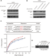
Phosphorylation of conserved Serine at the central linker region of Lgl2 is required for Dlg4/Lgl2 interaction. (A, B) GST pull-down-based assay of the binding of GST-tagged Dlg4 GK to various Lgl2 mutants. All Lgl2 mutants were co-expressed with Flag-tagged aPKC. (C) Fluorescence polarization-based measurement of the binding affinities of Dlg4 GK to the synthetic phosphor-Lgl2 peptides. The sequences of the peptides are shown at the top of the panel. (D) p-Lgl2 peptides block the binding of Lgl2 to Dlg4 GK. Equal amount of p-Lgl2 peptides (p-Lgl2a, p-Lgl2b and p-Lgl2c) were individually added into the mixture of HA-Lgl2 lysate and GST-Dlg4 GK.
Figure 3
Figure 3
The overall structures of Dlg4 GK in complex with the p-Lgl2a and p-Lgl2c peptides. (A, B) Ribbon diagram representation of the Dlg4 GK/p-Lgl2a complex (A), and the Dlg4 GK/p-Lgl2c complex (B). Phosphate group of each peptide is presented in the stick and ball model. (C, D) 2Fc-Fo simulated omit map of the p-Lgl2a peptide (C) and the p-Lgl2c peptide (D) in the complexes countered at the level of 1.0 σ. (E) Superposition of the structures of Dlg4 GK from GK/p-Lgl2a (deep green), GK/p-Lgl2c (light green) and the SH3-GK tandem (orange, PDB code: 1KJW).
Figure 4
Figure 4
Detailed interactions between Dlg4 and Lgl2. (A, B) The interaction interfaces between Dlg4 GK and p-Lgl2a (A), and p-Lgl2c (B). (C) Summary of the quantitative binding constants between various Dlg4 GK mutants and the p-Lgl2a peptide. All quantitative binding data were derived from fluorescence-based titration assays. (D) Comparison of the binding modes of p-Lgl2a, p-Lgl2c and p-LGN to Dlg GK. The structure-based sequence alignment of these three phosphor-peptides is shown below. (E) Structure-based sequence alignment of Dlg GK from different species (rDlg, rat Dlg; hDlg, human Dlg; dDlg, Drosophila Dlg; cDlg, C. elegans Dlg). In GK domains, the residues in the phosphor-site, the hydrophobic groove and the hydrophobic cradle are highlighted in yellow, green and blue, respectively. Residues involved in binding to p-LGN, p-Lgl2a and p-Lgl2c are annotated below as triangles, dots and stars, respectively.
Figure 5
Figure 5
Lgl2 3A mutant cannot rescue the tight junction formation defect caused by the loss of endogenous Lgl2 in MDCK cells. (A) Lgl2 3A mutant did not rescue the tight junction formation defects caused by loss of Lgl2 during a calcium switch. MDCK II cells with or without stable knockdown of Lgl2 were transfected with HA-tag only vector, HA-WT-Lgl2 or HA-Lgl2-3A. Cells were subjected to a calcium switch and stained for ZO-1 at 1 h after re-addition of calcium. (B) The expression of ectopic Lgl2 or Lgl2-3A in A are shown by immunofluorescence with the anit-HA antibody. (C) Quantification of percentage of consecutive ZO-1 staining in A. Error bars indicate SD values. *P < 0.001 when compared with the control.
Figure 6
Figure 6
A schematic model showing the phosphorylation-dependent recruitment of Lgl by basal-lateral localized Dlg. (A) Lgl contains three possible aPKC phosphorylation sites (S645, S649 and S653). Among the five possible phosphorylation patterns, Dlg GK can only bind to one phosphor-Ser, forming a 1:1 Dlg/Lgl complex. (B) In the unphosphorylated state, Lgl can form a complex with Par-6/aPKC at the apical cortex and inhibits the activity of aPKC. Once phosphorylated by aPKC, Lgl is released from the apical cell cortex. Phosphorylated Lgl is then recruited to the basal-lateral membranes via direct binding to Dlg, forming the basal-lateral Dlg/Lgl complex.

References

    1. Knoblich JA. Asymmetric cell division: recent developments and their implications for tumour biology. Nat Rev Mol Cell Biol. 2010;11:849–860. - PMC - PubMed
    1. St Johnston D, Ahringer J. Cell polarity in eggs and epithelia: parallels and diversity. Cell. 2010;141:757–774. - PubMed
    1. Bilder D. Epithelial polarity and proliferation control: links from the Drosophila neoplastic tumor suppressors. Genes Dev. 2004;18:1909–1925. - PubMed
    1. Humbert PO, Grzeschik NA, Brumby AM, Galea R, Elsum I, Richardson HE. Control of tumourigenesis by the Scribble/Dlg/Lgl polarity module. Oncogene. 2008;27:6888–6907. - PubMed
    1. Humbert PO, Dow LE, Russell SM. The Scribble and Par complexes in polarity and migration: friends or foes. Trends Cell Biol. 2006;16:622–630. - PubMed

Publication types

MeSH terms

LinkOut - more resources