. 2014 Mar 18;33(6):594-604.
doi: 10.1002/embj.201386523. Epub 2014 Feb 10.

Structural and mechanistic insights into MICU1 regulation of mitochondrial calcium uptake

Affiliations

Structural and mechanistic insights into MICU1 regulation of mitochondrial calcium uptake

Lele Wang et al. EMBO J. .

Abstract

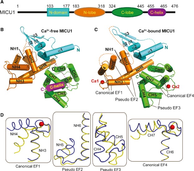
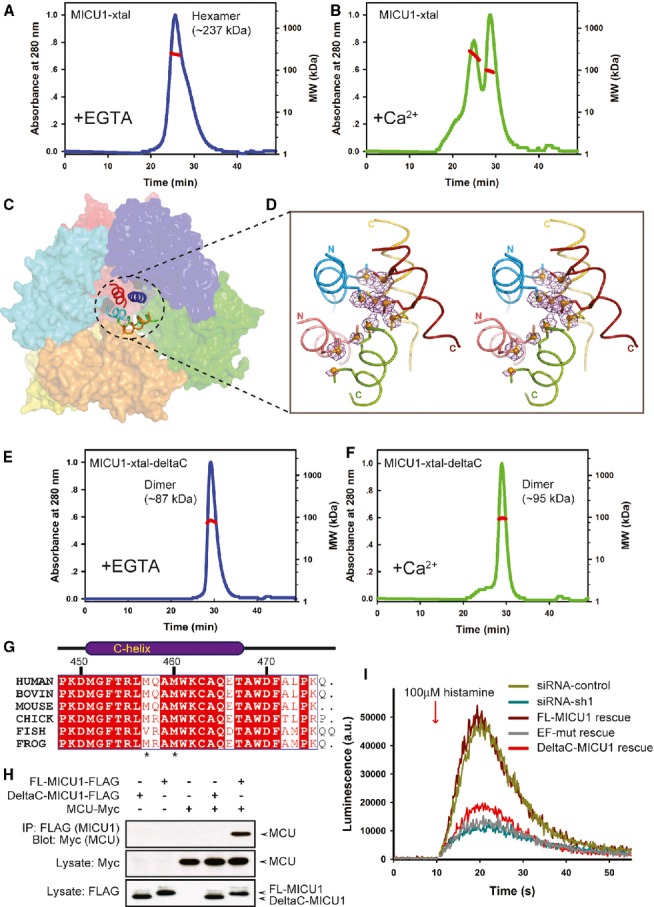
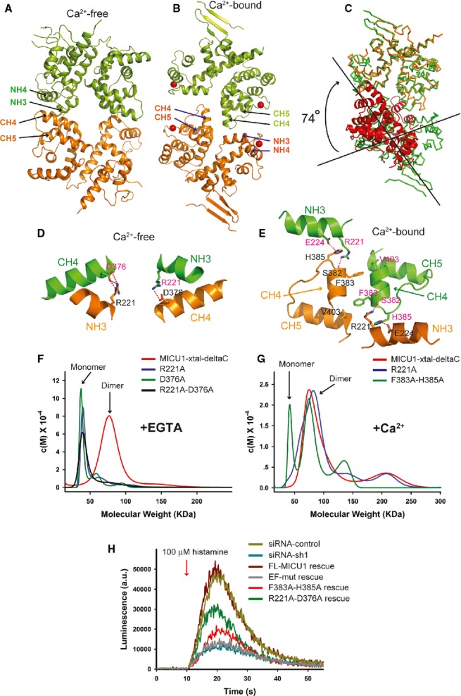
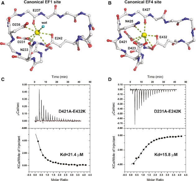
Mitochondrial calcium uptake is a critical event in various cellular activities. Two recently identified proteins, the mitochondrial Ca(2+) uniporter (MCU), which is the pore-forming subunit of a Ca(2+) channel, and mitochondrial calcium uptake 1 (MICU1), which is the regulator of MCU, are essential in this event. However, the molecular mechanism by which MICU1 regulates MCU remains elusive. In this study, we report the crystal structures of Ca(2+)-free and Ca(2+)-bound human MICU1. Our studies reveal that Ca(2+)-free MICU1 forms a hexamer that binds and inhibits MCU. Upon Ca(2+) binding, MICU1 undergoes large conformational changes, resulting in the formation of multiple oligomers to activate MCU. Furthermore, we demonstrate that the affinity of MICU1 for Ca(2+) is approximately 15-20 μM. Collectively, our results provide valuable details to decipher the molecular mechanism of MICU1 regulation of mitochondrial calcium uptake.


Figures

Figure 1
Figure 1
Crystal structures of Ca2+-free and Ca2+-bound human MICU1. A A schematic drawing of human MICU1. B, C Cartoon representation of the overall structure of MICU1 in the Ca2+-free and the Ca2+-bound state. The N-domain, N-lobe, C-lobe, C-helix, and Ca2+ are colored cyan, orange, green, purple, and red, respectively. D Structural comparisons of four helix-loop-helix structural units before and after Ca2+ binding. The Ca2+-free form is colored yellow, and the Ca2+-bound form is colored blue. The calcium ions are shown as red spheres.
Figure 2
Figure 2
The C-helix is critical for mitochondrial calcium uptake. A, B Multi-angle laser scattering analysis of MICU1-xtal in the absence or presence of Ca2+. MALS-determined molecular weights were calculated over prominent peaks (red line). C The hexameric packing of Ca2+-free MICU1. The surface map of six molecules in the asymmetric unit is shown. A cartoon model of the C-helix is presented in the center of the hexamer. D The anomalous scattering map of the selenium-substituted Ca2+-free MICU1-xtal structure. The map colored in magenta was contoured at 3.0σ. Cartoon representations of the five C-helices from different Ca2+-free MICU1 molecules are shown. One C-helix is not included in the final model due to poor electron density. The methionines are depicted as sticks, and Se atoms are depicted as spheres. E, F Multi-angle laser scattering analysis of MICU1-xtal-deltaC in the absence or presence of Ca2+. G Sequence alignment of the MICU1 protein from human (Q9BPX6), bovine (Q0IIL1), mouse (Q8VCX5), chick (E1BWC6), fish (A4IG32), and frog (B1H2N3). The residues that are conserved among the species are highlighted in red. The residue numbers of human MICU1 are shown in black. H Western blot analysis of co-immunoprecipitated wild-type FL-MICU1 and mutant DeltaC-MICU1 with the MCU channel in the absence of Ca2+. I cDNA rescue of the RNAi mitochondrial calcium uptake phenotype. Source data are available online for this figure.
Figure 3
Figure 3
The MICU1 dimer. A, B A cartoon representation of the MICU1 dimer in the absence or presence of Ca2+. Each molecule in one dimer is colored differently. Calcium is shown as a red sphere. C Superposition of Ca2+-free and Ca2+-bound MICU1. The Ca2+-bound MICU1 dimer is colored green. One molecule of Ca2+-free MICU1 (orange) superposed well with Ca2+-bound MICU1. However, the second molecule of Ca2+-free MICU1 (red) required a 74° rotation for superposition onto Ca2+-bound MICU1. D, E Detailed interactions within interfaces of Ca2+-free and Ca2+-bound MICU1 dimers. Each molecule in the dimer is colored green and orange, respectively. The oxygen and nitrogen atoms are colored red and blue, respectively. Hydrogen bonds are depicted as red dotted lines. F, G Sedimentation velocity analysis of the MICU1-xtal-deltaC wild-type and mutants in the absence or presence of Ca2+. H cDNA rescue of the RNAi mitochondrial calcium uptake phenotype.
Figure 4
Figure 4
MICU1 calcium-binding sites. A, B Detailed interactions of two calcium-binding sites of Ca2+-bound MICU1. Calcium ions are colored yellow. The oxygen and nitrogen atoms are colored red and blue, respectively. A water molecule is denoted as “wat.” The side chains of several residues are not shown for clarity. C, D The dissociation constants of MICU1-xtal-deltaC mutants (D421A-E432K and D231A-E242K) in the presence of calcium ions were determined using ITC.

References

    1. Adams PD, Afonine PV, Bunkoczi G, Chen VB, Davis IW, Echols N, Headd JJ, Hung LW, Kapral GJ, Grosse-Kunstleve RW, McCoy AJ, Moriarty NW, Oeffner R, Read RJ, Richardson DC, Richardson JS, Terwilliger TC, Zwart PH. PHENIX: a comprehensive Python-based system for macromolecular structure solution. Acta Crystallogr D Biol Crystallogr. 2010;66:213–221. - PMC - PubMed
    1. Aichberger KJ, Mittermann I, Reininger R, Seiberler S, Swoboda I, Spitzauer S, Kopp T, Stingl G, Sperr WR, Valent P, Repa A, Bohle B, Kraft D, Valenta R. Hom s 4, an IgE-reactive autoantigen belonging to a new subfamily of calcium-binding proteins, can induce Th cell type 1-mediated autoreactivity. J Immunol. 2005;175:1286–1294. - PubMed
    1. Alba de E, Tjandra N. Structural studies on the Ca2+-binding domain of human nucleobindin (calnuc) Biochemistry. 2004;43:10039–10049. - PubMed
    1. Babcock DF, Herrington J, Goodwin PC, Park YB, Hille B. Mitochondrial participation in the intracellular Ca2+ network. J Cell Biol. 1997;136:833–844. - PMC - PubMed
    1. Balaban RS. The role of Ca(2 + ) signaling in the coordination of mitochondrial ATP production with cardiac work. Biochim Biophys Acta. 2009;1787:1334–1341. - PMC - PubMed

Publication types

MeSH terms