

doi: 10.1534/genetics.114.161224.
Epub 2014 Feb 10.
Degradation of centromeric histone H3 variant Cse4 requires the Fpr3 peptidyl-prolyl Cis-Trans isomerase
Affiliations
- PMID: 24514906
- PMCID: PMC3982699
- DOI: 10.1534/genetics.114.161224
Item in Clipboard
Degradation of centromeric histone H3 variant Cse4 requires the Fpr3 peptidyl-prolyl Cis-Trans isomerase
Genetics.
2014 Apr.
Abstract
The centromeric histone H3 variant Cse4 in Saccharomyces cerevisiae is polyubiquitylated and degraded in a proteasome-dependent manner. We report here that the proline isomerase Fpr3 regulates Cse4 proteolysis. Structural change in Cse4 by Fpr3 might be important for the interaction between Cse4 and the E3 ubiquitin ligase Psh1.
Keywords: Cse4; Fpr3; Psh1; isomerization; protein degradation.
Figures
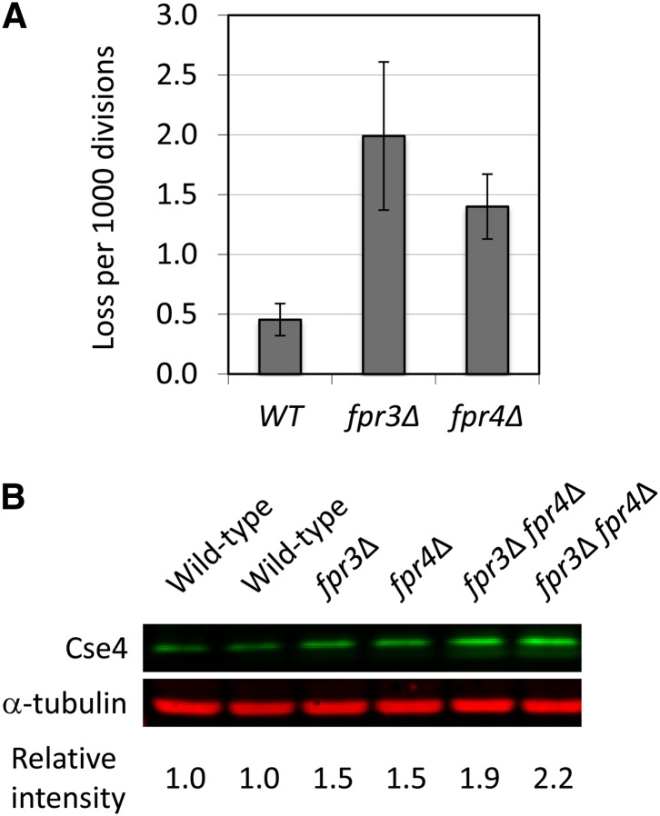
Fpr3 regulates Cse4 protein level in vivo. (A) The fpr3∆ or fpr4∆ strain displays a moderate chromosome missegregation phenotype. Chromosome loss rate in null mutants was determined by half sector analysis, as previously described (Ohkuni et al. 2008). Wild-type (Y14): 3 half-sectored colonies/6,421 total colonies; fpr3∆ (Y2249, Y2250, and Y2251): 17/8,598; and fpr4∆ (Y2252, Y2253, and Y2254): 14/10,105. P-value (chi-squared test): WT vs.
fpr3∆, 0.012; WT vs.
fpr4∆, 0.072. (B) Increased protein level of Cse4 in fpr∆ cells. Equal cell numbers of log phase cells, grown in SRaf medium, were visualized by Western blot analysis with anti-Cse4 and anti-α-tubulin antibodies. Cse4 and α-tubulin protein levels were overlayed and quantitated by the Odyssey Imaging System. Cse4 protein levels were normalized by the amount of α-tubulin. Isogenic yeast strains were wild type (YPH499 and YPH500), fpr3∆ (Y2243), fpr4∆ (Y2245), and fpr3∆ fpr4∆ (Y2247 and Y2248).
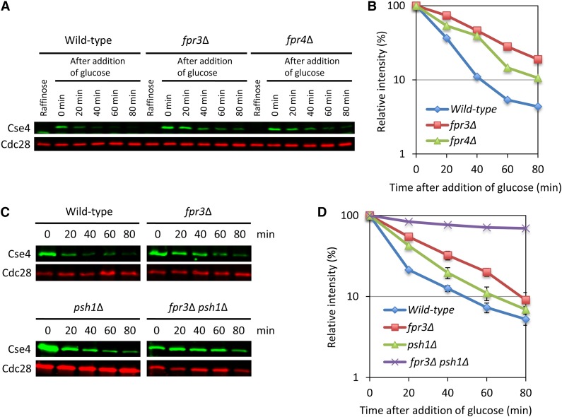
Deletion of FPR3 stabilizes Cse4 protein level in vivo. Cse4 was induced from a GAL1 promoter by the addition of galactose for 2 hr. Glucose was added and cells were collected at the time point. Equal cell numbers were visualized by Western blot analysis with anti-Cse4, or anti-Cdc28. We used the Odyssey Imaging System to detect and quantify the signals. (In detail, see File S1 ) (A and B) Isogenic yeast strains were wild type (Y2255), fpr3∆ (Y2256), and fpr4∆ (Y2257). (C and D) Isogenic yeast strains were wild type (Y2255), fpr3∆ (Y2256), psh1∆ (Y2258), and fpr3∆ psh1∆ (Y2340). Error bars represent SE of two independent experiments.
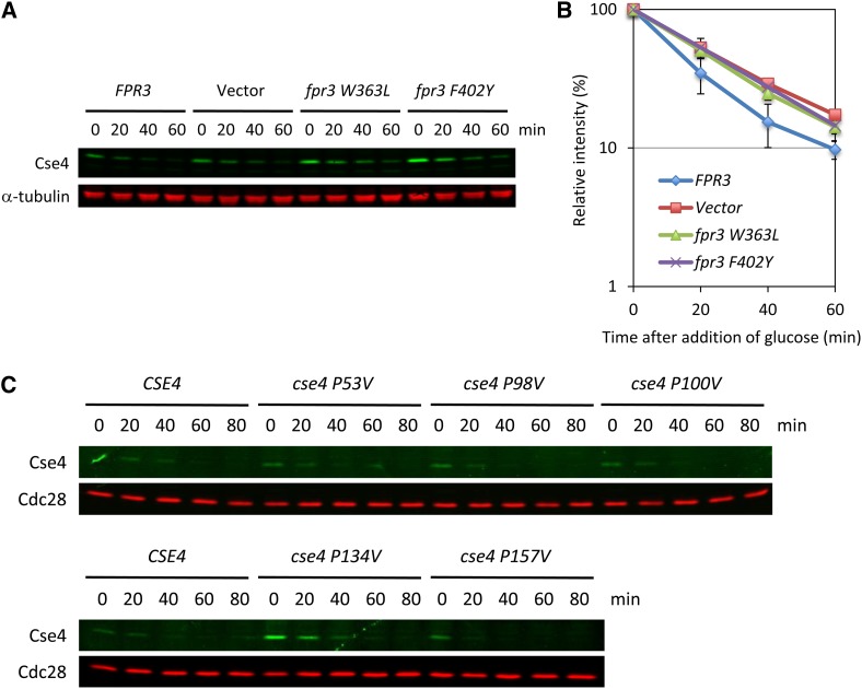
Fpr3 isomerization activity is necessary for the Cse4 proteolysis. (A and B) PPIase dead mutants stabilize the Cse4 protein level in vivo. We constructed plasmids harboring PPIase dead mutations (W363L and F402Y) (Table S2 ). The protein stability assay was performed as described in Figure 2. Isogenic yeast strains were FPR3 (Y2259), Vector (Y2260), fpr3 W363L (Y2261), and fpr3 F402Y (Y2262). Error bars represent SE of two or three independent experiments. (C) Mutation of P134 does stabilize the Cse4 protein level in vivo. There are five proline sites in Cse4 (P53, P98, P100, P134, and P157). We constructed plasmids harboring proline-to-valine mutation (Table S2 ). The protein stability assay was performed as described in Figure 2. Isogenic yeast strains were Cse4 (Y2255), cse4 P53V (Y2263), cse4 P98V (Y2264), cse4 P100V (Y2265), cse4 P134V (Y2266), and cse4 P157V (Y2267).
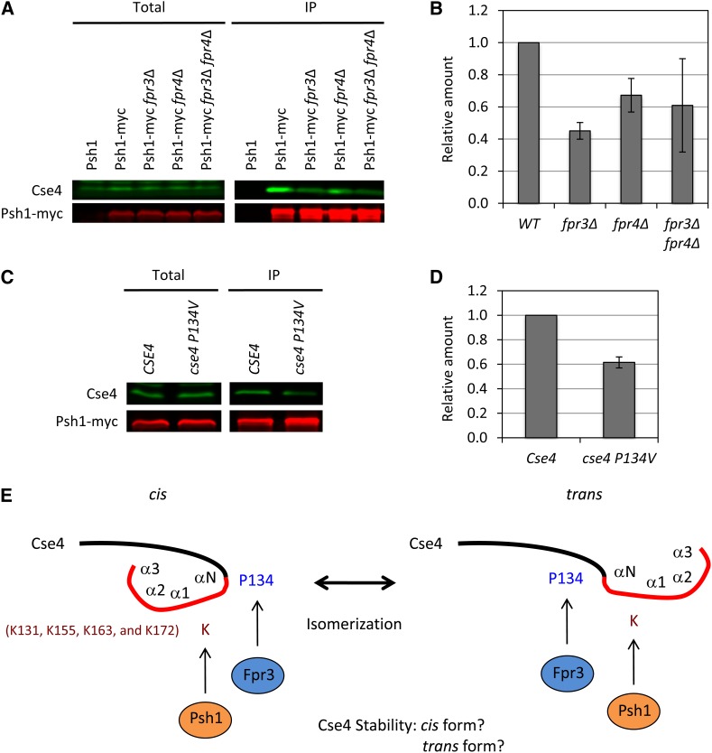
Fpr3 regulates the Cse4–Psh1 interaction. (A and B) Interaction between Cse4 and Psh1 was diminished in fpr3∆ cells. The indicated strains were grown to log phase, lysed, and anti-myc immunoprecipitations were performed as previously described (Ohkuni et al. 2008). Total and the immunoprecipitated fraction (IP) were subjected to SDS–PAGE, and Western blots were used to detect Cse4 and myc-tagged Psh1. We used the Odyssey Imaging System to detect and quantify the signals. Isogenic yeast strains were untagged (YPH500), Psh1–myc (Y2280), Psh1–myc fpr3∆ (Y2281), Psh1–myc fpr4∆ (Y2282), and Psh1–myc fpr3∆ fpr4∆ (Y2283). Error bars represent SE of two independent experiments. Significant difference, P = 0.0089 (WT vs.
fpr3∆). (C and D) P134V mutation in Cse4 diminishes the Psh1 interaction. Anti-myc immunoprecipitation assay and the quantification were performed as described in Figure 4, A and B. Isogenic yeast strains were Cse4 (Y2284) and cse4 P134V (Y2285). Error bars represent SE of two independent experiments. Significant difference, P = 0.0134. (E) A model for the role of Fpr3 in the Cse4 proteolysis. Psh1 is the E3 ubiquitin ligase that targets Cse4. Four lysine sites (K131, K155, K163, and K172) were ubiquitinated by Psh1. P134 close to the αN-helix (136–147) (Keith et al. 1999) might be the target of Fpr3 isomerization. We propose that the structural change in Cse4 from cis to trans or from trans to cis is important for the Cse4 degradation by Psh1. It is not known which form of Cse4 is ubiquitinated. The N-terminal domain (black) and the histone fold domain (red) of the α-N, α-1, α-2, and α-3 helices are indicated (Keith et al. 1999).
Similar articles
-
Molecular basis for the selective recognition and ubiquitination of centromeric histone H3 by yeast E3 ligase Psh1.J Genet Genomics. 2021 Jun 20;48(6):463-472. doi: 10.1016/j.jgg.2021.04.007. Epub 2021 May 26. J Genet Genomics. 2021. PMID: 34217622
-
Phosphorylation by casein kinase 2 facilitates Psh1 protein-assisted degradation of Cse4 protein.J Biol Chem. 2014 Oct 17;289(42):29297-309. doi: 10.1074/jbc.M114.580589. Epub 2014 Sep 2. J Biol Chem. 2014. PMID: 25183013 Free PMC article.
-
Psh1 is an E3 ubiquitin ligase that targets the centromeric histone variant Cse4.Mol Cell. 2010 Nov 12;40(3):444-54. doi: 10.1016/j.molcel.2010.10.014. Mol Cell. 2010. PMID: 21070970 Free PMC article.
-
Protein kinases in mitotic phosphorylation of budding yeast CENP-A.Curr Genet. 2019 Dec;65(6):1325-1332. doi: 10.1007/s00294-019-00997-5. Epub 2019 May 22. Curr Genet. 2019. PMID: 31119371 Review.
-
Dynamic ubiquitin signaling in cell cycle regulation.J Cell Biol. 2017 Aug 7;216(8):2259-2271. doi: 10.1083/jcb.201703170. Epub 2017 Jul 6. J Cell Biol. 2017. PMID: 28684425 Free PMC article. Review.
Cited by
-
Recent insights into mechanisms preventing ectopic centromere formation.Open Biol. 2021 Sep;11(9):210189. doi: 10.1098/rsob.210189. Epub 2021 Sep 8. Open Biol. 2021. PMID: 34493071 Free PMC article. Review.
-
Reduced gene dosage of histone H4 prevents CENP-A mislocalization and chromosomal instability in Saccharomyces cerevisiae.Genetics. 2021 May 17;218(1):iyab033. doi: 10.1093/genetics/iyab033. Genetics. 2021. PMID: 33751052 Free PMC article.
-
Conformational flexibility of histone variant CENP-ACse4 is regulated by histone H4: A mechanism to stabilize soluble Cse4.J Biol Chem. 2018 Dec 28;293(52):20273-20284. doi: 10.1074/jbc.RA118.004141. Epub 2018 Oct 31. J Biol Chem. 2018. PMID: 30381395 Free PMC article.
-
Cdc7-mediated phosphorylation of Cse4 regulates high-fidelity chromosome segregation in budding yeast.Mol Biol Cell. 2021 Nov 1;32(21):ar15. doi: 10.1091/mbc.E21-06-0323. Epub 2021 Aug 25. Mol Biol Cell. 2021. PMID: 34432494 Free PMC article.
-
CENP-A overexpression promotes aneuploidy with karyotypic heterogeneity.J Cell Biol. 2021 Apr 5;220(4):e202007195. doi: 10.1083/jcb.202007195. J Cell Biol. 2021. PMID: 33620383 Free PMC article.
References
-
- Arevalo-Rodriguez M., Wu X., Hanes S. D., Heitman J., 2004. Prolyl isomerases in yeast. Front. Biosci. 9: 2420–2446. - PubMed
-
- Collins S. R., Miller K. M., Maas N. L., Roguev A., Fillingham J., et al. , 2007. Functional dissection of protein complexes involved in yeast chromosome biology using a genetic interaction map. Nature 446: 806–810. - PubMed
Publication types
MeSH terms
Substances
Grants and funding
LinkOut - more resources
Full Text Sources
Other Literature Sources
Molecular Biology Databases
