. 2014 Jun;15(3):620-9.
doi: 10.1208/s12249-014-0086-y. Epub 2014 Feb 12.

Liposomal oxymatrine in hepatic fibrosis treatment: formulation, in vitro and in vivo assessment

Affiliations

Liposomal oxymatrine in hepatic fibrosis treatment: formulation, in vitro and in vivo assessment

Shujuan Zhang et al. AAPS PharmSciTech. 2014 Jun.

Abstract

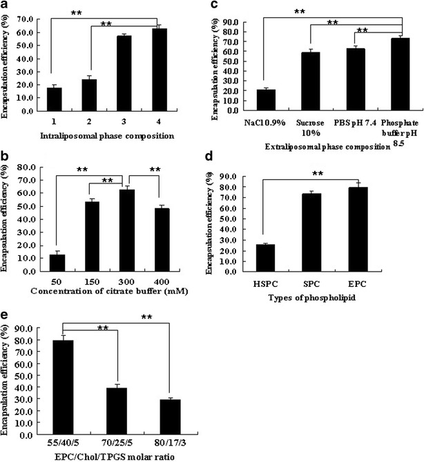
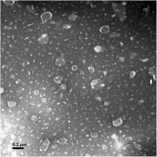
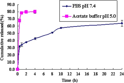
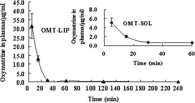
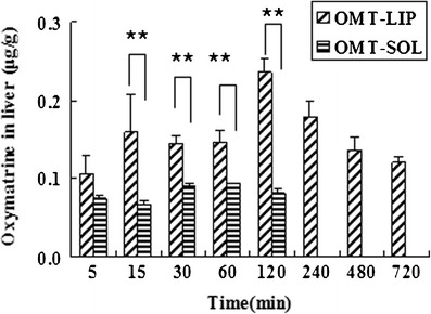
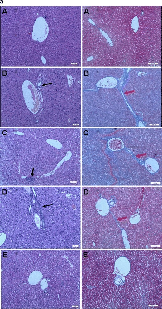
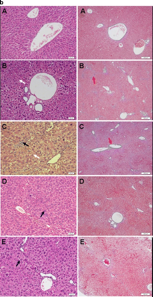
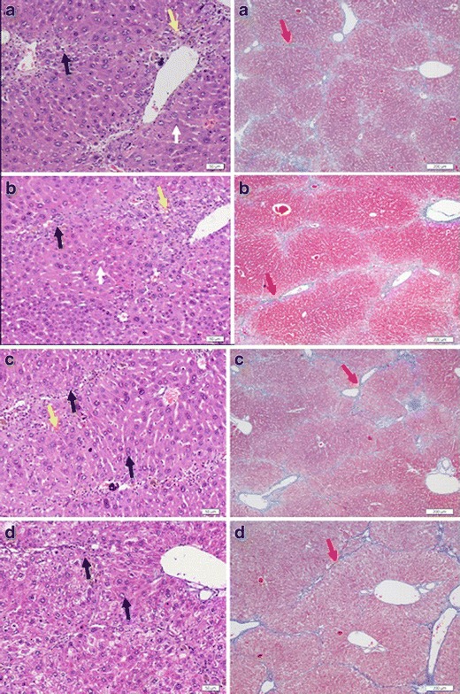
The aim was to develop a liposomal oxymatrine conjugating D-alpha tocopheryl polyethylene glycol 1000 succinate (OMT-LIP) for enhanced therapeutics of hepatic fibrosis. OMT-LIP was prepared using the remote loading method. The influences of formulation compositions on the encapsulation efficiency of OMT-LIP were investigated. Mean particle size, zeta potential, morphology, in vitro release, fibrotic liver targeting, and therapeutics of OMT-LIP were thoroughly assessed. The intraliposomal buffer composition and concentration, extraliposomal phase composition and pH, types of phospholipid, lipid molar ratio composition, and theoretical drug loading are crucial factors to entrap OMT into liposomes. The optimum OMT-LIP presented spherically unilamellar microstructures with entrapment efficiency of 79.7 ± 3.9%, mean particle size of 121.6 ± 52.9 nm, and zeta potential of -5.87 mV. OMT-LIP significantly increased the accumulation of OMT in the fibrotic liver with an 11.5-fold greater AUC than OMT solution in the dimethylnitrosamine (DMN)-induced hepatic fibrosis animals. OMT-LIP could be a potential strategy to improve treatment outcomes for hepatic fibrosis, showing the protective effects to mice given CCl4 and the enhanced therapeutics to mice with either DMN or CCl4-induced hepatic fibrosis.


Figures

Fig. 1
Fig. 1
Factors influencing the encapsulation efficiency of OMT-LIP using the remote loading method. a Intraliposomal phase composition with a concentration of 300 mM, pH 4.0 (1, (NH4)2SO4; 2, NH3–H3PO4; 3, NH3-citric buffer; 4, citrate buffer). b Intraliposomal citrate buffer concentrations. c Extraliposomal phase compositions. d Types of phospholipid. e EPC/Chol/TPGS molar ratio. **P < 0.05. Error bars refer to standard deviations (n = 3)
Fig. 2
Fig. 2
TEM image of OMT-LIP by negative staining with sodium phosphotungstate solution (×50,000)
Fig. 3
Fig. 3
In vitro release of OMT-LIP in PBS (pH 7.4) and acetate buffer (pH 5.0) media. Error bars refer to standard deviations (n = 3)
Fig. 4
Fig. 4
OMT plasma concentration versus time after intravenous injection of OMT-LIP to hepatic fibrosis mice in comparison with OMT-SOL. Error bars refer to standard deviations (n = 3)
Fig. 5
Fig. 5
Biodistribution of OMT-LIP in the fibrotic liver post-intravenous injection in comparison with OMT-SOL. **P < 0.05. At 240, 480, and 720 min, the OMT levels were below the detection limit in OMT-SOL. Error bars refer to standard deviations (n = 3)
Fig. 6
Fig. 6
Representative photographs of cross-sections of liver in mice with liver fibrosis induced by DMN (a) and in mice with liver fibrosis induced by CCl4 (b) (A, healthy group; B, saline control group; C, empty liposome group; D, OMT-SOL group; E, OMT-LIP group). Black arrow, inflammatory cells; white arrow, necrotic hepatocytes; blue arrow, swollen hepatocytes; yellow arrow, fatty degeneration; red arrow, deposited collagens. Left panel Hematoxylin–eosin staining. Bar, 50 μm. Right panel Masson’s trichrome staining. Bar, 200 μm
Fig. 6
Fig. 6
Representative photographs of cross-sections of liver in mice with liver fibrosis induced by DMN (a) and in mice with liver fibrosis induced by CCl4 (b) (A, healthy group; B, saline control group; C, empty liposome group; D, OMT-SOL group; E, OMT-LIP group). Black arrow, inflammatory cells; white arrow, necrotic hepatocytes; blue arrow, swollen hepatocytes; yellow arrow, fatty degeneration; red arrow, deposited collagens. Left panel Hematoxylin–eosin staining. Bar, 50 μm. Right panel Masson’s trichrome staining. Bar, 200 μm
Fig. 7
Fig. 7
Representative photographs of cross-sections of liver in the prophylactic study against liver fibrosis induced by CCl4: saline control group (a), empty liposome group (b), OMT-SOL group (c), OMT-LIP group (d). Black arrow, inflammatory cells; white arrow, necrotic hepatocytes; blue arrow, swollen hepatocytes; yellow arrow, fatty degeneration; red arrow, deposited collagens. Left panel Hematoxylin–eosin staining. Bar, 50 μm. Right panel Masson’s trichrome staining. Bar, 200 μm

References

    1. Iavarone M, Colombo M. HBV-related HCC, clinical issues and therapy. Dig Liver Dis. 2011;43(Suppl 1):S32–9. doi: 10.1016/S1590-8658(10)60690-1. - DOI - PubMed
    1. Pinzani M, Rosselli M, Zuckermann M. Liver cirrhosis. Best Pract Res Clin Gastroenterol. 2011;25:281–90. doi: 10.1016/j.bpg.2011.02.009. - DOI - PubMed
    1. Bonis PAL, Friedman SL, Kaplan MM. Is liver fibrosis reversible? N Engl J Med. 2001;344:452–4. doi: 10.1056/NEJM200102083440610. - DOI - PubMed
    1. Friedman SL. Cytokines and fibrogenesis. Semin Liver Dis. 1999;19:129–40. doi: 10.1055/s-2007-1007105. - DOI - PubMed
    1. Li D, Friedman SL. Liver fibrogenesis and the role of hepatic stellate cells: new insights and prospects for therapy. J Gastroenterol Hepatol. 1999;14:618–33. doi: 10.1046/j.1440-1746.1999.01928.x. - DOI - PubMed

Publication types

MeSH terms

LinkOut - more resources