. 2014 Feb 17;204(4):487-95.
doi: 10.1083/jcb.201308032. Epub 2014 Feb 10.

A bidirectional antagonism between aPKC and Yurt regulates epithelial cell polarity

Affiliations

A bidirectional antagonism between aPKC and Yurt regulates epithelial cell polarity

Clémence L Gamblin et al. J Cell Biol. .

Abstract

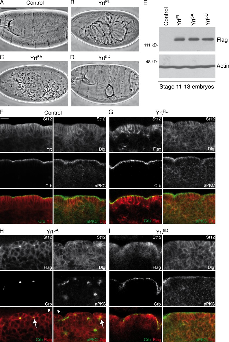
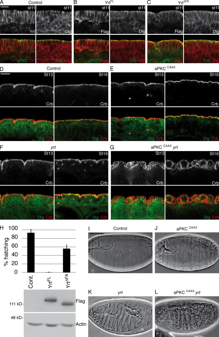
During epithelial cell polarization, Yurt (Yrt) is initially confined to the lateral membrane and supports the stability of this membrane domain by repressing the Crumbs-containing apical machinery. At late stages of embryogenesis, the apical recruitment of Yrt restricts the size of the apical membrane. However, the molecular basis sustaining the spatiotemporal dynamics of Yrt remains undefined. In this paper, we report that atypical protein kinase C (aPKC) phosphorylates Yrt to prevent its premature apical localization. A nonphosphorylatable version of Yrt dominantly dismantles the apical domain, showing that its aPKC-mediated exclusion is crucial for epithelial cell polarity. In return, Yrt counteracts aPKC functions to prevent apicalization of the plasma membrane. The ability of Yrt to bind and restrain aPKC signaling is central for its role in polarity, as removal of the aPKC binding site neutralizes Yrt activity. Thus, Yrt and aPKC are involved in a reciprocal antagonistic regulatory loop that contributes to segregation of distinct and mutually exclusive membrane domains in epithelial cells.


Figures

Figure 1.
Figure 1.
Yrt directly binds to aPKC via its FA domain. (A) Endogenous Yrt was immunoprecipitated from a wild-type embryo lysate (Yrt immunoprecipitation [IP]). Guinea pig IgG (IgG) purified from a nonimmune serum was used as a negative control. Western blot using anti-Yrt and anti-aPKC antibodies revealed that the immunoprecipitation was effective and that aPKC coprecipitated with Yrt. A portion of each homogenate was kept to monitor expression of Yrt and aPKC (input). (B and C) Upper portion of Fig. 1 B displays a schematic representation of the GST fusion proteins generated to investigate the Yrt–aPKC interaction. Full-length (FL) Yrt contains a FERM domain at its N terminus followed by a FA domain and a region with no defined domain referred to as the variable region (VR; Laprise et al., 2006). Numbers in brackets indicate the amino acids of Yrt comprised in each construct. GST pull-down experiments were performed on wild-type embryo lysates using these GST chimeric proteins. GST alone was used as a negative pull-down control. Pulled down aPKC was detected by Western blotting, and an anti-GST was used to control the amount of GST or GST fusion proteins used in each experiment. The asterisk indicates full-length Yrt. (D) Purified His-tagged aPKC and GST fused to the FA domain of Yrt (FA) were incubated together, and glutathione-coupled beads were used to pull down the protein complex. Western blotting detected aPKC and controlled the amount of GST or GST-FA used. F, FERM.
Figure 2.
Figure 2.
Yrt is a substrate of aPKC. (A) Control embryos (daughterless [da]-GAL4) or embryos overexpressing Par-6 and aPKCCAAX (da-GAL4/UAS–Par-6 and UAS-aPKCCAAX) were homogenized at different developmental stages. Samples were treated or not treated with the λ phosphatase (λ PPase) and processed for SDS-PAGE. Western blotting using anti-Yrt antibodies showed the migration profile of Yrt, whereas Actin was used as loading control. (B) Western blot showing the migration profile of Yrt extracted from control (wild type) or aPKC maternal and zygotic mutant embryos (we used the allele aPKCpsu265 that encodes a kinase inactive protein; Kim et al., 2009). Actin was used as a loading control. (C and D) Radioactive kinase assays in which purified aPKC was incubated with GST coupled to full-length Yrt (FL; C) or an extended version of the FA domain of Yrt (FA; aa 330–415) and a mutant version of it in which S348, S358, T379, S387, and S392 were mutagenized to A residues (FA5A; D). Proteins were separated on a polyacrylamide gel, which was exposed to monitor protein phosphorylation and then colored with Coomassie blue to control the amount of substrate used in each sample. (E) Anti–Par-6 antibodies were used to immunoprecipitate Par-6 from wild-type embryo extracts (stages 10–13; immunoprecipitate [IP] Par-6), whereas normal guinea pig IgG (IgG) was used as a negative control. GST or GST-FA was added to precipitate along with radiolabeled ATP in the absence or presence of PKCtide, which is a high affinity substrate of aPKC. Proteins were separated by SDS-PAGE, and the gel was exposed to monitor protein phosphorylation. Then, proteins were transferred on a membrane to validate immunoprecipitation of Par-6 and coimmunoprecipitation of aPKC and to monitor the amount of substrate used in each reaction. (F) GST pull-down experiments were performed on wild-type embryo lysates using GST-FA (FA), the nonphosphorylatable GST-FA5A (FA5A), or the phosphomimetic GST-FA5D (FA5D). GST alone was used as a negative control. Western blotting detected pulled down aPKC and monitored the amount of GST or GST fusion proteins used in each experiment. (G) Alignment of the FA domain of mouse Lulu2 and Drosophila Yrt. Numbers indicate amino acid positions in the Yrt sequence. Arrows point to amino acids previously shown to be phosphorylated by aPKC in Lulu2 (Nakajima and Tanoue, 2011). Three of these residues are conserved in Yrt (black rectangles). Black circles indicate phosphorylated residues identified by MS in the FA domain of Yrt. Three of these residues are conserved in Lulu2 (orange rectangles). As per ClustalW nomenclature (Larkin et al., 2007), asterisks indicate positions that have fully conserved residues. Colons designate conservation between groups of strongly similar properties, and periods indicate conservation between groups of weakly similar properties.
Figure 3.
Figure 3.
aPKC-dependent phosphorylation of Yrt is crucial for epithelial cell polarity. (A–D) Cuticle preparation of embryos of the following genotypes: wild type (A), da-GAL4/UAS-Flag-yrtFL (ubiquitous expression of Flag-tagged YrtFL; B), da-GAL4/UAS-Flag-yrt5A (C), and da-GAL4/UAS-Flag-yrt5D (D). Bar, 100 µm (also applies to B–D). (E) Western blot using an anti-Flag antibody showing that all constructs were expressed at similar levels. Actin was used as a loading control. (F) Portion of the ventral ectoderm of control (da-GAL4; driver line used to express Yrt constructs, this line has a wild-type phenotype) stage 12 (St12) embryos costained with Yrt and Crb (left) or with Dlg and aPKC (right). (G–I) Left images show costaining of Flag and Crb, whereas right images depict costaining of Dlg and aPKC in the ectoderm of an embryo expressing Flag-tagged YrtFL (G), an embryo expressing Flag-Yrt5A (H), or an embryo expressing Flag-Yrt5D (I). Arrows in H point to cysts of cells with contracted apexes, whereas arrowheads show cells with reduced Crb and aPKC levels. Bar, 10 µm (also applies to G–I).
Figure 4.
Figure 4.
Yrt limits aPKC-dependent apicalization of epithelial cells. (A) Portion of the ventral ectoderm of stage 11 control (da-GAL4) embryos stained for Yrt and aPKC (left) or Dlg and Crb (right). Bar, 10 µm (also applies to B and C). (B and C) Left images illustrate a portion of the ventral ectoderm costained with Flag and aPKC, whereas right images show a costaining of Dlg and Crb in stage 11 embryos expressing Flag-tagged YrtFL (B) or embryos expressing Flag-YrtΔFA (C). (D–G) Ventral ectoderm of stage 13 (St13) embryos (left) or ventral epidermis of stage 16 (St16) embryos (right) costained for Dlg and Crb. The embryonic genotypes were da-GAL4 (D), UAS-aPKCCAAX; da-GAL4 (ubiquitous expression of membrane-targeted aPKC; E), yrt75a (zygotic mutants; F), and UAS-aPKCCAAX; da-GAL4, yrt75a/yrt75a (G). Bar, 20 µm (also applies to E–G). (H) Histogram showing the hatching percentage of control (da-GAL4) embryos, embryos expressing YrtFL, or embryos expressing YrtΔFA. Error bars represent standard deviation. Below the histogram, a Western blot using an anti-Flag antibody shows that Flag-YrtFL and Flag-YrtΔFA are expressed at equivalent levels. Blotting for Actin validated equal loading. Cont., control. (I–L) Cuticle preparation of embryos of the following genotypes: da-GAL4 (I), UAS-aPKCCAAX; da-GAL4 (J), yrt75a (K), and UAS-aPKCCAAX; da-GAL4, yrt75a/yrt75a (L). Bar, 100 µm (also applies to J–L).

References

    1. Baines A.J. 2006. A FERM-adjacent (FA) region defines a subset of the 4.1 superfamily and is a potential regulator of FERM domain function. BMC Genomics. 7:85 10.1186/1471-2164-7-85 - DOI - PMC - PubMed
    1. Beausoleil S.A., Villén J., Gerber S.A., Rush J., Gygi S.P. 2006. A probability-based approach for high-throughput protein phosphorylation analysis and site localization. Nat. Biotechnol. 24:1285–1292 10.1038/nbt1240 - DOI - PubMed
    1. Betschinger J., Mechtler K., Knoblich J.A. 2003. The Par complex directs asymmetric cell division by phosphorylating the cytoskeletal protein Lgl. Nature. 422:326–330 10.1038/nature01486 - DOI - PubMed
    1. Bilder D., Li M., Perrimon N. 2000. Cooperative regulation of cell polarity and growth by Drosophila tumor suppressors. Science. 289:113–116 10.1126/science.289.5476.113 - DOI - PubMed
    1. Bisson N., James D.A., Ivosev G., Tate S.A., Bonner R., Taylor L., Pawson T. 2011. Selected reaction monitoring mass spectrometry reveals the dynamics of signaling through the GRB2 adaptor. Nat. Biotechnol. 29:653–658 10.1038/nbt.1905 - DOI - PubMed

Publication types

MeSH terms